. 2016 Jul 1;25(13):2712-2727.
doi: 10.1093/hmg/ddw129. Epub 2016 May 9.

The contribution of mutant GBA to the development of Parkinson disease in Drosophila

Affiliations

The contribution of mutant GBA to the development of Parkinson disease in Drosophila

Gali Maor et al. Hum Mol Genet. .

Abstract

Gaucher disease (GD) results from mutations in the acid β-glucocerebrosidase (GCase) encoding gene, GBA, which leads to accumulation of glucosylceramides. GD patients and carriers of GD mutations have a significantly higher propensity to develop Parkinson disease (PD) in comparison to the non-GD population. In this study, we used the fruit fly Drosophila melanogaster to show that development of PD in carriers of GD mutations results from the presence of mutant GBA alleles. Drosophila has two GBA orthologs (CG31148 and CG31414), each of which has a minos insertion, which creates C-terminal deletion in the encoded GCase. Flies double heterozygous for the endogenous mutant GBA orthologs presented Unfolded Protein Response (UPR) and developed parkinsonian signs, manifested by death of dopaminergic cells, defective locomotion and a shorter life span. We also established transgenic flies carrying the mutant human N370S, L444P and the 84GG variants. UPR activation and development of parkinsonian signs could be recapitulated in flies expressing these three mutant variants.UPR and parkinsonian signs could be partially rescued by growing the double heterozygous flies, or flies expressing the N370S or the L444P human mutant GCase variants, in the presence of the pharmacological chaperone ambroxol, which binds and removes mutant GCase from the endoplasmic reticulum (ER). However flies expressing the 84GG mutant, that does not express mature GCase, did not exhibit rescue by ambroxol. Our results strongly suggest that the presence of a mutant GBA allele in dopaminergic cells leads to ER stress and to their death, and contributes to development of PD.


Figures

Figure 1.
Figure 1.
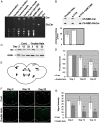
Decrease in amount of TH in flies double heterozygous for the fly GBA homologs. (A) TLC plate showing no GlcCer accumulation in heads and bodies of 22-day-old flies in comparison to its accumulation in type 2 GD fibroblasts or in RAW 264.7 mouse macrophages, both treated with 200 μM CBE for 10 days. (B) Activity assay using the synthetic fluorescent GCase substrate C6-NBD showing activity level of GCase in double heterozygous flies in comparison to control flies. For a positive control, control flies were treated with 3 μM of the GCase inhibitor CBE. (C) Protein lysates, prepared from heads of 10 w1118 (cont.) or double heterozygous flies (double hets.) at days 2, 12 and 22 post-eclosion, were subjected to western blotting. The corresponding blots were interacted with anti TH antibody. As a loading control, the blots were interacted with anti-actin antibody. (D) Intensities of the corresponding bands were quantified by densitometry and the value obtained for control flies was considered 1. Results represent the mean ± SEM of four independent experiments. (E) Schematic representation of dopaminergic neuronal clusters in the posterior region of the Drosophila brain. (F) Representative confocal images of brains that were isolated from w1118 (cont.) or double heterozygous (double hets.) flies after staining with anti-TH antibody, at days 2, 12 and 22 post-eclosion. G. Quantification of signal intensity obtained from brains at 2, 12 and 22 days post-eclosion. Results represent the mean ± SEM of 15 brains (n = 15). **P < 0.01.
Figure 2.
Figure 2.
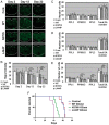
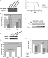
Degeneration of dopaminergic neurons and decreased survival in flies double heterozygous for the fly GBA homologs. Quantitative representation of the average number of cells in dopaminergic clusters in the brains at days 2 (A), 12 (B) or 22 (C) post-eclosion. Results represent the mean ± SEM of 15 brains. (D) Kaplan Meier curve showing the overall survival rates of control and double heterozygote flies tested on 100 flies. Flies were grown as 10 flies per vial and were transferred to fresh food every other day. ***P < 0.005.
Figure 3.
Figure 3.
Parkinsonian signs in transgenic flies expressing mutant versions of human GCase. (A) Protein lysates were prepared from 10 flies expressing the human UAS-mycHisWTGCase or human UAS-mycHisN370SGCase, driven by daughterless (Da) - GAL4. Samples, containing 100 μg of protein, were subjected to overnight Endo-H digestion, after which they were electrophoresed through SDS-PAGE and the corresponding blot was interacted with anti-GCase and anti-actin antibodies. lys-lysosomal, endo-H resistant, fraction. (B) Confocal images of flies expressing mycHis WT, N370S or L444P human GCase transgenes under the Ddc- GAL4 driver. Brains were stained at day 2 using anti-TH antibody (green) and anti-myc-antibody (red). The images show co-expression of TH and GCase in one confocal plane. (C) Protein lysates were prepared from heads of 10 control flies, or 10 flies expressing normal human GCase variants, at days 2, 12 and 22 days post-eclosion, and were subjected to western blotting. The corresponding blots were interacted with anti-TH antibody. As a loading control, the blots were interacted with anti-actin antibody. (D) Intensities of the corresponding bands were quantified by densitometry and the value obtained for control flies was considered 1. Results represent the mean ± SEM of four independent experiments. **P < 0.01.
Figure 4.
Figure 4.
Degeneration of dopaminergic neurons and decreased survival in transgenic flies expressing mutant versions of human GCase. (A) Representative confocal images of brains isolated from control flies (w118, cont.) or flies expressing the different human GCase transgenes, following their staining with anti-TH antibody, at days 2, 12 and 22 post-eclosion. (B) Quantification of signal intensity obtained from the tested brains described in (A). Quantitative representation of the average number of cells in dopaminergic clusters at days 2 (C), 12 (D) or 22 (E) post-eclosion. Results represent the mean ± SEM of 15 brains. (F) Kaplan Meier curve showing the overall survival rates of control (Ddc-GAL4), or flies expressing wild-type, N370S or L444P human GCase flies tested on 100 flies. Flies were grown as 10 flies per vial and were transferred to fresh food. ***P < 0.005.
Figure 5.
Figure 5.
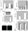
Rescue of the parkinsonian phenotype in flies double heterozygous for the fly GBA homologs. (A) RNA was isolated from ambroxol treated or untreated double heterozygous flies, and the cDNA prepared from it was subjected to quantitative RT-PCR with primers specific for Drosophila Hsc-70-3 or for the spliced form of Drosophila Xbp1. The results (three different experiments) were quantified, and the values obtained for untreated flies were considered 1. RP49 was used as a normalizing control. (B) Protein lysates prepared from heads of ten double heterozygous flies, treated or untreated with 1 mM ambroxol for 22 days, were subjected to western blotting and interaction with anti-TH antibody. As a loading control, the blots were interacted with anti-actin antibody. (C) Intensities of the corresponding bands were quantified by densitometry and the value obtained for untreated flies was considered 1. Results represent the mean ± SEM of four independent experiments. (D) Representative confocal images of adult brains from treated and untreated double heterozygous flies, stained with anti-TH antibody at day 22 post-eclosion. (E) Quantitative representation of the average number of cells in dopaminergic clusters. Results represent the mean ± SEM of 15 brains. (F) Quantification of signal intensity obtained from the brains described in (D). Results represent the mean ± SEM of 15 brains. (G) Five vials, each containing 10 double heterozygous flies were analyzed for locomotion behavior at days 2, 12 and 22 post-eclosion. *P < 0.05; **P < 0.01; ***P < 0.05.
Figure 6.
Figure 6.
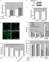
Rescue of parkinsonian phenotype in transgenic flies expressing mutant human GCase. (A) RNA was isolated from ambroxol treated or untreated transgenic flies, expressing the normal or the N370S or L444P human GCase variants, and the cDNA prepared from it was subjected to quantitative RT-PCR with primers specific for Drosophila Hsc-70-3 or for the spliced form of Drosophila Xbp1. The results (three different experiments) were quantified, and the values obtained for untreated flies were considered 1. RP49 was used as a normalizing control. (B) Lysates prepared from heads of transgenic flies, treated or untreated with 1mM ambroxol, were analyzed by western blotting, as described in the legend to Figure 5. (C) Intensities of the corresponding bands were quantified by densitometry and the value obtained for untreated flies was considered 1. Results represent the mean ± SEM of four independent experiments. (D) Representative confocal images of adult brains of transgenic flies, expressing the human normal (mycHisWTGCase), the N370S (mycHisN370SGCase) or the L444P (mycHisL444PGCase) variants, untreated or ambroxol treated, following their staining with anti-TH antibody at day 22 post-eclosion. (E) Quantitative representation of the average number of cells in dopaminergic clusters. Results represent the mean ± SEM of 15 brains. (F) Quantification of TH signal intensity (Signal intensity) obtained from tested brains at day 22 post-eclosion. Results represent the mean ± SEM of 15 brains. (G) Flies expressing wild-type, N370S or L444P human GCase transgenes in their dopaminergic cells were treated with 1mM ambroxol and analyzed for climbing ability. **P < 0.01.
Figure 7.
Figure 7.
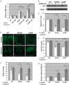
Activation of the UPR pathways and development of parkinsonian signs in 84GG transgenic flies. (A) RNA was isolated from Ddc-GAL4 or from flies expressing the human wild-type or the 84GG GBA transgenes under the Ddc-GAL4 driver. cDNA was prepared and subjected to PCR using primers specific for an 1200 bp fragment of the human GBA mRNA. Primers for the CG31414 Drosophila gene were used as a DNA loading control. Products were separated through a 1% agarose gel. (B) RNA was isolated from the above-mentioned flies, and the cDNA prepared from it was subjected to quantitative RT-PCR with the appropriate primers. RP49 was used as a normalizing control. The results (three different experiments) were quantified and the values obtained for flies expressing normal human GCase were considered 1. (C) Protein lysates, prepared from Da-GAL4 (Cont.), or flies expressing WT and 84GG human GCase using the Da-GAL4 driver, were subjected to western blotting and interaction with anti-phosphorylated eIF2α antibodies (p-eIF2α). As a loading control, the blot was interacted with anti-IF2α antibodies. (D) The results (three different experiments) were quantified and the values obtained for control flies were considered 1. (E) Kaplan Meier curve showing the overall survival rates of control (Ddc-GAL4), or flies expressing wild-type, or 84GG human GCase tested on 100 flies. Flies were grown as 10 flies per vials and were transferred to fresh food every other day. (F) Protein lysates were prepared from heads of 10 transgenic flies with the genotypes: Ddc-GAL4; wild-type GCase (cont.) or Ddc-GAL4; 84GG GCase, at days 2, 12 and 22 post-eclosion. They were subjected to western blotting and the corresponding blots were interacted with anti-TH antibody. As a loading control, the blots were interacted with anti-actin antibody. (G) Intensities of the corresponding bands were quantified by densitometry and the value obtained for control flies was considered 1. Results represent the mean ± SEM of four independent experiments. **P < 0.01.
Figure 8.
Figure 8.
Development of parkinsonian signs in 84GG transgenic flies. (A) Transcription levels of Drosophila Hsc-70-3 or of the spliced form of Drosophila Xbp1 in ambroxol treated or untreated flies, expressing the human 84GG GBA transgene, were analyzed by quantitative RT PCR as described in the legend to Figure 5. The results (three different experiments) were quantified, and the values obtained for untreated flies were considered 1. RP49 was used as a normalizing control. (B) Protein lysates prepared from heads of ten Ddc-GAL4 flies or flies expressing the 84GG human GBA transgene, treated or untreated with 1 mM ambroxol for 22 days, were subjected to western blotting and interaction with anti-TH antibody. As a loading control, the blots were interacted with anti-actin antibody. (C) Intensities of the corresponding bands were quantified by densitometry and the value obtained for untreated flies was considered 1. Results represent the mean ± SEM of four independent experiments. (D) Representative confocal images of the posterior region of brains isolated from Ddc-GAL4 flies or flies expressing the human 84GG GBA transgene, stained with anti-TH antibody at days 2, 12, 22 post-eclosion and treatment with 1 mM ambroxol. (E) Quantitative representation of the average number of cells in dopaminergic clusters. Results represent the mean ± SEM of 15 brains. (F) Quantification of TH signal intensity obtained from tested brains at days 2, 12, 22 post-eclosion and treatment with 1 mM ambroxol. Results represent the mean ± SEM of 15 brains. (G) Five vials, each containing 10 control flies or control, or flies expressing the wild-type or the 84GG human GBA transgenes were analyzed for locomotion behavior. Climbing ability was tested at days 2, 12 and 22 post-eclosion. For each fly line, locomotion at day 2 was considered 100%. **P < .01, ***P < 0.005.

Similar articles

Cited by

References

    1. Beutler E. (1980) Gaucher’s disease. Compr Ther, 6, 65–68. - PubMed
    1. Beutler E. (1999) Gaucher disease. Arch. Intern. Med., 159, 881–882. - PubMed
    1. Brady R.O., Kanfer J.N., Shapiro D. (1965) Metabolism of Glucocerebrosides. Ii. Evidence of an Enzymatic Deficiency in Gaucher's Disease. Biochem. Biophys. Res. Commun., 18, 221–225. - PubMed
    1. Hruska K.S., LaMarca M.E., Scott C.R., Sidransky E. (2008) Gaucher disease: mutation and polymorphism spectrum in the glucocerebrosidase gene (GBA). Hum. Mutat., 29, 567–583. - PubMed
    1. Tsuji S., Martin B.M., Barranger J.A., Stubblefield B.K., LaMarca M.E., Ginns E.I. (1988) Genetic heterogeneity in type 1 Gaucher disease: multiple genotypes in Ashkenazic and non-Ashkenazic individuals. Proc. Natl. Acad. Sci. U S A, 85, 2349–2352. - PMC - PubMed

Publication types

LinkOut - more resources