

Three-dimensional models for studying development and disease: moving on from organisms to organs-on-a-chip and organoids
- PMID: 27156572
- PMCID: PMC4905804
- DOI: 10.1039/c6ib00039h
Three-dimensional models for studying development and disease: moving on from organisms to organs-on-a-chip and organoids
Abstract
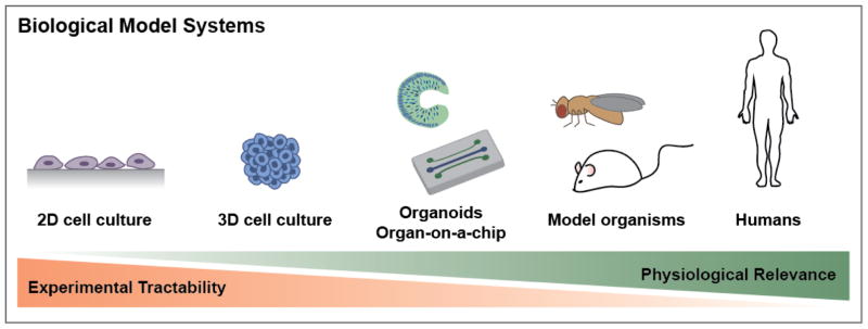
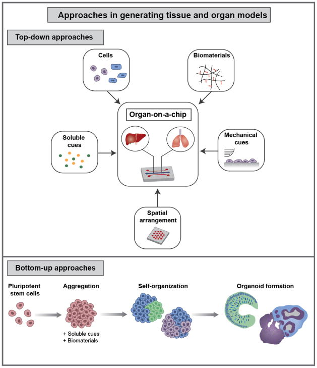
Human development and disease are challenging to study because of lack of experimental accessibility to in vivo systems and the complex nature of biological processes. For these reasons researchers turn to the use of model systems, ranging in complexity and scale from single cells to model organisms. While the use of model organisms is valuable for studying physiology and pathophysiology in an in vivo context and for aiding pre-clinical development of therapeutics, animal models are costly, difficult to interrogate, and not always equivalent to human biology. For these reasons, three-dimensional (3D) cell cultures have emerged as an attractive model system that contains key aspects of in vivo tissue and organ complexity while being more experimentally tractable than model organisms. In particular, organ-on-a-chip and organoid models represent orthogonal approaches that have been able to recapitulate characteristics of physiology and disease. Here, we review advances in these two categories of 3D cultures and applications in studying development and disease. Additionally, we discuss development of key technologies that facilitate the generation of 3D cultures, including microfluidics, biomaterials, genome editing, and imaging technologies.
Figures
Similar articles
-
Patient-Specific Organoid and Organ-on-a-Chip: 3D Cell-Culture Meets 3D Printing and Numerical Simulation.Adv Biol (Weinh). 2021 Jun;5(6):e2000024. doi: 10.1002/adbi.202000024. Epub 2021 Apr 15. Adv Biol (Weinh). 2021. PMID: 33856745 Free PMC article. Review.
-
Developmentally inspired human 'organs on chips'.Development. 2018 May 18;145(16):dev156125. doi: 10.1242/dev.156125. Development. 2018. PMID: 29776965 Free PMC article.
-
3D culture models for studying branching morphogenesis in the mammary gland and mammalian lung.Biomaterials. 2019 Apr;198:135-145. doi: 10.1016/j.biomaterials.2018.08.043. Epub 2018 Aug 23. Biomaterials. 2019. PMID: 30174198 Free PMC article. Review.
-
A brief history of organoids.Am J Physiol Cell Physiol. 2020 Jul 1;319(1):C151-C165. doi: 10.1152/ajpcell.00120.2020. Epub 2020 May 27. Am J Physiol Cell Physiol. 2020. PMID: 32459504 Free PMC article. Review.
-
A closed 3D printed microfluidic device for automated growth and differentiation of cerebral organoids from single-cell suspension.Biotechnol J. 2024 Aug;19(8):e2400240. doi: 10.1002/biot.202400240. Biotechnol J. 2024. PMID: 39212189
Cited by
-
Tackling Current Biomedical Challenges With Frontier Biofabrication and Organ-On-A-Chip Technologies.Front Bioeng Biotechnol. 2021 Sep 16;9:732130. doi: 10.3389/fbioe.2021.732130. eCollection 2021. Front Bioeng Biotechnol. 2021. PMID: 34604190 Free PMC article. Review.
-
Electroactive 4D Porous Scaffold Based on Conducting Polymer as a Responsive and Dynamic In Vitro Cell Culture Platform.ACS Appl Mater Interfaces. 2024 Feb 7;16(5):5613-5626. doi: 10.1021/acsami.3c16686. Epub 2024 Jan 26. ACS Appl Mater Interfaces. 2024. PMID: 38278772 Free PMC article.
-
Generation of Pancreatic Ductal Organoids and Whole-Mount Immunostaining of Intact Organoids.Curr Protoc Cell Biol. 2019 Jun;83(1):e82. doi: 10.1002/cpcb.82. Epub 2018 Dec 12. Curr Protoc Cell Biol. 2019. PMID: 30548444 Free PMC article.
-
Pannexin1 Channel-Mediated Inflammation in Acute Ischemic Stroke.Aging Dis. 2024 May 7;15(3):1296-1307. doi: 10.14336/AD.2023.0303. Aging Dis. 2024. PMID: 37196132 Free PMC article. Review.
-
Biomedical Applications of Microfluidic Devices: A Review.Biosensors (Basel). 2022 Nov 16;12(11):1023. doi: 10.3390/bios12111023. Biosensors (Basel). 2022. PMID: 36421141 Free PMC article. Review.
References
-
- Elsea SH, Lucas RE. ILAR journal/National Research Council, Institute of Laboratory Animal Resources. 2002;43:66–79. - PubMed
Publication types
MeSH terms
Grants and funding
LinkOut - more resources
Full Text Sources
Other Literature Sources
