

Tracking Transitions in Spider Wrapping Silk Conformation and Dynamics by (19)F Nuclear Magnetic Resonance Spectroscopy
- PMID: 27153372
- PMCID: PMC5770200
- DOI: 10.1021/acs.biochem.6b00429
Tracking Transitions in Spider Wrapping Silk Conformation and Dynamics by (19)F Nuclear Magnetic Resonance Spectroscopy
Abstract
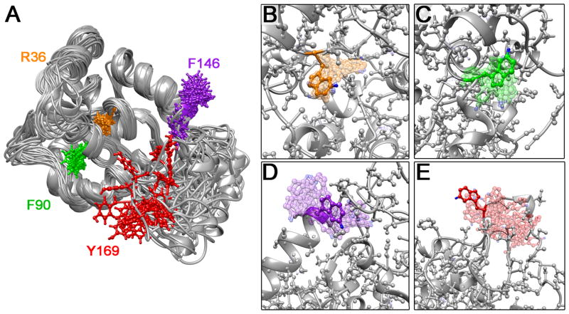
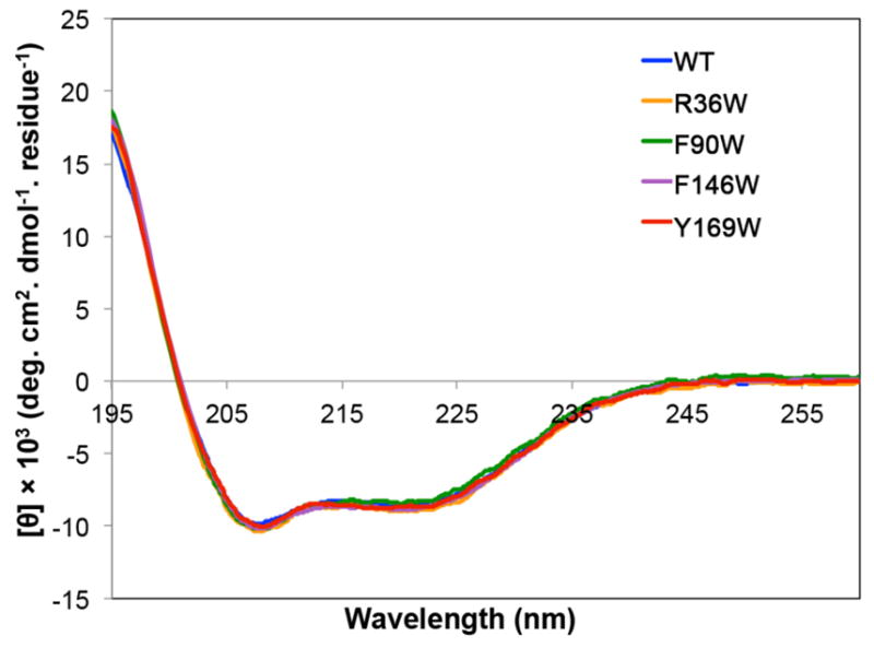
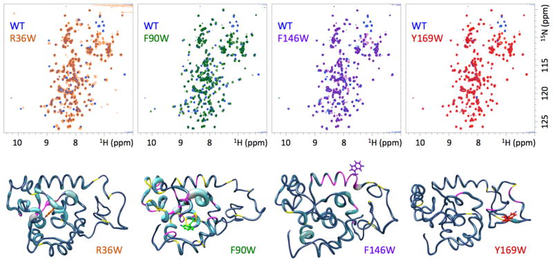
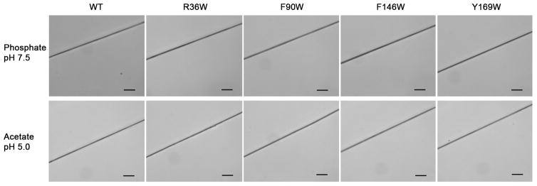
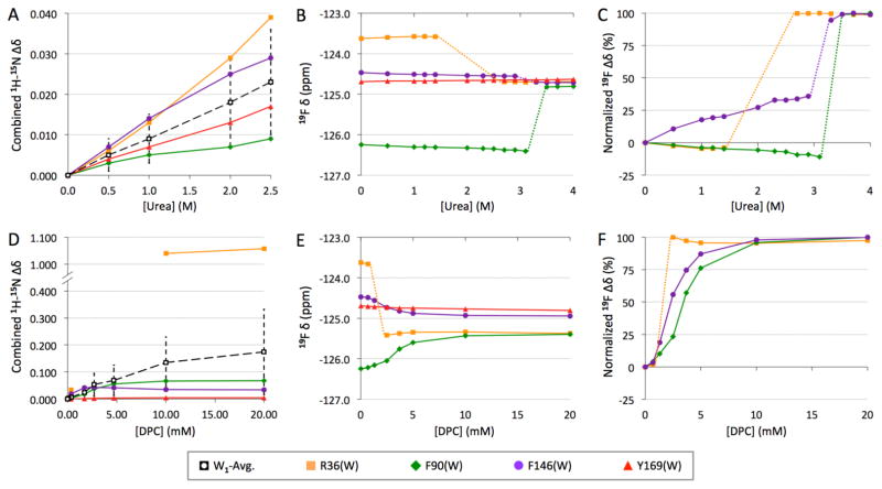
Aciniform silk protein (AcSp1) is the primary component of wrapping silk, the toughest of the spider silks because of a combination of high tensile strength and extensibility. Argiope trifasciata AcSp1 contains a core repetitive domain with at least 14 homogeneous 200-amino acid units ("W" units). Upon fibrillogenesis, AcSp1 converts from an α-helix-rich soluble state to a mixed α-helical/β-sheet conformation. Solution-state nuclear magnetic resonance (NMR) spectroscopy allowed demonstration of variable local stability within the W unit, but comprehensive characterization was confounded by spectral overlap, which was exacerbated by decreased chemical shift dispersion upon denaturation. Here, (19)F NMR spectroscopy, in the context of a single W unit (W1), is applied to track changes in structure and dynamics. Four strategic positions in the W unit were mutated to tryptophan and biosynthetically labeled with 5-fluorotryptophan (5F-Trp). Simulated annealing-based structure calculations implied that these substitutions should be tolerated, while circular dichroism (CD) spectroscopy and (1)H-(15)N chemical shift displacements indicated minimal structural perturbation in W1 mutants. Fiber formation by W2 concatemers containing 5F-Trp substitutions in both W units demonstrated retention of functionality, a somewhat surprising finding in light of sequence conservation between species. Each 5F-Trp-labeled W1 exhibited a unique (19)F chemical shift, line width, longitudinal relaxation time constant (T1), and solvent isotope shift. Perturbation to (19)F chemical shift and nuclear spin relaxation parameters reflected changes in the conformation and dynamics at each 5F-Trp site upon addition of urea and dodecylphosphocholine (DPC). (19)F NMR spectroscopy allowed unambiguous localized tracking throughout titration with each perturbant, demonstrating distinct behavior for each perturbant not previously revealed by heteronuclear NMR experiments.
Figures

Similar articles
-
Characterizing Aciniform Silk Repetitive Domain Backbone Dynamics and Hydrodynamic Modularity.Int J Mol Sci. 2016 Aug 10;17(8):1305. doi: 10.3390/ijms17081305. Int J Mol Sci. 2016. PMID: 27517921 Free PMC article.
-
Spider wrapping silk fibre architecture arising from its modular soluble protein precursor.Sci Rep. 2015 Jun 26;5:11502. doi: 10.1038/srep11502. Sci Rep. 2015. PMID: 26112753 Free PMC article.
-
Nanoparticle self-assembly by a highly stable recombinant spider wrapping silk protein subunit.FEBS Lett. 2013 Oct 1;587(19):3273-80. doi: 10.1016/j.febslet.2013.08.024. Epub 2013 Aug 28. FEBS Lett. 2013. PMID: 23994530
-
Structure and Dynamics of Spider Silk Studied with Solid-State Nuclear Magnetic Resonance and Molecular Dynamics Simulation.Molecules. 2020 Jun 5;25(11):2634. doi: 10.3390/molecules25112634. Molecules. 2020. PMID: 32517041 Free PMC article. Review.
-
Structure of Silk I (Bombyx mori Silk Fibroin before Spinning) -Type II β-Turn, Not α-Helix.Molecules. 2021 Jun 17;26(12):3706. doi: 10.3390/molecules26123706. Molecules. 2021. PMID: 34204550 Free PMC article. Review.
Cited by
-
Observing enzyme ternary transition state analogue complexes by 19F NMR spectroscopy.Chem Sci. 2017 Dec 1;8(12):8427-8434. doi: 10.1039/c7sc04204c. Epub 2017 Oct 23. Chem Sci. 2017. PMID: 29619190 Free PMC article.
-
Identification of Wet-Spinning and Post-Spin Stretching Methods Amenable to Recombinant Spider Aciniform Silk.Biomacromolecules. 2016 Aug 8;17(8):2737-46. doi: 10.1021/acs.biomac.6b00857. Epub 2016 Jul 20. Biomacromolecules. 2016. PMID: 27387592 Free PMC article.
-
Characterizing Aciniform Silk Repetitive Domain Backbone Dynamics and Hydrodynamic Modularity.Int J Mol Sci. 2016 Aug 10;17(8):1305. doi: 10.3390/ijms17081305. Int J Mol Sci. 2016. PMID: 27517921 Free PMC article.
-
Assessing the applicability of 19F labeled tryptophan residues to quantify protein dynamics.J Biomol NMR. 2023 Apr;77(1-2):55-67. doi: 10.1007/s10858-022-00411-2. Epub 2023 Jan 14. J Biomol NMR. 2023. PMID: 36639431 Free PMC article.
-
Fluorotryptophan Incorporation Modulates the Structure and Stability of Transthyretin in a Site-Specific Manner.Biochemistry. 2017 Oct 17;56(41):5570-5581. doi: 10.1021/acs.biochem.7b00815. Epub 2017 Sep 28. Biochemistry. 2017. PMID: 28920433 Free PMC article.
References
-
- Gosline JM, Guerette PA, Ortlepp CS, Savage KN. The mechanical design of spider silks: From fibroin sequence to mechanical function. J Exp Biol. 1999;202:3295–3303. - PubMed
-
- Vollrath F, Porter D. Spider silk as a model biomaterial. Appl Phys A Mater Sci. 2006;82:205–212.
Publication types
MeSH terms
Substances
Grants and funding
LinkOut - more resources
Full Text Sources
Other Literature Sources
