. 2016 May 1;57(6):2509-21.
doi: 10.1167/iovs.15-18680.

Calpain-5 Expression in the Retina Localizes to Photoreceptor Synapses

Affiliations

Calpain-5 Expression in the Retina Localizes to Photoreceptor Synapses

Kellie A Schaefer et al. Invest Ophthalmol Vis Sci. .

Abstract

Purpose: We characterize calpain-5 (CAPN5) expression in retinal and neuronal subcellular compartments.

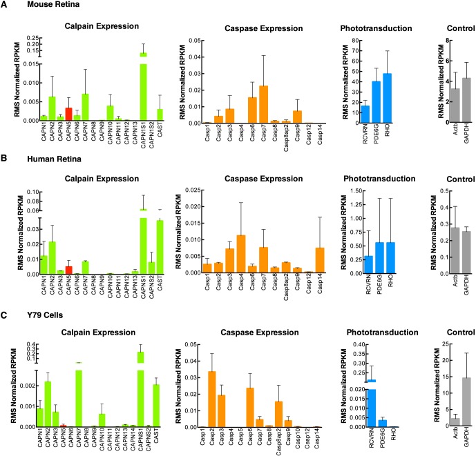
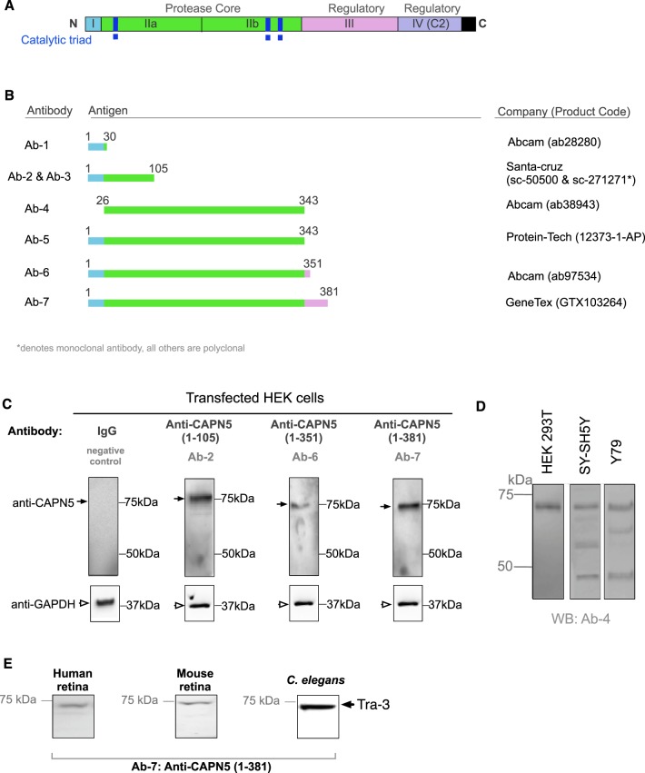
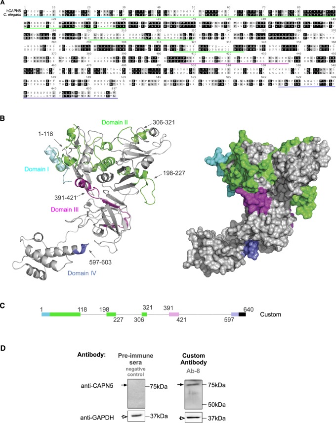
Methods: CAPN5 gene variants were classified using the exome variant server, and RNA-sequencing was used to compare expression of CAPN5 mRNA in the mouse and human retina and in retinoblastoma cells. Expression of CAPN5 protein was ascertained in humans and mice in silico, in mouse retina by immunohistochemistry, and in neuronal cancer cell lines and fractionated central nervous system tissue extracts by Western analysis with eight antibodies targeting different CAPN5 regions.

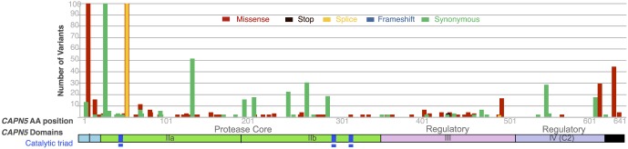
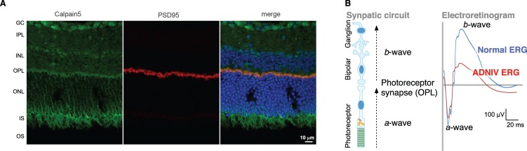
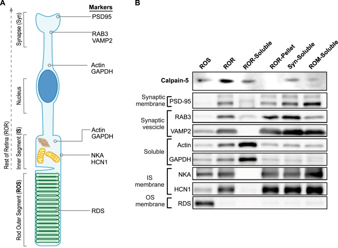
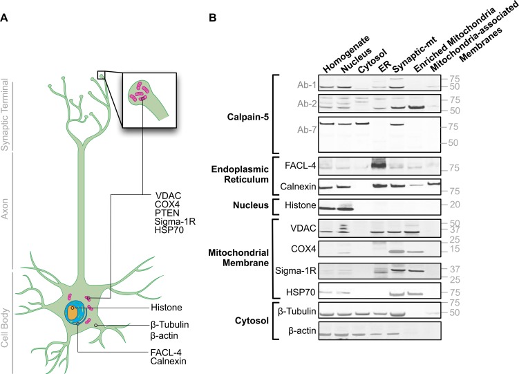
Results: Most CAPN5 genetic variation occurs outside its protease core; and searches of cancer and epilepsy/autism genetic databases found no variants similar to hyperactivating retinal disease alleles. The mouse retina expressed one transcript for CAPN5 plus those of nine other calpains, similar to the human retina. In Y79 retinoblastoma cells, the level of CAPN5 transcript was very low. Immunohistochemistry detected CAPN5 expression in the inner and outer nuclear layers and at synapses in the outer plexiform layer. Western analysis of fractionated retinal extracts confirmed CAPN5 synapse localization. Western blots of fractionated brain neuronal extracts revealed distinct subcellular patterns and the potential presence of autoproteolytic CAPN5 domains.

Conclusions: CAPN5 is moderately expressed in the retina and, despite higher expression in other tissues, hyperactive disease mutants of CAPN5 only manifest as eye disease. At the cellular level, CAPN5 is expressed in several different functional compartments. CAPN5 localization at the photoreceptor synapse and with mitochondria explains the neural circuitry phenotype in human CAPN5 disease alleles.


Figures

Figure 1
Figure 1
CAPN5 EVS Variants. Public exome databases show common variants in CAPN5 by amino acid position and type. The CAPN5 gene is 59 kilobases, contains 13 exons, and generates a 2791 nucleotide transcript (encompassing all purported isoforms) that encodes a 640 amino acid protein. Shown here, this data set serves as a normative database for CAPN5 variants, and depicts the relationship of these variants to functional residues and domains within CAPN5. Variants are rare within the proteolytic core, where retinal disease alleles are located. The EVS sequenced over 6476 individuals, with high CAPN5 coverage, and found no frameshift variants, two premature stop-codon variants, 843 missense variants (of which 237 were splice-site mutations), and 681 synonymous variants. Of these variants, the majority was scored as either “unknown” or “benign” by PolyPhen. PolyPhen predicted that 1% and 0.83% of the population has “Possibly Damaging” and “Probably Damaging” alleles, respectively.
Figure 2
Figure 2
RNA sequence analysis of human and mouse retina. (A) Calpain expression in the mouse retina, as compared to levels of caspase expression, phototransduction genes, and housekeeping genes as controls (n = 3). Results show CAPN5 expression is approximately half that of the most abundant calpains, CAPN2 and CAPN7. Calpain expression is comparable to caspase expression in the mouse retina. Expression of CAPN5 is several orders of magnitude less than phototransduction genes and control genes. (B) Slightly different patterns of expression are seen in the human retina (n = 3). (C) Calpain expression observed in Y79 cells as compared to levels of caspase expression, phototransduction genes, and controls (n = 3). Similar to human and mouse retina, CAPN2 and CAPN7 are the most abundant calpains in Y79 cells, but CAPN5 expression is lower.
Figure 3
Figure 3
CAPN5 Western blot shows specificity of multiple CAPN5 antibodies. (A) Schematic diagram of human CAPN5 protein domain structure. CAPN5 is composed of 640 amino acids that comprise four domains (A). Domain-I spans amino acid residues 1 to 25. Domain-II is the protease core and spans amino acid residues 26 to 343; it consists of two subdomains that together contain the catalytic triad and calcium binding sites. Domain II also contains the three different ADNIV-causing mutations. Domain-III spans amino acid residues 344 to 496 and contains a calcium-binding site leading to speculation that it is a regulatory region. Domain-IV (C2) also is believed to be regulatory; it spans amino acid residues 518 to 619, and contains three calcium-binding sites. (B) A diagram showing the epitopes of the seven different commercially available anti-CAPN5 antibodies tested, as well as the companies they were obtained from. Antibody Ab-1 (ab28280) mainly targeted domain I; Ab-2 (sc-50500) and Ab-3 (sc271271) targeted all of domain I, and part of domain II (catalytic domain); Ab-4 (ab38943) targeted the entire domain II and part of domain III; Ab-5 (12373-1-AP) and Ab-6 (ab97534) targeted domains I, II, and part of III. All antibodies were rabbit polyclonal antibodies, except for Ab-3, which was a mouse monoclonal antibody. (C) Immunoblots of HEK-293 cells transfected with full-length CAPN5 (tagged with c-myc and flag tag) and probed separately with three unique anti-CAPN5 antibodies. Anti-GAPDH antibody was used as a protein loading control. Immunoglobulin G (IgG) was used as a negative control. The closed arrows indicate CAPN5 bands at approximately 75 kDa, while the open arrowheads indicate GAPDH bands at approximately 37 kDa. Immunoblot images were captured with an ECL imager. Antibody Ab-7 consistently gave the best bands with the least amount of background, indicating higher specificity for CAPN5. (D) Immunoblots of HEK-293, SY-SH5Y, and Y79 cells transfected with full length CAPN5 and probed with Ab-4. (E) Immunoblots of retinas belonging to human and mice were probed separately with CAPN5 specific Ab-7. Immunoblot of protein obtained from wildtype (N2) C. elegans. 10 μg of protein were loaded.
Figure 4
Figure 4
Customized antigen raises highly specific CAPN5 antibodies. (A) Alignment of CAPN5 and Tra-3 sequences. Regions of homology are shown in black. Regions corresponding to the customized CAPN5 antigen are underlined with colors corresponding to the different CAPN5 domains. (B) A structural model of CAPN5. The highlighted regions correspond to those recognized by our custom antibody. (C) A diagram showing the epitopes of our custom anti-CAPN5 antibody. (D) Immunoblots of HEK-293 cells transfected with full-length CAPN5 and probed with our custom anti-CAPN5 antibody. We used GAPDH as a protein loading control. Preimmune-serum obtained from the same animal was used as a negative control. The closed arrows indicate CAPN5 bands at approximately 75 kDa, while the open arrowheads indicate GAPDH bands at approximately 37 kDa. Immunoblot images were captured with an ECL imager.
Figure 5
Figure 5
CAPN5 Immunohistochemistry in the retina synapse. (A) CAPN5 expression was present along a region corresponding to the OPL of the retina, where photoreceptors form synaptic connections with bipolar cells. PSD95 expression, a marker of neural postsynaptic densities, colocalized with CAPN5 expression along the synapses forming the outer plexiform layer. (B) The retinal synaptic circuit begins with light depolarization (a-wave) at the photoreceptor. Synaptic transmission from the photoreceptor to the bipolar cell and then to the RGC generates the hyperpolarized b-wave. This is shown in human ERG that trace the a- and b-waves in an ADNIV patient and a wildtype individual. Early in ADNIV patients and transgenic mouse models, the a-wave is intact but the b-wave is reduced (electronegative), indicating a defect in synaptic signaling between the photoreceptor and bipolar cell. The unusual phenotype is explained by CAPN5 localization to this synaptic junction. Scale bar: 10 μm. Blue = DAPI; Red = PSD95; green = CAPN5.
Figure 6
Figure 6
CAPN5 co-fractionates with both soluble and synaptic vesicle proteins. (A) A schematic illustration of a photoreceptor cell maps the location of subcellular marker proteins. (B) Antibodies against CAPN5 were used to probe retinal protein fractions alongside protein markers for specific retinal compartments. CAPN5 was detected in the ROS, but most of it remained in the ROR fraction. After the ROR was fractionated to enrich for soluble (ROR-S1), membrane (ROR-P1), and synaptic (Syn-S2) proteins, CAPN5 was found in the soluble and synaptic vesicle pools (along with Actin and GAPDH, or RAB3 and VAMP2, respectively). NKA, sodium potassium ATPase; HCN1, hyperpolarization-activated and cyclic nucleotide-gated channel, family member 1.
Figure 7
Figure 7
CAPN5 co-fractionates with specific cellular compartments. (A) A schematic illustration of a neuron maps the location of subcellular marker proteins. (B) Antibodies against CAPN5 were used to probe neuronal protein fractions alongside protein markers for specific cellular compartments. CAPN5 was detected in all compartments probed, depending on the antibody used. Most striking is the high levels of CAPN5 found in mitochondrial fractions, and the crude synaptosomal/mitochondrial fraction.

Similar articles

Cited by

References

    1. Mahajan VB,, Skeie JM,, Bassuk AG,, et al. Calpain-5 mutations cause autoimmune uveitis, retinal neovascularization, and photoreceptor degeneration. PLoS Genet. 2012; 8: e1003001. - PMC - PubMed
    1. Mahajan VB,, Lin JH. Lymphocyte infiltration in CAPN5 autosomal dominant neovascular inflammatory vitreoretinopathy. Clin Ophthalmol. 2013; 7: 1339–1345. - PMC - PubMed
    1. Wert KJ,, Skeie JM,, Bassuk AG,, Olivier AK,, Tsang SH,, Mahajan VB. Functional validation of a human CAPN5 exome variant by lentiviral transduction into mouse retina. Hum Mol Genet. 2014; 23: 2665–2677. - PMC - PubMed
    1. Bassuk AG,, Yeh S,, Wu S,, et al. Structural modeling of a novel CAPN5 mutation that causes uveitis and neovascular retinal detachment. PLoS One. 2015; 10: e0122352. - PMC - PubMed
    1. Wert KJ,, Bassuk AG,, Wu WH,, et al. CAPN5 mutation in hereditary uveitis: the R243L mutation increases calpain catalytic activity and triggers intraocular inflammation in a mouse model. Hum Mol Genet. 2015; 24: 4584–4598. - PMC - PubMed

Publication types

MeSH terms