. 2017 Jun;18(5):635-648.
doi: 10.1111/mpp.12422. Epub 2016 Jul 27.

Analysis of the interacting partners eIF4F and 3'-CITE required for Melon necrotic spot virus cap-independent translation

Affiliations

Analysis of the interacting partners eIF4F and 3'-CITE required for Melon necrotic spot virus cap-independent translation

Manuel Miras et al. Mol Plant Pathol. 2017 Jun.

Abstract

We have shown previously that the translation of Melon necrotic spot virus (MNSV, family Tombusviridae, genus Carmovirus) RNAs is controlled by a 3'-cap-independent translation enhancer (CITE), which is genetically and functionally dependent on the eukaryotic translation initiation factor (eIF) 4E. Here, we describe structural and functional analyses of the MNSV-Mα5 3'-CITE and its translation initiation factor partner. We first mapped the minimal 3'-CITE (Ma5TE) to a 45-nucleotide sequence, which consists of a stem-loop structure with two internal loops, similar to other I-shaped 3'-CITEs. UV crosslinking, followed by gel retardation assays, indicated that Ma5TE interacts in vitro with the complex formed by eIF4E + eIF4G980-1159 (eIF4Fp20 ), but not with each subunit alone or with eIF4E + eIF4G1003-1092 , suggesting binding either through interaction with eIF4E following a conformational change induced by its binding to eIF4G980-1159 , or through a double interaction with eIF4E and eIF4G980-1159 . Critical residues for this interaction reside in an internal bulge of Ma5TE, so that their mutation abolished binding to eIF4E + eIF4G1003-1092 and cap-independent translation. We also developed an in vivo system to test the effect of mutations in eIF4E in Ma5TE-driven cap-independent translation, showing that conserved amino acids in a positively charged RNA-binding motif around amino acid position 228, implicated in eIF4E-eIF4G binding or belonging to the cap-recognition pocket, are essential for cap-independent translation controlled by Ma5TE, and thus for the multiplication of MNSV.

Keywords: CITE; Carmovirus; RNA structure; Tombusviridae; cucurbit; translation enhancer; translation initiation.


Figures

Figure 1
Figure 1
Mapping the minimal sequence of the Melon necrotic spot virus (MNSV)‐Mα5 3′‐untranslated region (3′‐UTR) required for cap‐independent translation. (A) Schematic representation of reporter constructs consisting of the luciferase gene (LUC) flanked by 5′‐UTR and 3′‐UTR or 5′‐UTR and stem‐loop structure (SLC) variants SLC‐53, SLC‐45 and SLC‐30 of MNSV‐Mα5. (B) M‐fold prediction of SLC showing the fragments SLC‐53 [53 nucleotides (nt)], SLC‐45 and SLC‐30 corresponding to the different sequences added at the 3′‐end of the luciferase gene. The GC clamp added at the end of these variants is shown in a box. (C) Relative luciferase activity (%) in melon protoplasts (y‐axis) obtained with different constructs (x‐axis) differing in their 3′‐ends. The luciferase activity obtained with the construct 5′‐UTR‐luc‐3′‐UTR was set as 100% (second bar). 3′‐ends: 3′pl, plasmid sequence; 3′‐UTR, 3′‐UTR; SLC‐30, 30 nt from SLC (4085–4114); SLC‐45, 45 nt from SLC (4079–4122); SLC‐53, SLC (53 nt; 4074–4126). SLC‐30c, SLC‐45c and SLC‐53c correspond to constructs with the clamp added to the SLC fragments analysed. The last column corresponds to the activity obtained with a construct with SLC‐45c at the 3′‐end and plasmid sequence instead of the 5′‐UTR at the 5′‐end. Error bars are ± Standard deviation (SD). (D) MNSV‐Mα5 3′‐UTR secondary structure as predicted by M‐fold; the box marks SLC.
Figure 2
Figure 2
Chemical solution structure probing of Ma5TE. (A) Structure probing by SHAPE (selective 2′‐hydroxyl acylation analysed by primer extension) of SLC‐45 [nucleotides 4079–4112 in the Melon necrotic spot virus (MNSV)‐Mα5 sequence]. Primer extension products from RNAs modified with benzoyl cyanide (BzCN) using increasing Mg2+ concentrations (0–3 mm) third to fifth lanes. ‘–’, untreated RNA; ‘C’, sequencing ladder generated with dideoxy‐CTP on unmodified RNA. Positions marking nucleotides C4092 and C4118 are indicated on the left. The regions of modified nucleotides corresponding to the internal loops (IL1/2) and final loop (L) are marked on the right side of the polyacrylamide gel. (B) Secondary structure of Ma5TE according to the SHAPE reactivity data on the best‐fitting predicted MC‐Fold secondary structure. Levels of BzCN modification are indicated by a colour‐coded scale where red indicates the strongest modification. The pentanucleotide proposed to be involved in long‐distance interaction with the 5′‐untranslated region (5′‐UTR) (V. Truniger, unpublished data) is marked in L and arrows point to nucleotide variations found with respect to the stem‐loop structure (SLC) sequence of other MNSV isolates. Asterisks indicate the nucleotides whose accessibility changes with changing Mg2+ concentrations, as they are increasingly modified at 1 mm Mg2+, but appear to be less modified at 3 mm Mg2+. (C) Alignment of the Ma5TE sequences conserved in the 3′‐UTR of all MNSV isolates. Nucleotide variations are shaded in light blue. GenBank accession numbers of MNSV sequences included in the alignment are as follows: Mα5, AY122286; Dutch, NC001504; 17A/01A, D12536.2; HM, GU480022.1; Mα71, EU589619; MNSV‐N, KF060715; ABCA13‐01, KR094068; Mα24, EU589616; Pα58, EU589620; Nagasaki, AB250686; Yamaguchi, AB250687; Chiba, AB250684; Kochi, AB250685; Kouchi CP gene (KS), AB189943; ISR (Israel), DQ922807; Al, DQ339157; Pα57, EU589621.
Figure 3
Figure 3
Identification of the interaction between eIF4F and Ma5TE. (A) Schematic representation of melon eIF4E and subunits of eIF4G. Factor‐binding domains in eIF4G predicted by Pfam are shaded in grey: eIF4E‐binding, MIF4G and MA3 domains. The eIF4E‐binding domain was retained in the two truncated eIF4G proteins (eIF4Gp20 and eIF4Gp10). The region of eIF4Gp20 crucial for the binding of eIF4F to Ma5TE is striped. (B) UV crosslinking followed by 12% sodium dodecylsulfate‐polyacrylamide gel electrophoresis (SDS‐PAGE) separation of labelled Ma5TE (SLC‐45c) in the absence (–) or presence of 400 nm of each protein: bovine serum albumin (BSA), eIF4E, eIF4G980–1159 (4Gp20), eIF4Fp10 (4Fp10) and eIF4Fp20 (4F20). The retarded Ma5TE as a result of the complex formed with eIF4Fp20 is marked on the right. (C) eIF4Fp20/Ma5TE complex formation using increasing eIF4Fp20 concentrations (nm, as indicated).
Figure 4
Figure 4
Mapping of the eIF4F‐binding sites on the Ma5TE sequence. (A) Benzoyl cyanide (BzCN) modification analysis of Ma5TE in buffer (+ lane) and incubated with eIF4Fp20 (4Fp20) or non‐binding protein BSA (bovine serum albumin). Proteins were added to a final concentration of 1 µm. ‘C’ corresponds to the sequencing ladder obtained using dideoxy‐CTP. The second lane shows unmodified RNA (–). Nucleotides protected in the presence of eIF4Fp20 are marked with a blue arrow, whereas red arrows mark residues with increased accessibility. (B) SHAPE (selective 2′‐hydroxyl acylation analysed by primer extension) reactivity profiles (bars) in the absence (‘Buffer’) or presence of eIF4Fp20. Values correspond to the mean SHAPE reactivity (±SD) of four independent experiments. SHAPE reactivity is measured on a scale from ‘0’ (unreactive nucleotides) to ‘2’ (maximum reactivity). Nucleotide positions are shown on the y‐axis. Nucleotides that were protected or strongly modified are boxed in blue or red, respectively.
Figure 5
Figure 5
Identification of nucleotides of Ma5TE involved in eIF4F binding. (A) SHAPE (selective 2′‐hydroxyl acylation analysed by primer extension) Ma5TE secondary structure analysis showing the nucleotides protected in the presence of eIF4Fp20 in blue and those with increased accessibility in red. The Ma5TE point mutations studied are boxed. (B) Relative luciferase activity (%) in susceptible melon protoplasts of the constructs with the luciferase gene flanked by the 5′‐untranslated region (5′‐UTR) and Ma5TE [wild‐type (WT) and mutated]. The activity obtained with WT Ma5TE was set as 100% (column 1). Ma5TE mutations analysed: A4109C, A4109G and G4093C without and with the complementary mutation C4105G. Error bars are ± Standard deviation (SD). (C) UV crosslinking of labelled Ma5TE and mutant Ma5TE A4109C with eIF4Fp20, followed by sodium dodecylsulfate‐polyacrylamide gel electrophoresis (SDS‐PAGE) separation. The eIF4Fp20/Ma5TE complex was visible only with WT Ma5TE. Factor concentration (nm) is indicated. BSA, bovine serum albumin.
Figure 6
Figure 6
Effect of substitutions in eIF4E on cap‐independent Ma5TE‐mediated translation. (A) Relative luciferase activity (%) obtained with luciferase constructs (5′‐UTR‐luc‐3′‐UTR) in resistant melon protoplasts expressing transiently eIF4E from susceptible melon (H228 and mutants). For normalization of the different protoplast preparations, luciferase activity obtained with construct 5′‐UTR‐luc‐3′‐UTR from resistant‐breaking Melon necrotic spot virus (MNSV) (MNSV‐264) was set to 100% for each protoplast preparation. Translation of MNSV‐264 has been shown to be eIF4E independent (Rodríguez‐Hernández et al., 2012). The transiently expressed eIF4E mutants are indicated below each bar: ‘–’, silencing suppressor P19 alone; H228, susceptible eIF4E; H228L, resistance allele; engineered eIF4E mutations: H228R; K230R (in residues proposed to be involved in RNA binding); W82L (in cap‐binding pocket); Y154H, Q163A, W99A, L157G and Y154H‐W99A (in residues proposed to be involved in eIF4G interaction). Error bars are ± Standard deviation (SD). The top blot shows the expression of each eIF4E mutant in resistant melon cotyledon visualized by western blot using antibodies against melon eIF4E. Low expression of endogenous eIF4E (‘–’) can be detected. The bottom blot shows the loading control visualized by Coomassie blue staining. (B) Predicted structure of melon eIF4E (dark blue) based on Pisum sativum eIF4E crystal structure complexed with eIF4G602–638 from Drosophila melanogaster (light blue). The residues of eIF4E suggested to be involved in the interaction with eIF4G (W99, Y154, L157 and Q163, underlined in bold) are drawn with red sticks. Residue W82, located in the cap‐binding pocket, and residues H228 and K230 are coloured in yellow, green and orange, respectively. Residues in eIF4G of the conserved canonical eIF4E‐binding motif YxxxxLϕ (YSRDFLL in melon, Y1049, L1054 and L1055) are shown in pink.
Figure 7
Figure 7
Mutations in eIF4E and eIF4Gp20 involved in their interaction. Maltose‐binding protein (MBP) pulldown assays showing the interaction of eIF4E (H228 and mutants) and MBP‐eIF4G980‐1159 (4Gp20 and mutant). (A) Pulldown of eIF4EH228 and mutant proteins through their interactions with eIF4Gp20. Top gel shows pulled down eIF4E (MBP pulldown) visualized by western blot (WB) with melon eIF4E‐specific antibody. The same gel was stained with Coomassie to compare a similar amount of eluted eIF4Gp20 in each experiment. The third gel visualizes the amount of eIF4E present in the input by western blot. The factors interacting in each experiment are described below the gels, as pulldown protein (PDprot: either MBP or eIF4Gp20) and as partner of the eIF4E proteins (H228 or mutant L157G, W99A or double mutant W99A/Y154H). (B) Interaction of eIF4Gp20 and the triple mutant in the canonical 4E‐binding domain (4Gp20TC) with purified eIF4EH228 protein. The input (lanes 2 and 3) and bound (lanes 5 and 6) fractions were analysed by 12% sodium dodecylsulfate‐polyacrylamide gel electrophoresis (SDS‐PAGE), followed by Coomassie blue staining. As negative control for the specificity of the interaction, we incubated eIF4Gp20 alone or MBP with lysate (A) or purified eIF4E (B).
Figure 8
Figure 8
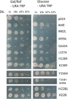
Complementation of translation by eIF4E mutants in eIF4E‐deficient yeast. Yeast strain JO55 was transformed with plasmid p424 expressing Arabidopsis thaliana eIF4E (At4E) or melon eIF4E (H228 or mutant). Yeast cultures were spotted either undiluted (1×) or diluted (10×, 102× and 103×) on Galactose+raffinose and glucose‐selective media [both media without uracil and tryptophan (–Ura/–Trp)]. The negative control with the empty p424 and the cap‐binding pocket mutant W82L were unable to complement translation, and thus to grow on glucose.
Figure 9
Figure 9
Comparison of sequence and structure of I‐shaped structured 3′‐cap‐independent translation enhancers (3′‐CITEs). (A) Ma5TE and Maize necrotic streak virus (MNeSV) I‐shaped structure (ISS) secondary structures. (A) The Ma5TE (i) and MNeSV (ii) ISS secondary structures were probed by SHAPE (selective 2′‐hydroxyl acylation analysed by primer extension) analysis, whereas the MNeSV ISS alternative conformation (iii) was predicted by M‐fold based on in vivo evolution studies (Nicholson et al., 2013). (B) Maize white line mosaic virus (MWLMV) (i), Johnsongrass chlorotic stripe mosaic virus (JCSMV) (ii), Cucumber Bulgarian virus (CBV) (iii) and Melon necrotic spot virus (MNSV)−264 (iv) M‐fold‐predicted secondary structures of non‐characterized ISSs. The adenosine residue involved in the interaction of Ma5TE with eIF4F is shaded in red and the guanosine residue important for MNeSV 3′‐CITE interaction with eIF4F is marked in light blue. Nucleotides shaded in orange are conserved in all ISSs and those in grey correspond to those conserved between Ma5TE and MNeSV ISSs. The nucleotides shaded in green are conserved between Ma5TE and non‐characterized ISSs.

Similar articles

Cited by

References

    1. Aitken, C.E. and Lorsch, J.R. (2012) A mechanistic overview of translation initiation in eukaryotes. Nat. Struct. Mol. Biol. 19, 568–576. - PubMed
    1. Altmann, M. , Muller, P.P. , Pelletier, J. , Sonenberg, N. and Trachsel, H. (1989) A mammalian translation initiation factor can substitute for its yeast homologue in vivo. J. Biol. Chem. 264, 12 145–12 147. - PubMed
    1. Ashby, J.A. , Stevenson, C.E.M. , Jarvis, G.E. , Lawson, D.M. and Maule, A.J. (2011) Structure‐based mutational analysis of eIF4E in relation to sbm1 resistance to Pea seed‐borne mosaic virus in pea. PLoS One, 6, e15873. - PMC - PubMed
    1. Carstens, E.B. (2010) Ratification vote on taxonomic proposals to the international committee on taxonomy of viruses (2009). Arch. Virol. 155, 133–146. - PMC - PubMed
    1. Charron, C. , Nicolaï, M. , Gallois, J.L. , Robaglia, C. , Moury, B. , Palloix, A. and Caranta, C. (2008) Natural variation and functional analyses provide evidence for co‐evolution between plant eIF4E and potyviral VPg. Plant J. 54, 56–68. - PubMed

Publication types

MeSH terms