. 2016 May 4;12(5):e1005617.
doi: 10.1371/journal.ppat.1005617. eCollection 2016 May.

Mnn10 Maintains Pathogenicity in Candida albicans by Extending α-1,6-Mannose Backbone to Evade Host Dectin-1 Mediated Antifungal Immunity

Affiliations

Mnn10 Maintains Pathogenicity in Candida albicans by Extending α-1,6-Mannose Backbone to Evade Host Dectin-1 Mediated Antifungal Immunity

Shi Qun Zhang et al. PLoS Pathog. .

Abstract

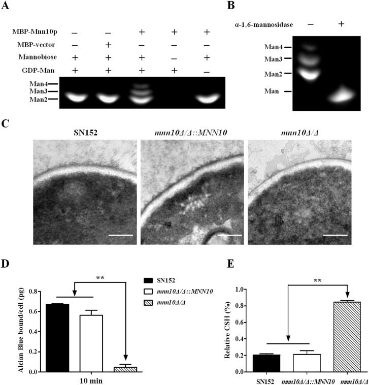
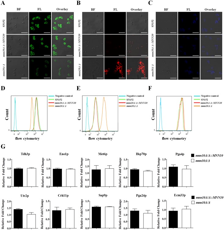
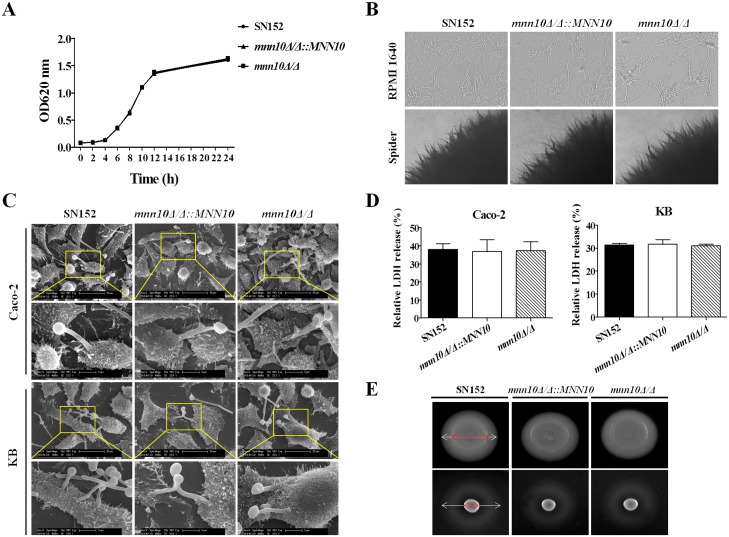
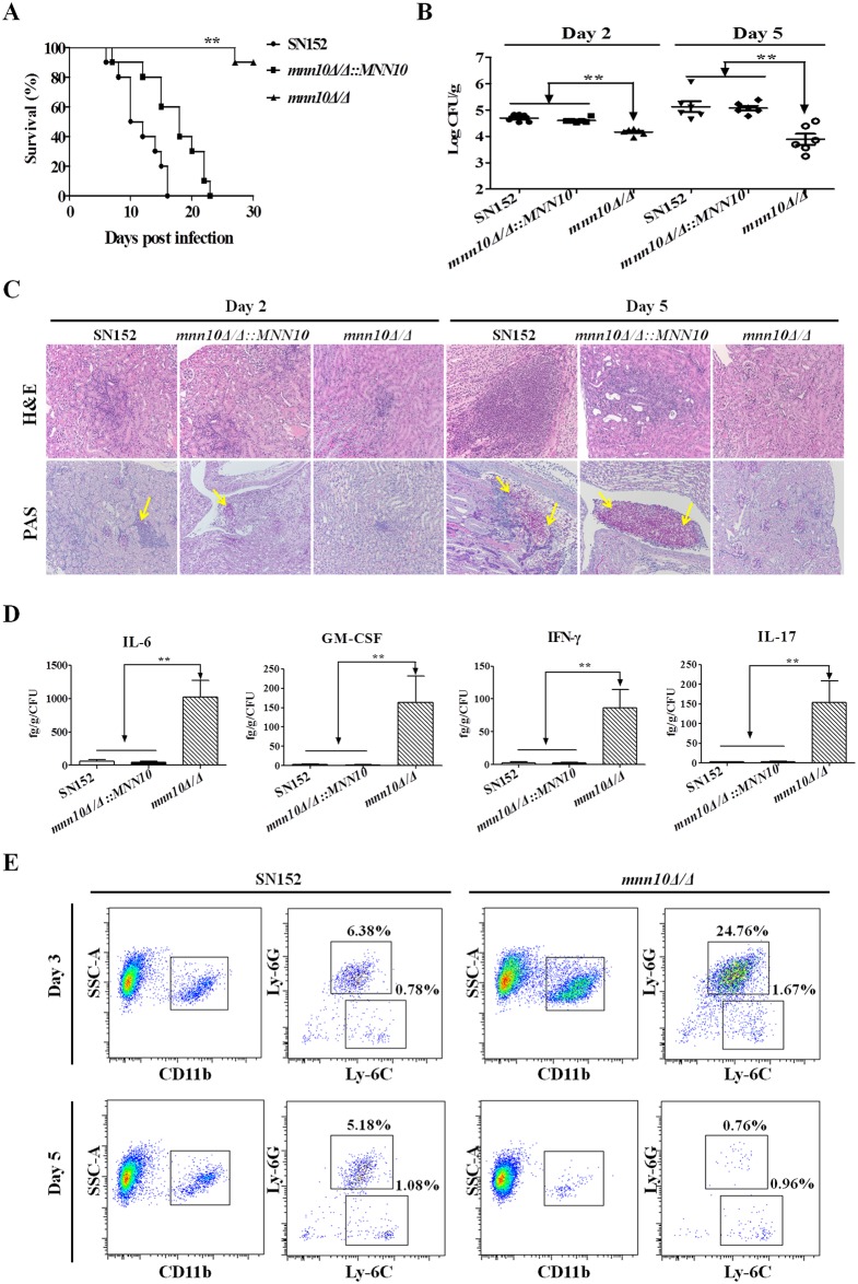
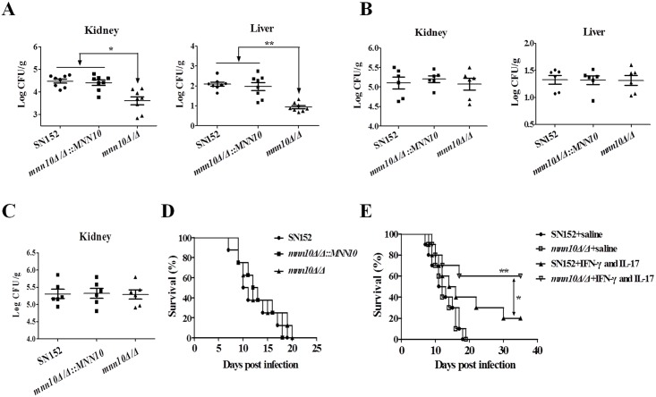
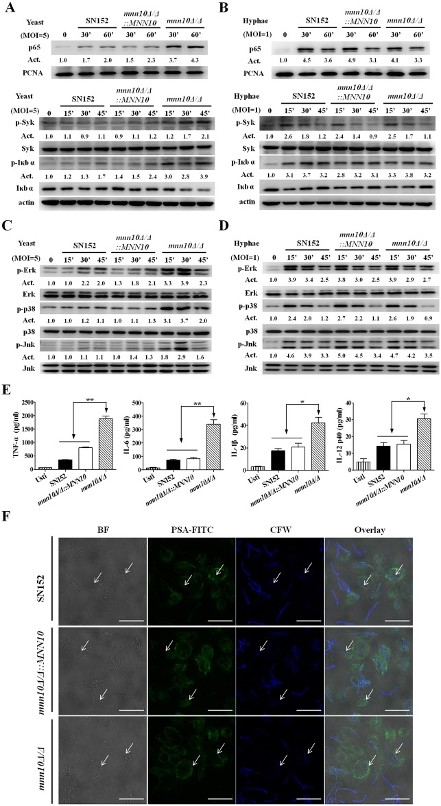
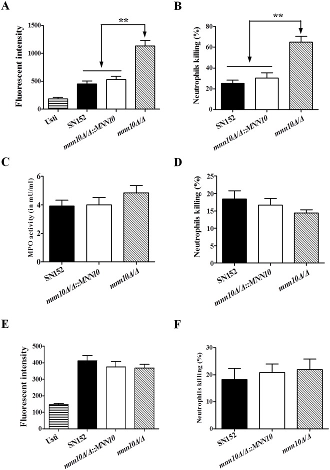
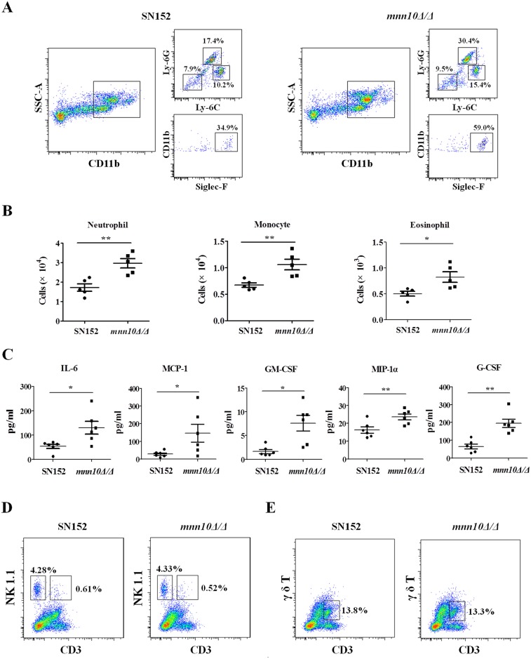
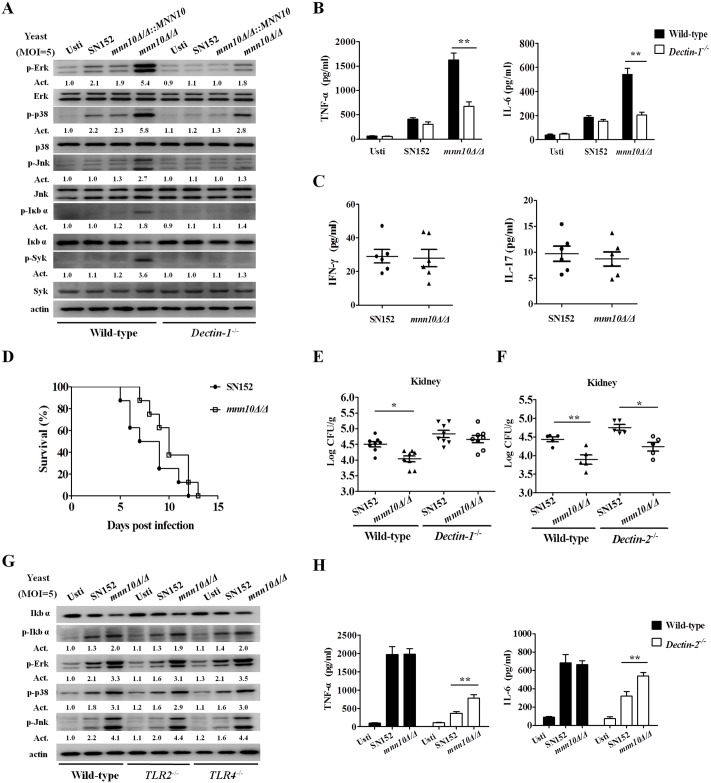
The cell wall is a dynamic structure that is important for the pathogenicity of Candida albicans. Mannan, which is located in the outermost layer of the cell wall, has been shown to contribute to the pathogenesis of C. albicans, however, the molecular mechanism by which this occurs remains unclear. Here we identified a novel α-1,6-mannosyltransferase encoded by MNN10 in C. albicans. We found that Mnn10 is required for cell wall α-1,6-mannose backbone biosynthesis and polysaccharides organization. Deletion of MNN10 resulted in significant attenuation of the pathogenesis of C. albicans in a murine systemic candidiasis model. Inhibition of α-1,6-mannose backbone extension did not, however, impact the invasive ability of C. albicans in vitro. Notably, mnn10 mutant restored the invasive capacity in athymic nude mice, which further supports the notion of an enhanced host antifungal defense related to this backbone change. Mnn10 mutant induced enhanced Th1 and Th17 cell mediated antifungal immunity, and resulted in enhanced recruitment of neutrophils and monocytes for pathogen clearance in vivo. We also demonstrated that MNN10 could unmask the surface β-(1,3)-glucan, a crucial pathogen-associated molecular pattern (PAMP) of C. albicans recognized by host Dectin-1. Our results demonstrate that mnn10 mutant could stimulate an enhanced Dectin-1 dependent immune response of macrophages in vitro, including the activation of nuclear factor-κB, mitogen-activated protein kinase pathways, and secretion of specific cytokines such as TNF-α, IL-6, IL-1β and IL-12p40. In summary, our study indicated that α-1,6-mannose backbone is critical for the pathogenesis of C. albicans via shielding β-glucan from recognition by host Dectin-1 mediated immune recognition. Moreover, our work suggests that inhibition of α-1,6-mannose extension by Mnn10 may represent a novel modality to reduce the pathogenicity of C. albicans.


Conflict of interest statement

The authors have declared that no competing interests exist.

Figures

Fig 1
Fig 1. Mnn10 has mannosyltransferase activity and is required for α-1,6-mannose backbone length.
(A) Mannosyltransferase activity assay of Mnn10. The reaction of expressed MBP-fused Mnn10 protein or MBP protein incubated with α-1,6-mannobiose (Man 2), GDP-mannose (GDP-Man) or controls. The reaction products were labeled with ANTS and separated by fluorophore-assisted carbohydrate gel electrophoresis (FACE). Man 3, mannotriose; Man 4, mannotetraose. (B) α-1,6-mannose assay. The reaction products of (A) were treated with or without α-1,6-mannosidase treatment, and then subjected to FACE. (C) Representative cell wall ultrastructures of SN152, mnn10Δ/Δ::MNN10 and mnn10Δ/Δ strains were observed by transmission electron microscopy. The scale bar represents 0.2 μm. (D) Alcian Blue binding assay. The cells were incubated with Alcian Blue for 10 min and the amount of dye bound to the cell wall were calculated. Data represent the mean amount of dye bound per cell ± SD from triplicates of one representative experiment of three. (E) Cell surface hydrophobicity of the indicated C. albicans strains was measured by water-hydrocarbon two-phase assay. Data are means ± SD of triplicates of one representative experiment of three. **, P < 0.01 [One-way ANOVA with Bonferroni post-test (D, E)].
Fig 2
Fig 2. Mnn10 is required for cell wall polysaccharides organization, but not virulence factors attachment.
Representative fluorescence micrographs of three cell wall carbohydrate layers from SN152, mnn10Δ/Δ::MNN10 and mnn10Δ/Δ, which were stained with ConA-FITC to visualise mannan (A), β-glucan antibody to visualize β-(1,3)-glucan (B) and calcofluor white to visualise chitin (C). Bright field (BF), fluorescence (FL), and overlay are shown individually. Scale bar represents 10 μm. The fluorescence intensity was quantified by flow cytometry [mannan (D), β-(1,3)-glucan (E), chitin (F)]. Data are representative of three independent experiments. (G) Relative fold change of representative virulence related cell wall proteins (CWPs) in mnn10 mutant compared to mnn10Δ/Δ::MNN10. The cell wall proteins of SN152, mnn10Δ/Δ::MNN10, and mnn10Δ/Δ strains were analyzed by LC-MS/MS on high-resolution instruments, respectively. Raw files were processed by Max Quant for peptide/protein identification and quantification. The relative data were changed to the ratio of mnn10Δ/Δ::MNN10 and mnn10Δ/Δ to SN152. Data are represented as means ± SEM of three independent experiments. P > 0.05 (Student’s t-test).
Fig 3
Fig 3. Inhibition of α-1,6-mannose backbone extension in C. albicans does not affect its invasive capacity in vitro.
(A) Growth curves of SN152, mnn10Δ/Δ::MNN10 and mnn10Δ/Δ strains. (B) Representative photomicrographs of indicated C. albicans growing in liquid RPMI 1640 culture for 3 h at 37°C or on spider agar media for 5 days at 37°C to induce hyphal form. (C) Representative micrographs of scanning electron microscope of Caco-2 and KB cells invaded or penetrated by SN152, mnn10Δ/Δ::MNN10 and mnn10Δ/Δ after 2 h co-incubation (MOI = 1). (D) Epithelial cells damage was determined by assaying LDH release. Relative LDH release from Caco-2 or KB cells was measured after 12 h incubation with C. albicans (MOI = 0.1). Data are represented as means ± SD from triplicates of one representative experiment of three. P > 0.05 (One-way ANOVA with Bonferroni post-test). (E) Extracellular phospholipase and hemolytic activity assays. The phospholipase activity was examined by spotted C. albicans on egg yolk agar at 37°C for 3 days and observed the width of the zone of precipitation around each colony (top panel). The hemolytic activity was assessed by growing the indicated C. albicans strains on sugar-enriched sheep blood agar at 37°C for 3 days to observe the presence of a distinct translucent halo around the colonies (lower panel). Representative images are shown from one of three independent experiments.
Fig 4
Fig 4. Mnn10 is required for C. albicans systemic infection.
C57BL/6 mice were infected with 5×10⁵ CFU SN152, mnn10Δ/Δ::MNN10 or mnn10Δ/Δ strain in 200 μl sterile saline via lateral tail vein. (A) Survival of C57BL/6 mice infected with the indicated strains was monitored for over 30 days (n = 10 per group). Data are representative of three independent experiments. (B) Quantification of the fungal burden in kidney tissues of C57BL/6 mice (n = 6 per group) infected with indicated C. albicans strains at day 2 and day 5. Data are representative of three independent experiments. (C) Representative H&E (for the inflammatory cells influx and the extent of tissue necrosis) and PAS (for C. albicans) staining of kidneys from C57BL/6 mice infected with indicated strains at day 2 and 5. Arrows indicate C. albicans filaments in the tissues. Magnification = 200 ×. (D) ELISA assays for IL-6, GM-CSF, IFN-γ and IL-17 in homogenized kidneys from C57BL/6 mice infected with indicated C. albicans at day 5 (n = 8 per group). The cytokine levels were normalized to burden of infection in each individually kidney as fg/g tissue/CFU. Data are means ± SD and are representative of three independent experiments. (E) The cellular inflammation in the kidneys of SN152 or mnn10Δ/Δ infected mice. SSChighCD11b+Ly-6C+Ly-6G+ neutrophils and SSChighCD11b+Ly-6C+Ly-6G- monocytes in the kidneys were detected at Day 3 and Day 5 by flow cytometry. Data are representative images of five mice. *, P < 0.05; **, P < 0.01 [Log-rank test (A) and Kruskal-Wallis nonparametric One-way ANOVA with Dunns post-test (B, D)].
Fig 5
Fig 5. Mnn10 is not required for C. albicans systemic infection in athymic nude mice.
BALB/c mice or athymic nude mice (BALB/c background) were infected with 3×10⁵ CFU of SN152, mnn10Δ/Δ::MNN10 or mnn10Δ/Δ strain via lateral tail vein, respectively. The kidney and liver fungal burdens of BALB/c mice (n = 8 per group) (A) and athymic nude mice (n = 6 per group) (B) 5 days after infection. *, P < 0.05; **, P < 0.01 (Kruskal-Wallis nonparametric One-way ANOVA with Dunns post-test). (C) The kidney fungal burdens of athymic nude mice (n = 6 per group) 10 days after infection. (D) Survival of athymic nude mice infected with the indicated C. albicans (n = 8 per group). (E) Survival of SN152 and mnn10Δ/Δ infected athymic nude mice treated with combination of IFN-γ (100 ng) and IL-17 (100 ng) or the same volume of sterile saline. Data shown are representative of three independent experiments. **, P < 0.01 (mnn10Δ/Δ+IFN-γ and IL-17 versus mnn10Δ/Δ+saline); *, P < 0.05 (mnn10Δ/Δ+IFN-γ and IL-17 versus SN152+IFN-γ and IL-17) [Kruskal-Wallis nonparametric One-way ANOVA with Dunns post-test (A, B, C) and Log-rank test (D, E)].
Fig 6
Fig 6. α-1,6-mannose backbone inhibition in C. albicans yeasts, but not hyphae, could induce an increased immune responses.
(A and B) Thioglycollate-elicited peritoneal macrophages were stimulated with UV-inactivated SN152, mnn10Δ/Δ::MNN10 and mnn10Δ/Δ yeasts (MOI = 5) (A) or hyphae (MOI = 1) (B) for the indicated times. The nuclear extracts (top panel) and total cell lysates (lower panel) were subjected to immunoblotting analysis with the indicated antibodies of NF-κB signaling. (C and D) Thioglycollate-elicited peritoneal macrophages were challenged with UV-inactivated SN152, mnn10Δ/Δ::MNN10 and mnn10Δ/Δ yeasts (MOI = 5) (C) or hyphae (MOI = 1) (D) for the indicated times. The total cell lysates were subjected to immunoblotting with the indicated antibodies of MAPK signaling. Numbers between blots indicate activity (Act) of phosphorylation of MAPK or NF-κB pathways, as measured by densitometry. (E) ELISA results for cytokines TNF-α, IL-6, IL-1β and IL-12p40 in cell supernatants of thioglycollate-elicited peritoneal macrophages, which were stimulated with the indicated C. albicans yeasts (MOI = 5) for 6 h. Usti, unstimulated. Data are means ± SD of triplicates from one representative experiment of three. Usti, unstimulated. *, P < 0.05; **, P < 0.01 (One-way ANOVA with Bonferroni post-test). (F) Phagocytosis of C. albicans by thioglycollate-elicited peritoneal macrophages. Live C. albicans was co-cultured with the macrophages grown on coverslips in multiwell plates for 90 min. After staining with CFW (1 μg/ml) and PSA-FITC (20 μg/ml) for 10 min, the samples were viewed by confocal laser scanning microscope directly. Scale bar represents 10 μm. Arrows indicate the internalized C. albicans cells inaccessible to staining with CFW. Bright field (BF), fluorescein isothiocyanate-conjugated pisum sativum agglutinin (PSA-FITC), calcofluor white (CFW) and overlay are shown individually.
Fig 7
Fig 7. Neutrophils killed mnn10 mutant more efficiently with an augmented respiratory burst.
(A, E) Neutrophils respiratory burst assay. The cellular reactive oxygen species production of thioglycollate-elicited peritoneal neutrophils were measured after incubation with C. albicans for 1 h (MOI = 1) with (E) or without (A) 2 mM ascorbic acid. (B, D) Neutrophils killing assay. Neutrophils (6×10⁵ cells) were incubated with 3×10⁴ CFU viable C. albicans for 1 h with (D) or without (B) 2 mM ascorbic acid. Then the suspension was plated on SDA agar for 48 h to count live C. albicans colonies. (C) Neutrophils were incubated with the indicated C. albicans for 1 h (MOI = 1). The cellular level of MPO in neutrophils was measured after incubation with C. albicans. (F) Neutrophils (6×10⁵ cells) were incubated with 3×10⁴ CFU C. albicans hyphae for 1 h. Then the suspension was plated on SDA agar to count live C. albicans colonies. Data shown are means ± SD of triplicates from one representative experiment of three. Usti, unstimulated. **, P < 0.01 (One-way ANOVA with Bonferroni post-test).
Fig 8
Fig 8. Mnn10 mutant stimulated enhanced inflammatory responses in vivo.
(A, B, C) C57BL/6 mice were intraperitoneal infected with 5×10⁵ UV-inactivated C. albicans of SN152 or mnn10Δ/Δ strain for 4 h. (A) Flow cytometry for SSChighCD11b+Ly-6C+Ly-6G+ neutrophils, SSChighCD11b+Ly-6C+Ly-6G- monocytes and SSChighCD11b+Siglec-F+ eosinophils in the peritoneum. Data are representative images of five mice. (B) Scatter plots of myeloid cell subsets in the peritoneum cavities (n = 5 per group). (C) ELISA assays for cytokines, chemokines and growth factors in lavage fluid from the inflamed peritoneal cavities (n = 6 per group). Data are representative of three independent experiments. (D, E) C57BL/6 mice were intraperitoneal infected with 5×10⁵ live SN152 or mnn10Δ/Δ strain for 2 days. CD3-NK1.1+ NK cells (D), CD3+NK1.1+ NKT cells (D), and CD3+γ/δ T+ cells (E) in the peritoneum were detected by flow cytometry. Data are representative images of five mice. MCP-1, chemokine CCL2; MIP-1α, chemokine CCL3; GM-CSF, granulocyte-monocyte colony-stimulating factor; G-CSF, granulocyte colony-stimulating factor. *, P < 0.05; **, P < 0.01 [Mann-Whitney nonparametric t-test (B, C)].
Fig 9
Fig 9. Inflammatory responses, stimulated by C. albicans mnn10 mutant, were Dectin-1 dependent.
(A) Thioglycollate-elicited peritoneal macrophages from wild-type or Dectin-1-deficient mice were stimulated with UV-inactivated C. albicans yeast SN152, mnn10Δ/Δ::MNN10 or mnn10Δ/Δ (MOI = 5) for 30 minutes. The total cell lysates were then analyzed by immunoblotting with the indicated antibodies. Numbers between blots indicate activity (Act) of phosphorylation of MAPK or NF-κB pathways, as measured by densitometry. (B) Thioglycollate-elicited peritoneal macrophages from wild-type or Dectin-1-deficient mice were stimulated with the UV-inactivated C. albicans yeasts (MOI = 5) for 6 h. The amount of TNF-α and IL-6 in supernatants was determined by ELISA. Data are means ± SD of triplicates. (C) ELISA assays for IFN-γ and IL-17 in homogenized kidney from infected Dectin-1-deficient mice at day 5 (n = 6 per group). (D) Survival of Dectin-1-deficient mice infected with 5×10⁵ CFU C. albicans of SN152 or mnn10Δ/Δ strain (n = 8 per group). (E and F) The kidneys fungal burden of Dectin-1-deficient mice (E) or Dectin-2-deficient mice (F) infected with 3×10⁵ C. albicans at day 5. (G) Wild-type, TLR2-, and TLR4-deficient thioglycollate-elicited peritoneal macrophages were stimulated with UV-inactivated SN152 or mnn10Δ/Δ strain of C. albicans yeast cells (MOI = 5) for 30 min for preparing cell lysates. Samples were subjected to immunoblotting analysis using indicated antibodies. (H) Thioglycollate-elicited peritoneal macrophages from wild-type or Dectin-2-deficient mice were stimulated with the UV-inactivated C. albicans hyphae (MOI = 1) for 6 h. The amount of TNF-α and IL-6 in supernatants was determined using ELISA. Data shown are representative of three independent experiments. Usti, unstimulated. *, P < 0.05; **, P < 0.01 [Two-way ANOVA with Bonferroni post-test (B, H); Mann-Whitney nonparametric t-test (C); Log-rank test (D); Kruskal-Wallis nonparametric One-way ANOVA with Dunns post-test (E, F)].

Similar articles

Cited by

References

    1. Romani L. Immunity to fungal infections. Nat Rev Immunol. 2011; 11: 275–288. 10.1038/nri2939 - DOI - PubMed
    1. Gow NA, van de Veerdonk FL, Brown AJ, and Netea MG. Candida albicans morphogenesis and host defence: discriminating invasion from colonization. Nat Rev Microbiol. 2011; 10: 112–122. 10.1038/nrmicro2711 - DOI - PMC - PubMed
    1. Netea MG, Brown GD, Kullberg BJ, Gow NA. An integrated model of the recognition of Candida albicans by the innate immune system. Nat Rev Microbiol. 2008; 6: 67–78. - PubMed
    1. Leroy O, Gangneux JP, Montravers P, Mira JP, Gouin F, Sollet JP, et al. Epidemiology, management, and risk factors for death of invasive Candida infections in critical care: a multicenter, prospective, observational study in France (2005–2006). Crit Care Med. 2009; 37: 1612–1618. 10.1097/CCM.0b013e31819efac0 - DOI - PubMed
    1. Wisplinghoff H, Ebbers J, Geurtz L, Stefanik D, Major Y, Edmond MB, et al. Nosocomial bloodstream infections due to Candida spp. in the USA: species distribution, clinical features and antifungal susceptibilities. Int J Antimicrob Agents. 2014; 43: 78–81. 10.1016/j.ijantimicag.2013.09.005 - DOI - PubMed

MeSH terms

Grants and funding

This study was financially supported by the National Natural Science Foundation of China (No 81330083, www.nsfc.gov.cn/, YYJ); the National Natural Science Foundation of China (No 81471924, 81202563, www.nsfc.gov.cn/, MMA); the National Key Basic Research Program of China (No 2013CB531602, www.973.gov.cn/, YYJ); the Natural Science Foundation of Science and Technology Commission of Shanghai Municipality (No 13ZR1437900, www.stcsm.gov.cn/, XH); the Fundamental Research Funds for the Central Universities, MMA; the Shanghai Science and Technology Support Program (No 14401902200, www.stcsm.gov.cn/, MMA); the Shanghai Science and Technology Support Program (No 14431902200, www.stcsm.gov.cn/, YYJ). The funders had no role in study design, data collection and analysis, decision to publish, or preparation of the manuscript.