. 2016 Jun 7;7(23):35208-23.
doi: 10.18632/oncotarget.9106.

Nobiletin inhibits human osteosarcoma cells metastasis by blocking ERK and JNK-mediated MMPs expression

Affiliations

Nobiletin inhibits human osteosarcoma cells metastasis by blocking ERK and JNK-mediated MMPs expression

Hsin-Lin Cheng et al. Oncotarget. .

Abstract

Nobiletin, a polymethoxyflavone, has a few pharmacological activities, including anti-inflammation and anti-cancer effects. However, its effect on human osteosarcoma progression remains uninvestigated. Therefore, we examined the effectiveness of nobiletin against cellular metastasis of human osteosarcoma and the underlying mechanisms. Nobiletin, up to 100 μM without cytotoxicity, significantly decreased motility, migration and invasion as well as enzymatic activities, protein levels and mRNA expressions of matrix metalloproteinase (MMP)-2 and MMP-9 in U2OS and HOS cells. In addition to inhibition of extracellular signal-regulated kinase (ERK) and c-Jun N-terminal kinase (JNK), the inhibitory effect of nobiletin on the DNA-binding activity of the transcription factor nuclear factor-kappa B (NF-κB), cAMP response element-binding protein (CREB), and specificity protein 1 (SP-1) in U2OS and HOS cells. Co-treatment with ERK and JNK inhibitors and nobiletin further reduced U2OS cells migration and invasion. These results indicated that nobiletin inhibits human osteosarcoma U2OS and HOS cells motility, migration and invasion by down-regulating MMP-2 and MMP-9 expressions via ERK and JNK pathways and through the inactivation of downstream NF-κB, CREB, and SP-1. Nobiletin has the potential to serve as an anti-metastatic agent for treating osteosarcoma.

Keywords: CREB; MMP; SP-1; metastasis; nobiletin.


Conflict of interest statement

The authors declare that no conflicts of interest exist.

Figures
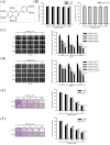
Figure 1
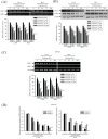
Figure 1. Nobiletin inhibits in vitro wound closure, migration and invasion in the U2OS and HOS cells
A. The structure of nobiletin. B. U2OS and HOS human osteosarcoma cells and MC3T3-E1 mouse osteoblast cells were treated with nobiletin (0-100 μM) in serum free medium for 24 h by MTT assay are illustrated. C. and D. U2OS and HOS cells were wounded and then treated with nobiletin (0–100 μM) for 24 h in a serum-containing medium. At 0 h, 6 h, 12 h and 24 h, phase-contrast pictures of the wounds at four different locations were taken. E. Cell migration was measured using a Boyden chamber for 24 h with polycarbonate filters. F. Cell invasion was measured using a Matrigel-coated Boyden chamber for 48 h with polycarbonate filters. Cytotoxicity effects: U2OS: F = 2.827, p = 0.083; HOS: F = 0.274, p = 0.888; MC3T3-E1: F = 0.121, p = 0.972. Concentration effects: wounding healing (U2OS: F = 279.878, p < 0.001; HOS: F = 109.236, p < 0.001); cell migration (U2OS: F = 296.344, p < 0.001; HOS: F = 464.672, p < 0.001); invasion (U2OS: F = 327.312, p < 0.001; HOS: F = 196.642, p < 0.001). aSignificantly different, p < 0.05, when compared with 0 μM. bSignificantly different, p < 0.05, when compared with 25 μM. cSignificantly different, p < 0.05, when compared with 50 μM. dSignificantly different, p < 0.05, when compared with 75 μM. Time effects: wound healing (U2OS: F = 1555.239, p < 0.001; HOS: F = 928.975, p < 0.001). †Significantly different, p < 0.05, when compared with the vehicle group. ‡Significantly different, p < 0.05, when compared with 6 h. #Significantly different, p < 0.05, when compared with 12 h.
Figure 2
Figure 2. Nobiletin inhibits MMP-2 and MMP-9 proteolytic activity, protein and mRNA expression
A. U2OS and HOS cells were treated with nobiletin (0–100 μM) for 24 h in a serum free medium and then subjected to gelatin zymography to analyze the activity of MMP-2 and MMP-9. B. Western blotting to analyze the protein levels of MMP-2 and MMP-9. Quantitative results of MMP-2 and MMP-9 protein levels, which were adjusted with β-actin protein level. C. RT-PCR and D. q-RT-PCR to analyze the mRNA expression of MMP-2 and MMP-9. Quantitative MMP-2 and MMP-9 mRNA levels were adjusted to GAPDH level. Concentration effects: gelatin zymography (MMP-2 of U2OS: F = 60.254, p < 0.001 and HOS: F = 160.282, p < 0.001; MMP-9 of U2OS: F = 133.253, p < 0.001 and HOS: F = 67.263, p < 0.001); western blot (MMP-2 of U2OS: F = 92.251, p < 0.001 and HOS: F = 25.151, p < 0.001; MMP-9 of U2OS: F = 25.156, p < 0.001 and HOS: F = 172.876, p < 0.001); RT-PCR (MMP-2 of U2OS: F = 68.584, p < 0.001 and HOS: F = 138.258, p < 0.001; MMP-9 of U2OS: F = 60.675, p < 0.001 and HOS: F = 63.82, p < 0.001); q-RT-PCR (MMP-2 of U2OS: F = 8.363, p = 0.019 and HOS: F = 116.834, p < 0.001; MMP-9 of U2OS: F = 186.999, p < 0.001 and HOS: F = 52.371, p < 0.001). aSignificantly different, p < 0.05, when compared with 0 μM. bSignificantly different, p < 0.05, when compared with 25 μM. cSignificantly different, p < 0.05, when compared with 50 μM. dSignificantly different, p < 0.05, when compared with 75 μM.
Figure 3
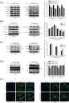
Figure 3. NF-κB and CREB are crucial to nobiletin-mediated transcriptional inhibition of MMP-2 and MMP-9
A-D. U2OS and HOS cells were treated with nobiletin (0–100 μM) for 24 h. The nuclear fraction and total cell lysates were prepared as described in Materials and Methods. Levels of NF-κB, SP-1, c-Jun and c-Fos from nuclear (B and D) and levels of p-IKKα/β, IKKβ, p-IκBα, IκBα, CREB and p-CREB from the total cell lysates (A and C) were determined by western blotting. Quantitative results of p-IKKα/β, p-IκBα, NF-κB, SP-1, c-Jun, c-Fos and p-CREB protein levels were adjusted with β-actin, C23 or α-tubulin protein level. Concentration effects: p-IKKα/β (U2OS: F = 15.419, p = 0.004; HOS: F = 28.397, p = 0.001); p-IκBα (U2OS: F = 194.788, p < 0.001; HOS: F = 21.407, p = 0.002); total-p-CREB (U2OS: F = 61.097, p < 0.001; HOS: F = 34.873, p < 0.001); cytosol-NF-κB (U2OS: F = 60.412, p < 0.001; HOS: F = 16.628, p = 0.007); nuclear-NF-κB (U2OS: F = 50.038, p < 0.001; HOS: F = 17.562, p = 0.003); SP-1 (U2OS: F = 58.954, p < 0.001; HOS: F = 49.681, p < 0.001); c-Jun (U2OS: F = 0.402, p =0.685; HOS: F = 0.097, p =0.909); c-Fos (U2OS: F = 0.124, p =0.885; HOS: F = 0.207, p =0.747). aSignificantly different, p < 0.05, when compared with 0 μM. bSignificantly different, p < 0.05, when compared with 25 μM. E. U2OS and HOS cells were stained for NF-κB by immunofluorescence. Pre-treatment with 100 μM nobiletin for 24 h significantly inhibited the nuclear translocation of NF-κB (White arrows were added to direct against distinct changes).
Figure 4
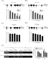
Figure 4. Nobiletin inhibits MMP-2 and MMP-9 promoter and DNA-binding activity
A-D. U2OS and HOS cells were treated with nobiletin (0–100 μM) for 24 h and then subjected to luciferase assay to analyze the promoter activity of MMP-2 and MMP-9. Upper: schematic of the wild type promoter region of the human MMP-2 and MMP-9 gene and the utilized mutant constructs. Lower: (A and B) U2OS and HOS cells were transfected with MMP-2 and MMP-9 promoter plasmid (wild type); (C and D) U2OS cells were transfected with MMP-2 (CREB-mut) and MMP-9 (NF-κB-mut) promoter plasmid. Transfected cells were treated with nobiletin (0–100 μM) for 24 h. Luciferaes activity was determined in triplicates, was normalized to the β-galactosidase. Concentration effects: promoter activity (wt-MMP-2 of U2OS: F = 13.634, p = 0.007 and HOS: F = 25.179, p = 0.002; wt-MMP-9 of U2OS: F = 14.477, p = 0.006 and HOS: F = 13.898, p = 0.006); CREB-mut MMP-2: F = 4.612, p = 0.062; NF-κB-mut MMP-9: F = 12.477, p = 0.008. aSignificantly different, p < 0.05, when compared with 0 μM. bSignificantly different, p < 0.05, when compared with 25 μM. E. ChIP assay was used to analyze the association of transcription factor CREB with MMP-2 and NF-κB with MMP-9 promoter region in U2OS and HOS cells. Concentration effects: CREB (MMP-2 of U2OS: F = 9.09, p = 0.015 and HOS: F = 72.564, p < 0.001); NF-κB (MMP-9 of U2OS: F = 14.909, p = 0.005 and HOS: F = 77.244, p < 0.001). aSignificantly different, p < 0.05, when compared with 0 μM. bSignificantly different, p < 0.05, when compared with 50 μM.
Figure 5
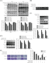
Figure 5. Effects of nobiletin on the MAPK pathway in U2OS and HOS cells
A. U2OS and HOS cells were treated with nobiletin (0–100 μM) for 24 h and the total cell lysates were then subjected to western blotting with anti-ERK, anti-JNK and anti-p38 antibodies. Quantitative results of ERK, JNK and p38 protein levels were adjusted to β-actin protein level. Concentration effects: p-ERK (U2OS: F = 22.386, p < 0.001; HOS: F = 25.074, p < 0.001); p-JNK (U2OS: F= 31.325, p < 0.001; HOS: F= 53.906, p < 0.001); p-p38 (U2OS: F = 1.694, p =0.227; HOS: F = 0.687, p =0.617). aSignificantly different, p < 0.05, when compared with control. bSignificantly different, p < 0.05, when compared with 25 μM. cSignificantly different, p < 0.05, when compared with 50 μM. B-E. U2OS cells were co-treated with specific protein inhibitor U0126 or SP600125, which incubated in the presence or absence of nobiletin (50 μM) for 24 h. (B) Gelatin zymography to analyze the activity of MMP-2 and MMP-9. (C and D) U2OS cells were co-treated with specific protein inhibitor U0126 or SP600125, which incubated in the presence or absence of nobiletin (50 μM) for 24 h and then the total cell lysates were subjected to western blotting with (C) anti-MMP-2, anti-MMP-9 and anti-ERK. MMP-2: F = 122.929, p < 0.001; MMP-9: F = 104.197, p < 0.001; p-ERK: F = 186.714, p < 0.001. (D) anti-MMP-2, anti-MMP-9 and anti-JNK. MMP-2: F = 56.897, p < 0.001; MMP-9: F = 50.368, p < 0.001; p-ERK: F = 26.765, p < 0.001. aSignificantly different, p < 0.05, when compared with 0 μM. bSignificantly different, p < 0.05, when compared with noibletin-treated group. cSignificantly different, p < 0.05, when compared with U0126 or SP600125 treated-group. (E) Cell migration and invasion ability of U2OS cells were measured by using a Boyden chamber assay. Migration: F = 68.12, p < 0.001; invasion: F = 102.991, p < 0.001. aSignificantly different, p < 0.05, when compared with 0 μM. bSignificantly different, p < 0.05, when compared with noibletin-treated group. cSignificantly different, p < 0.05, when compared with U0126 treated-group. dSignificantly different, p < 0.05, when compared with nobiletin plus U0126-treated group. eSignificantly different, p < 0.05, when compared with SP600125 treated-group.
Figure 6
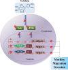
Figure 6. Propose signaling pathways by which nobiletin inhibits cell motility, migration and invasion of human osteosarcoma cells
⊝ indicates inhibition.

Similar articles

Cited by

References

    1. Kager L, Potschger U, Bielack S. Review of mifamurtide in the treatment of patients with osteosarcoma. Therapeutics and clinical risk management. 2010;6:279–286. - PMC - PubMed
    1. Mirabello L, Troisi RJ, Savage SA. Osteosarcoma incidence and survival rates from 1973 to 2004: data from the Surveillance, Epidemiology, and End Results Program. Cancer. 2009;115:1531–1543. - PMC - PubMed
    1. Yadav L, Puri N, Rastogi V, Satpute P, Ahmad R, Kaur G. Matrix metalloproteinases and cancer - roles in threat and therapy. Asian Pacific journal of cancer prevention: APJCP. 2014;15:1085–1091. - PubMed
    1. Hsieh YS, Chu SC, Yang SF, Chen PN, Liu YC, Lu KH. Silibinin suppresses human osteosarcoma MG-63 cell invasion by inhibiting the ERK-dependent c-Jun/AP-1 induction of MMP-2. Carcinogenesis. 2007;28:977–987. - PubMed
    1. Foroni C, Broggini M, Generali D, Damia G. Epithelial-mesenchymal transition and breast cancer: role, molecular mechanisms and clinical impact. Cancer treatment reviews. 2012;38:689–697. - PubMed

MeSH terms