. 2016 Aug;14(8):683-95.
doi: 10.1158/1541-7786.MCR-15-0479. Epub 2016 May 2.

AMPK Causes Cell Cycle Arrest in LKB1-Deficient Cells via Activation of CAMKK2

Affiliations

AMPK Causes Cell Cycle Arrest in LKB1-Deficient Cells via Activation of CAMKK2

Sarah Fogarty et al. Mol Cancer Res. 2016 Aug.

Abstract

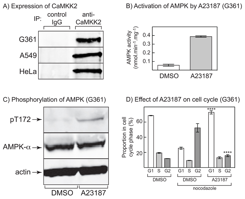
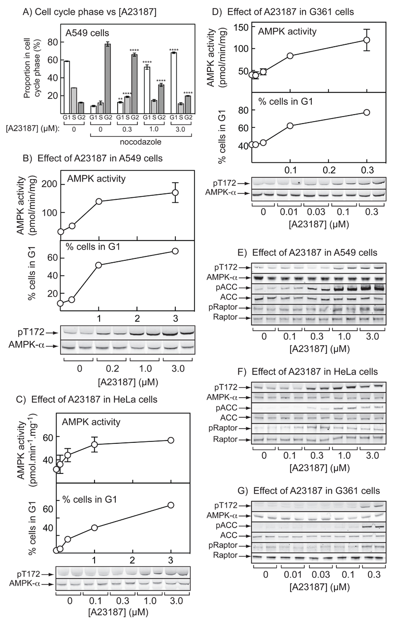
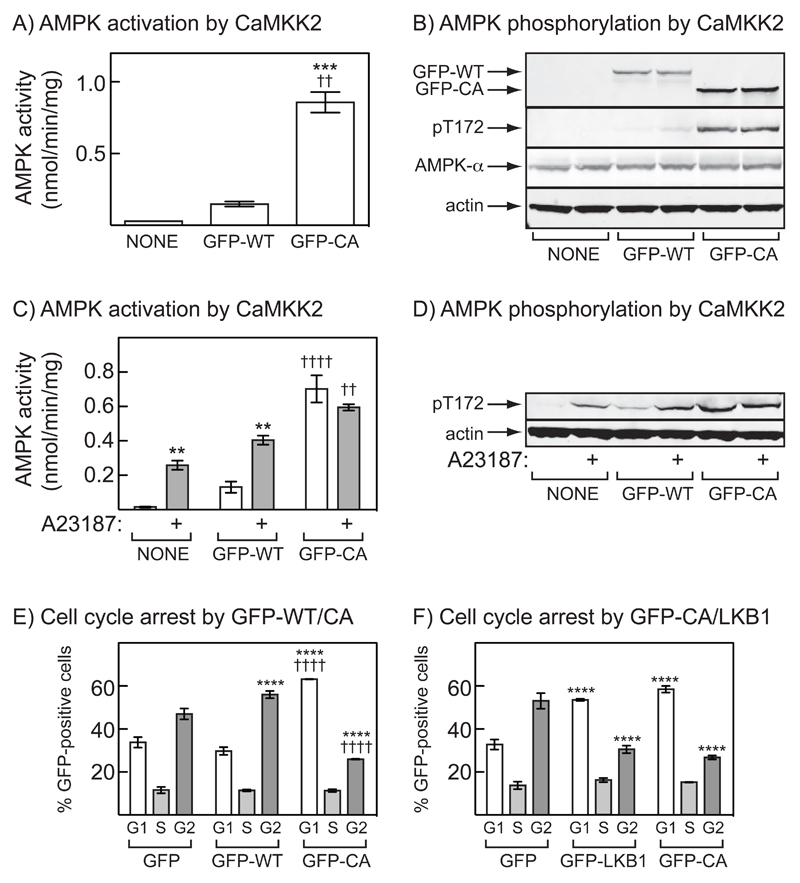
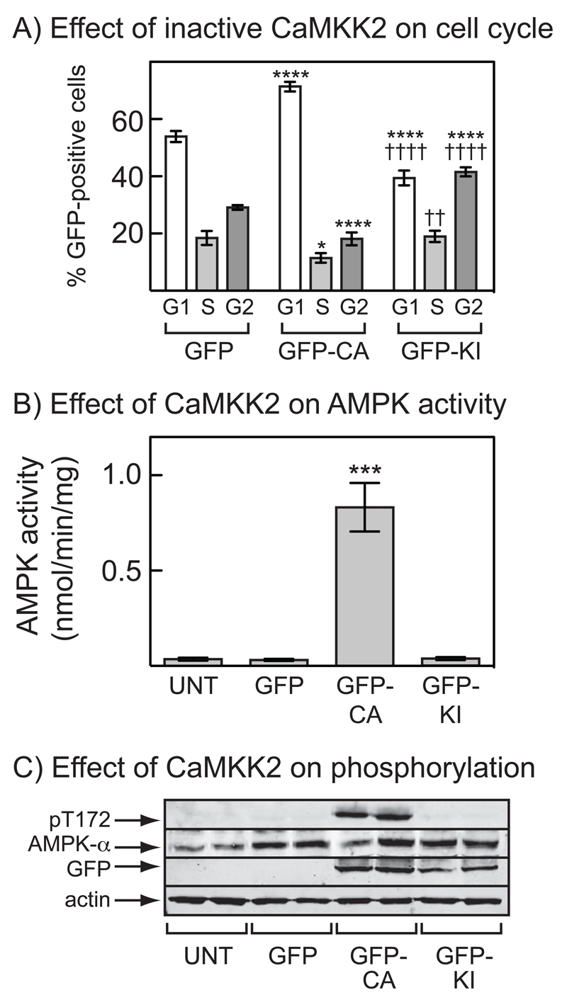
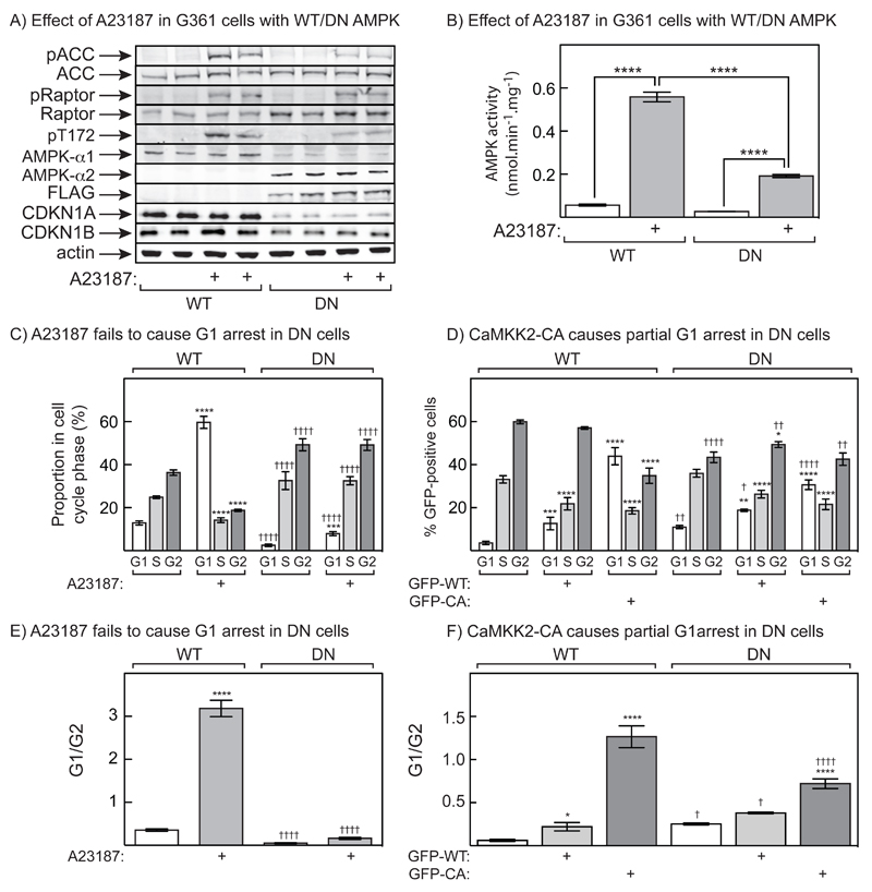
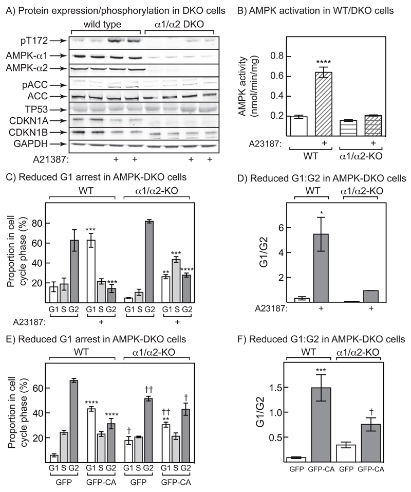
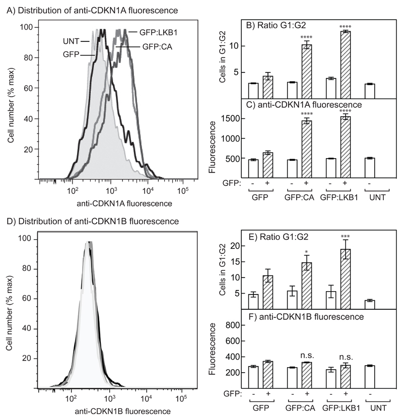
The AMP-activated protein kinase (AMPK) is activated by phosphorylation at Thr172, either by the tumor suppressor kinase LKB1 or by an alternate pathway involving the Ca(2+)/calmodulin-dependent kinase, CAMKK2. Increases in AMP:ATP and ADP:ATP ratios, signifying energy deficit, promote allosteric activation and net Thr172 phosphorylation mediated by LKB1, so that the LKB1-AMPK pathway acts as an energy sensor. Many tumor cells carry loss-of-function mutations in the STK11 gene encoding LKB1, but LKB1 reexpression in these cells causes cell-cycle arrest. Therefore, it was investigated as to whether arrest by LKB1 is caused by activation of AMPK or of one of the AMPK-related kinases, which are also dependent on LKB1 but are not activated by CAMKK2. In three LKB1-null tumor cell lines, treatment with the Ca(2+) ionophore A23187 caused a G1 arrest that correlated with AMPK activation and Thr172 phosphorylation. In G361 cells, expression of a truncated, Ca(2+)/calmodulin-independent CAMKK2 mutant also caused G1 arrest similar to that caused by expression of LKB1, while expression of a dominant-negative AMPK mutant, or a double knockout of both AMPK-α subunits, also prevented the cell-cycle arrest caused by A23187. These mechanistic findings confirm that AMPK activation triggers cell-cycle arrest, and also suggest that the rapid proliferation of LKB1-null tumor cells is due to lack of the restraining influence of AMPK. However, cell-cycle arrest can be restored by reexpressing LKB1 or a constitutively active CAMKK2, or by pharmacologic agents that increase intracellular Ca(2+) and thus activate endogenous CAMKK2.

Implications: Evidence here reveals that the rapid growth and proliferation of cancer cells lacking the tumor suppressor LKB1 is due to reduced activity of AMPK, and suggests a therapeutic approach by which this block might be circumvented. Mol Cancer Res; 14(8); 683-95. ©2016 AACR.


Conflict of interest statement

The authors have no conflicts of interest to declare.

Figures

Figure 1
Figure 1. Expression of CAMKK2 in LKB1-deficient tumor cells, and AMPK activation and cell cycle arrest in G361 melanoma cells.
(A) Extracts of G361, A549 and HeLa cells were immunoprecipitated with anti-CAMKK2 antibody or control immunoglobulin (IgG), and the precipitates analyzed by Western blotting using the same anti-CAMKK2 antibody. (B) AMPK activity in immunoprecipitates from G361 cells treated with 0.3 μM A23187 for 20 hr; results are mean ± SD, n = 2. (C) Western blotting using anti-pT172, anti-AMPK-α and anti-actin antibodies in extracts of G361 cells treated with A23187 as in (B). (D) G361 cells were treated with vehicle (DMSO) or A23187 (0.3 μM) for 20 hr with (where indicated) nocodazole (70 ng.ml-1) added for a further 18 hrs. The cells were then fixed, stained with propidium iodide and DNA content analyzed by flow cytometry. Bars show the percentages of cells with G1, S and G2/M phase DNA content (mean ± SD, n = 3); significant differences from DMSO control (with nocodazole) by 2-way ANOVA are shown, ****p<0.0001. Similar results were obtained in three identical experiments.
Figure 2
Figure 2. Effect of A23187 on AMPK activation and cell cycle arrest in A549, HeLa and G361 cells.
(A) A549 cells were treated with the indicated concentration of A23187 or vehicle (DMSO) for 20 hr with (where indicated) nocodazole (70 ng.ml-1) added for a further 18 hrs. The cells were then fixed, stained with propidium iodide and DNA content analyzed by flow cytometry. Bars show the percentages of cells with G1, S and G2/M phase DNA content (mean ± SD, n = 3); significant differences in the % of cells in the same cell cycle phase compared with DMSO control by 2-way ANOVA are shown: **p<0.01, ****p<0.0001. Similar results were obtained in three identical experiments. (B) AMPK activity measured in immunoprecipitates (top), percentage of cells in G1 phase (middle) (both mean ± SEM (n = 3)), and Thr172 phosphorylation (bottom) in A549 cells treated with various concentrations of A23187 for 20 hr;. (C) As (B), but in HeLa cells. (D) As (B), but in G361 cells (note different scale on x axis). (E) Phosphorylation of AMPK (Thr172), ACC (Ser79) and Raptor (Ser792) in A549 cells incubated as in (B). (F) As (E), but in HeLa cells incubated as in (C). (G) As (E), but in G361 cells incubated as in (D).
Figure 3
Figure 3. Effect of expression of constitutively active CAMKK2 on AMPK activation and the cell cycle in G361 cells.
(A) Cells were left untransfected, or were transfected with DNAs encoding GFP fused to full length (1-588, GFP-WT), or truncated (1-471, GFP-CA) CAMKK2. After 36 hr, AMPK activity was measured in immunoprecipitates; significant differences by 1-way ANOVA compared with untransfected control (***p<0.001) or GFP-WT (††p<0.01) are shown. (B) Analysis by Western blotting of extracts of untransfected cells and cells transfected with DNAs encoding GFP-WT or GFP-CA. Extracts were made after 36 hr and antibodies used were anti-GFP (top panel), anti-pt172, anti-AMPK-α or anti-actin. Results show samples from duplicate cell incubations: similar results were obtained in three independent experiments. (C) AMPK activity in immunoprecipitates from cells 36 hr after transfection with DNAs encoding GFP-WT or GFP-CA, or untransfected controls. Cells were then incubated with or without 10 μM A23187 for 1 hr; results are mean ± S.D. (n = 2); significant effects by two way ANOVA compared with vehicle controls, **p<0.01, or untransfected controls (††††p<0.0001, ††p<0.01) are shown. (D) Analysis by Western blotting of extracts of the same cells shown in (C). (E) Percentage of cells in G1, S or G2/M for cells transfected with DNAs encoding GFP, GFP-WT or GFP-CA (mean ± S.D., n = 3). After 36 hr, cells were treated with nocodazole (70 ng.ml-1) and 18 hrs later were fixed, stained with propidium iodide and the DNA content analyzed by flow cytometry. The flow cytometer was set-up to analyze only cells expressing GFP. Significant differences between the percentage of that cell cycle phase compared with GFP alone (****p<0.0001) or between GFP-WT and GFP-CA (††††p<0.0001) are shown (2-way ANOVA). Similar results were obtained in three identical experiments. (F) As (E), except that cells were transfected with DNAs encoding GFP, GFP-CA, or GFP-LKB1 plus STRADA and CAB39. Similar results were obtained in two identical experiments.
Figure 4
Figure 4. Cell cycle arrest and AMPK activation in G361 cells requires the kinase activity of CaMKKβ.
(A) Percentage of cells in G1, S or G2/M for cells transfected with DNAs encoding GFP, GFP-CA or GFP-KI (mean ± SD, n = 3). After 36 hr, cells were treated with nocodazole (70 ng.ml-1) and 18 hrs later were fixed, stained with propidium iodide and the DNA content analyzed by flow cytometry. The cytometer was set-up to analyze only cells expressing GFP. Significant differences by 2-way ANOVA compared with values for the same cell cycle phase with GFP alone (*p<0.05, ****p<0.001) or between GFP-CA and GFP-KI (††p<0.01, ††††p<0.0001) are shown. Similar results were obtained in two identical experiments. (B) AMPK activity measured in immunoprecipitates from untransfected cells or cells transfected with DNAs encoding GFP, GFP-CA or GFP-KI. Results are mean ± S.D. (n = 2); significant differences compared with untransfected cells are shown: ***p<0.001 (1-way ANOVA). (C) Analysis by Western blotting of extracts of the cells shown in (B) (n = 2).
Figure 5
Figure 5. Cell cycle arrest induced by A23187, or expression of constitutively active CAMKK2, in G361 cells is AMPK-dependent.
(A) G361 cells stably expressing a kinase-inactive (D157A) mutant of AMPK-α2 (DN), or control G361 cells (WT), were treated with A23187 (0.3 μM, 24 hr) and duplicate cell lysates analysed by Western blotting using the indicated antibodies. (B) AMPK activity was assessed in immunoprecipitates made with pan-α antibody. Significant differences by 2-way ANOVA are indicated (****p<0.0001). (C) Cells treated with A23187 (0.3 μM) for 24 hr and then nocodazole (70 ng.mL-1) for 24 hr were fixed, stained with propidium iodide and the proportions of cells in G1, S and G2 assessed by flow cytometry. Significant differences by 2-way ANOVA from controls without A23187: ***p<0.001, ****p<0.0001; significant differences between the same treatments in WT and DN cells: ††††p<0.0001. (D) Cell cycle arrest by transient expression of wild type or constitutively active CAMKK2 (GFP-WT or GFP-CA) is reduced in DN cells. DNAs encoding GFP-WT, GFP-CA or GFP alone (control) was transfected into G361 cells for 24 hr, and the cells then treated with nocodazole for a further 24 hr; cell cycle analysis was then performed only on transfected cells, i.e. those expressing GFP. Significant differences by 2-way ANOVA from GFP controls: **p<0.01, ***p<0.001, ****p<0.0001; significant differences between the same treatments in WT and DN cells: †p<0.05, ††p<0.01, ††††p<0.0001. (E) Same data as (C), but analysing G1:G2 ratio; statistical tests as in (C). (F) Same data as (D), but analysing G1:G2 ratio; statistical tests as in (D).
Figure 6
Figure 6. Cell cycle arrest by A23187 in G361 cells is AMPK-dependent.
(A) Expression and phosphorylation of proteins in wild type (WT) and α1/α2 double knockout (DKO) cells derived from G361 cells using the CRISPR/Cas9 system. Phosphorylation of AMPK, ACC and Raptor, and expression of AMPK-α1, -α2, ACC, Raptor, TP53, CDKN1A and CDKN1B, was assessed in cells treated with and without 0.3 μM A23187 for 24 hr. (B) AMPK activity in WT and DKO cells treated with and without 0.3 μM A23187 for 24 hr. Significant differences between A23187- and vehicle-treated samples, determined by 2-way ANOVA, are indicated: ****p<0.0001. (C) Cell cycle analysis in cells treated with 0.3 μM A23187 for 24 hr, then with nocodazole for a further 24 hr. The proportion of cells in each cell cycle phase was determined by flow cytometry of cells stained with propidium iodide. Significant differences in each cell cycle phase between A23187- and vehicle-treated samples, determined by 2-way ANOVA, are indicated: **p<0.01, ***p<0.001, ****p<0.0001. (D) Results from the experiment shown in (C) but expressed as ratios of G1:G2 phases. Significant differences between A23187- and vehicle-treated samples, determined by 2-way ANOVA, are indicated: p<0.05. (E) Cell cycle analysis in WT or DKO cells transfected with DNA encoding GFP or GFP-CA.for 24hr, then with nocodazole for 24 hr. Significant differences in each cell cycle phase between GFP and GFP-CA-transfected samples (**p<0.01; ****p<0.0001), and between WT and DKO samples (p<0.05, ††p<0.01) are indicated. (F) Same data as (E), but analysing G1:G2 ratios. Significant differences between GFP and GFP-CA-transfected samples (***p<0.001), and between WT and DKO samples (p<0.05) are indicated.
Figure 7
Figure 7. AMPK activation in G361 cells induces increased expression of CDKN1A but not CDKN1B, and cell cycle arrest.
G361 cells were transiently transfected with DNAs encoding GFP, GFP-LKB1 or GFP-CA. (A) Distribution of CDKN1A expression using anti-CDKN1A antibody in untransfected cells (UNT) and in cells expressing GFP, GFP-LKB1 or GFP-CA. (B) Ratios of G1:G2 phases in cells not expressing GFP (open bars), or in cells from the same dish expressing GFP, GFP-LKB1 or GFP-CA (hatched bars); significant differences by 2-way ANOVA from controls expressing GFP alone are shown: ****p<0.0001 (n = 4). (C) Expression of CDKN1A by flow cytometry in the same cells analysed in (B); significant differences by 2-way ANOVA from controls expressing GFP alone are shown: ****p<0.0001 (n = 4). (D) Distribution of CDKN1B expression using anti-CDKN1B antibody in untransfected cells (UNT) and in cells expressing GFP, GFP-LKB1 or GFP-CA; the grayscale coding is as in 1A and there are no significant differences. (E) Ratios of G1:G2 phases in untransfected cells (open bars), or in cells from the same dish expressing GFP, GFP-LKB1 or GFP-CA (hatched bars); significant differences by 2-way ANOVA from controls expressing GFP alone are shown: *p<0.05, ***p<0.001 (n = 4). (F) Expression of CDKN1B by flow cytometry in the same cells analysed in (E); there were no significant differences (n = 4).

Similar articles

Cited by

References

    1. Hardie DG. Molecular Pathways: Is AMPK a friend or a foe in cancer? Clin Cancer Res. 2015;21:3836–40. - PMC - PubMed
    1. Hardie DG. AMPK--sensing energy while talking to other signaling pathways. Cell Metab. 2014;20:939–52. - PMC - PubMed
    1. Hardie DG. AMPK: positive and negative regulation, and its role in whole-body energy homeostasis. Curr Opin Cell Biol. 2014;33C:1–7. - PubMed
    1. Hawley SA, Davison M, Woods A, Davies SP, Beri RK, Carling D, et al. Characterization of the AMP-activated protein kinase kinase from rat liver, and identification of threonine-172 as the major site at which it phosphorylates and activates AMP-activated protein kinase. J Biol Chem. 1996;271:27879–87. - PubMed
    1. Gowans GJ, Hawley SA, Ross FA, Hardie DG. AMP is a true physiological regulator of AMP-activated protein kinase by both allosteric activation and enhancing net phosphorylation. Cell Metab. 2013;18:556–66. - PMC - PubMed

MeSH terms

Substances