Emergence of undifferentiated colonies from mouse embryonic stem cells undergoing differentiation by retinoic acid treatment
- PMID: 27130680
- PMCID: PMC4884469
- DOI: 10.1007/s11626-016-0013-5
Emergence of undifferentiated colonies from mouse embryonic stem cells undergoing differentiation by retinoic acid treatment
Abstract
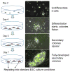
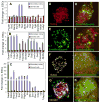
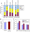
Retinoic acid (RA) is one of the most potent inducers of differentiation of mouse embryonic stem cells (ESCs). However, previous studies show that RA treatment of cells cultured in the presence of a leukemia inhibitory factor (LIF) also result in the upregulation of a gene called Zscan4, whose transient expression is a marker for undifferentiated ESCs. We explored the balance between these two seemingly antagonistic effects of RA. ESCs indeed differentiated in the presence of LIF after RA treatment, but colonies of undifferentiated ESCs eventually emerged from these differentiated cells - even in the presence of RA. These colonies, named secondary colonies, consist of three cell types: typical undifferentiated ESCs expressing pluripotency genes such as Pou5f1, Sox2, and Nanog; cells expressing Zscan4; and endodermal-like cells located at the periphery of the colony. The capacity to form secondary colonies was confirmed for all eight tested ESC lines. Cells from the secondary colonies - after transfer to the standard ESC medium - retained pluripotency, judged by their strong alkaline phosphatase (ALP) staining, typical colony morphology, gene expression profile, stable karyotype, capacity to differentiate into all three germ layers in embryoid body formation assays, and successful contribution to chimeras after injection into blastocysts. Based on flow cytometry analysis (FACS), the proportion of Zscan4-positive cells in secondary colonies was higher than in standard ESC colonies, which may explain the capacity of ESCs to resist the differentiating effects of RA and instead form secondary colonies of undifferentiated ESCs. This hypothesis is supported by cell-lineage tracing analysis, which showed that most cells in the secondary colonies were descendents of cells transiently expressing Zscan4.
Keywords: Mouse embryonic stem cells; Pluripotency; Retinoic acid; Zscan4.
Figures


Similar articles
-
Retinoic Acid Specifically Enhances Embryonic Stem Cell Metastate Marked by Zscan4.PLoS One. 2016 Feb 3;11(2):e0147683. doi: 10.1371/journal.pone.0147683. eCollection 2016. PLoS One. 2016. PMID: 26840068 Free PMC article.
-
Retinoic acid maintains self-renewal of murine embryonic stem cells via a feedback mechanism.Differentiation. 2008 Nov;76(9):931-45. doi: 10.1111/j.1432-0436.2008.00272.x. Epub 2008 Jul 1. Differentiation. 2008. PMID: 18637026
-
Global gene expression profiling reveals similarities and differences among mouse pluripotent stem cells of different origins and strains.Dev Biol. 2007 Jul 15;307(2):446-59. doi: 10.1016/j.ydbio.2007.05.004. Epub 2007 May 10. Dev Biol. 2007. PMID: 17560561 Free PMC article.
-
Heparan sulfate: a key regulator of embryonic stem cell fate.Biol Chem. 2013 Jun;394(6):741-51. doi: 10.1515/hsz-2012-0353. Biol Chem. 2013. PMID: 23370908 Free PMC article. Review.
-
Influence of Amino Acid Metabolism on Embryonic Stem Cell Function and Differentiation.Adv Nutr. 2016 Jul 15;7(4):780S-9S. doi: 10.3945/an.115.011031. Print 2016 Jul. Adv Nutr. 2016. PMID: 27422515 Free PMC article. Review.
Cited by
-
Short-term retinoic acid treatment sustains pluripotency and suppresses differentiation of human induced pluripotent stem cells.Cell Death Dis. 2018 Jan 5;9(1):6. doi: 10.1038/s41419-017-0028-1. Cell Death Dis. 2018. PMID: 29305588 Free PMC article.
-
Topographic Cues Impact on Embryonic Stem Cell Zscan4-Metastate.Front Bioeng Biotechnol. 2020 Mar 6;8:178. doi: 10.3389/fbioe.2020.00178. eCollection 2020. Front Bioeng Biotechnol. 2020. PMID: 32211397 Free PMC article.
-
Retinoic acid induces NELFA-mediated 2C-like state of mouse embryonic stem cells associates with epigenetic modifications and metabolic processes in chemically defined media.Cell Prolif. 2021 Jun;54(6):e13049. doi: 10.1111/cpr.13049. Epub 2021 May 7. Cell Prolif. 2021. PMID: 33960560 Free PMC article.
-
Enhanced self-renewal of human pluripotent stem cells by simulated microgravity.NPJ Microgravity. 2022 Jul 4;8(1):22. doi: 10.1038/s41526-022-00209-4. NPJ Microgravity. 2022. PMID: 35787634 Free PMC article.
-
Using high throughput screens to predict miscarriages with placental stem cells and long-term stress effects with embryonic stem cells.Birth Defects Res. 2022 Oct 1;114(16):1014-1036. doi: 10.1002/bdr2.2079. Epub 2022 Aug 18. Birth Defects Res. 2022. PMID: 35979652 Free PMC article. Review.
References
-
- Callicott RJ, Womack JE. Real-time PCR assay for measurement of mouse telomeres. Comp Med. 2006;56:17–22. - PubMed
-
- Colleoni S, Galli C, Gaspar JA, Meganathan K, Jagtap S, Hescheler J, Sachinidis A, Lazzari G. Development of a neural teratogenicity test based on human embryonic stem cells: response to retinoic acid exposure. Toxicol Sci. 2011;124:370–377. - PubMed
MeSH terms
Substances
Grants and funding
LinkOut - more resources
Full Text Sources
Other Literature Sources
Molecular Biology Databases
Research Materials

