. 2016 May 24;7(21):30804-19.
doi: 10.18632/oncotarget.9001.

A Toll-like receptor 2 agonist-fused antigen enhanced antitumor immunity by increasing antigen presentation and the CD8 memory T cells population

Affiliations

A Toll-like receptor 2 agonist-fused antigen enhanced antitumor immunity by increasing antigen presentation and the CD8 memory T cells population

Chiao-Chieh Wu et al. Oncotarget. .

Abstract

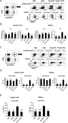
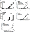
The induction of long-lived effector CD8+ T cells is key to the development of efficient cancer vaccines. In this study, we demonstrated that a Toll-like receptor 2 (TLR2) agonist-fused antigen increased antigen presentation via TLR2 signaling and induced effector memory-like CD8+ T cells against cancer after immunization. The N-terminus of ovalbumin (OVA) was biologically fused with a bacterial lipid moiety TLR2 agonist to produce a recombinant lipidated ovalbumin (rlipo-OVA). We demonstrated that rlipo-OVA activated bone marrow-derived dendritic cells (BM-DCs) maturation and increased antigen presentation by major histocompatibility complex (MHC) class I via TLR2. After immunization, rlipo-OVA skewed the immune response towards T helper (Th) 1 and induced OVA-specific cytotoxic T lymphocyte (CTL) responses. Moreover, immunization with rlipo-OVA induced higher numbers of effector memory (CD44+CD62L-) CD8+ T cells compared with recombinant ovalbumin (rOVA) alone or rOVA mixed with the TLR2 agonist Pam3CSK4. Accordingly, the CD27+CD43+ effector memory CD8+ T cells expressed high levels of the long-lived CD127 marker. The administration of rlipo-OVA could inhibit tumor growth, but the anti-tumor effects were lost after the depletion of CD8 or CD127 cells in vivo. These findings suggested that the TLR2 agonist-fused antigen induced long-lived memory CD8+ T cells for efficient cancer therapy.

Keywords: Toll-like receptor 2; antigen presentation; memory T cells; rlipo-immunogen; tumor regression.


Conflict of interest statement

No potential conflicts of interest were disclosed.

Figures

Figure 1
Figure 1. Construction, production and identification of rOVA and rlipo-OVA
(A) The plasmid maps of pOVA and pLOVA that express rOVA and rlipo-OVA, respectively. (B) The rOVA and rlipo-OVA protein purification process used 10% reducing SDS-PAGE followed by Coomassie Blue staining and anti-HisTag antibodies for immunoblotting. The recombinant rOVA was expressed in the E. coli strain BL21 (DE3). Lane 1, rOVA expression after IPTG induction; lane 2, protein expression in the absence of IPTG induction; lane 3, rOVA extracted fraction; lane 4, recombinant rOVA purified by Ni-NTA resin; and lane 5, polished recombinant rOVA by Q sepharose resin. Lanes 6–10 show immunoblotting to monitor the process of rOVA purification; these lanes are the same as lanes 1–5, respectively. The recombinant rlipo-OVA was expressed in the E. coli strain C43 (DE3). Lane 11, rlipo-OVA expression after IPTG induction; lane 12, protein expression in the absence of IPTG induction; lane 13, rlipo-OVA extracted fraction; and lane 14, rlipo-OVA protein purified by Ni-NTA resin. Lanes 15–18 show immunoblotting to monitor the rlipo-OVA purification process; the samples in these lanes are the same as those in lanes 11–14, respectively. The arrows indicate the electrophoretic positions of rOVA or rlipo-OVA in the SDS gels or blots. (C) N-terminal rlipo-OVA fragments were obtained and identified after 3 days of digestion. The digested sample was analyzed on a WatersR MALDI micro MX mass spectrometer. The MALDI-TOF MS spectra revealed lipid peptide signals with three m/z value peaks of 1452.09, 1466.10, and 1480.13.
Figure 2
Figure 2. rlipo-OVA stimulates immune cell activation via TLR2
(A) Splenocytes were isolated from wild-type C57BL/6 mice and seeded at a density of 2.5 × 10⁵ cells/well in 96-well cell culture plates. The cells were incubated with different concentrations of LPS (10–1000 ng/ml), Pam3 (10–1000 ng/ml), OVA (10–1000 ng/ml), rOVA (10–1000 ng/ml) or rlipo-OVA (10–1000 ng/ml) for 72 h. After 72 h, 10% CellTiter 96® AQueous One Solution Reagent was added to each well and the OD490 was measured. Stimulation index: (SI) = OD490 of the stimulated cells/ OD490 of the negative controls. The data represent the mean ± SD of six animals. BM-DCs from wild-type mice were cultured in medium supplemented with LPS (100 ng/ml), Pam3 (100 nM), OVA (2.5 μg/ml), rOVA (2.5 μg/ml) or rlipo-OVA (2.5 μg/ml) in the presence or absence of polymyxin B (50 μg/ml). After an 18-h incubation, the dendritic cells were pre-gated on the CD11c+ cell population to measure the expression of the cell surface markers CD40 (B) and CD80 (C) by flow cytometry. The mean fluorescence intensity (MFI)% for cells cultured in medium was defined as 100%, and the independent experiments were performed in triplicate. For the inflammatory cytokine secretion studies, the supernatants were collected and analyzed for TNF-α (D) and IL-12 (p40) (E) production by ELISA. Three independent experiments were performed, and the data are presented as the mean + SD. (F) BM-DCs from WT or TLR2KO mice were cultured either in medium alone or in medium supplemented with LPS (100 ng/ml), Pam3 (100 nM), OVA (2.5 μg/ml), rOVA (2.5 μg/ml) or rlipo-OVA (2.5 μg/ml). After an 18-h incubation, the supernatants were collected and analyzed for TNF-α production by ELISA. The data are presented as the mean + SD, n = 3.
Figure 3
Figure 3. rlipo-OVA increases BM-DCs presenting the OVA-H-2Kb molecule via TLR2 signaling
(A) BM-DCs from WT, TLR2KO and MyD88KO mice were incubated for 24 h with PBS, (100–25 nM) rOVA, or (100–25 nM) rlipo-OVA, and the OVA-H-2Kb OVA-peptide were assessed by flow-cytometry analysis. Cells were stained with the PE-labeled 25-D1.16 antibody that recognizes OVA-derived SIINFEKL (SII) assembled with BM-DCs H-2Kb. (B) Antigen presentation was determined by cell proliferation using a [3H]thymidine incorporation assay. The antigen-pulsed BM-DCs (1 × 10⁴) were cultured with 1 × 10⁵ CD8+ T cells of OT-1 mice for 72 h. Average cpm incorporated in triplicate samples are shown (+SD). (C) At 5 day after the antigen-pulsed BM-DCs were cultured with CD8+ T cells of OT-1 mice, supernatant was collected and IFN-γ was measured using ELISA. The data are expressed as the means + SD from three independent experiments.
Figure 4
Figure 4. Immunization with rlipo-OVA induces higher levels of OVA-specific cytotoxic T cell responses compared to immunization with rOVA
C57BL/6 mice were immunized twice by subcutaneous injection at one-week intervals. The experiments were performed after seven days of last immunization. (A) A density of 5 × 10⁵ splenocytes (per well) from immunized mice were incubated with 2.5 μg/ml of the irrelevant RAHYNIVTF (RAH) peptide or SII peptide for 48 h in an anti-IFN-γ-coated 96-well ELISPOT plate. The IFN-γ-secreting spots were calculated using an ELISPOT reader. The data are expressed as the mean + SD, n = 6 / per group. (B) The SII-specific IFN-γ production in CD8+ T cells. Splenocytes were isolated from each group of vaccinated mice. The cells were re-stimulated with 1 μg/ml of the SII peptides for 20 h; for the final 4 h, the cells were treated with 10 ng/ml phorbol 12-myristate 12-acetate (PMA), 1 μg/ml ionomycin, and Brefeldin A solution. The percentages of CD8+/IFN-γ+ T cells pre-gated on CD3+ cells were determined by flow cytometry. The data are presented as the mean + SD, n = 5. (C) The graphs show the specific lysis of SII peptide-pulsed targets (CFSEhigh) and the un-pulsed peptide control (CFSElow). The graph summarizes the results of the specific lysis in vivo (D). The following equation was used to analyze specific lysis:% Specific lysis = [(% non-peptide × A) −% SII peptide] / (% non-peptide × A). Adjustment factor A = SII peptide/non-peptide from the naïve controls. The data are expressed as the mean + SD of nine animals per group.
Figure 5
Figure 5. Immunization with rlipo-OVA increases memory CD8 T cells
5 × 10⁵ OT-I CD8+ T cells were adoptively transferred via the tail vein one day before once immunization. The mice were injected in each footpad with either PBS alone, rlipo-OVA (5 μg), rOVA (5 μg), or rOVA (5 μg) formulated with an equal molar amount of Pam3CSK4 in 50 ml of PBS. For the analysis of memory populations, the lymphocytes were pooled from the popliteal and inguinal lymph nodes and splenocytes were isolated from the spleens from the groups of immunized mice 28 days after once immunization. The cells were stained with Va2TCR-PE and CD8a-APC. (A) The antibodies CD44-FITC and CD62L-APC were used to evaluate TCM cells (CD44+CD62L+) and TEM cells (CD44+CD62L) (B). (C) The antibodies CD27-FITC and CD43 PE-Cy7 were used to assess effector CD8+ T cells in the memory population evaluated for the expression of CD27+CD43 and CD27+CD43+ (D). The CD127 marker was used to evaluate the CD27+CD43+ memory population in the lymph nodes and spleens (E). The data are expressed as the mean + SEM. ns = not significant, *p < 0.05, **p < 0.01 and ***p < 0.001 are significant differences compared to rlipo-OVA, n = 6.
Figure 6
Figure 6. Immunization with rlipo-OVA induces CD8-dependent anti-tumor effects and the functional requirement of CD127 for long term anti-tumor ability
(A) The mice were inoculated with 2 × 10⁴ EG7 cells in a total volume of 200 μl of PBS. After 3 days, tumor-bearing mice were subcutaneously injected once with rOVA (30 μg), rlipo-OVA (30 μg) or PBS (n = 12). (B) Three groups (rlipo-OVA/CD4, rlipo-OVA/CD8 and rlipo-OVA/Rat IgG) of mice were injected with anti-CD4, anti-CD8 and control antibodies one day before the injection of rlipo-OVA (30 μg/mouse). On day 3, these groups and two additional control groups (rlipo-OVA and PBS) were subcutaneously injected once with rlipo-OVA (30 μg/mouse) or PBS at the base of tail after inoculation with EG7 cells (2 × 10⁴/per mouse; n = 6). (C and D) Mice were immunized twice by subcutaneous injection of rOVA (30 μg), rlipo-OVA (30 μg) or PBS alone at one-week intervals, and the experiments were performed 4 weeks after the final immunization. (C) The splenocytes (5 × 10⁵ cells/well) from the groups of immunized mice were incubated with or without 5 μg/ml of the RAH peptide or SII peptide for 48 h in an anti-IFN-γ-coated 96-well ELISPOT plate. The IFN-γ-secreting spots were measured using an ELISPOT reader (n = 6). (D) The mice were subcutaneously injected with a density of 2 × 10⁴ EG7 cells in a total volume of 200 μl. Tumor growth was observed three times per week (n = 8). (E) Three groups of C57BL/6 mice were subcutaneously injected with rlipo-OVA (30 μg), and the control group was administered PBS at one-week intervals. On day 17, two groups of immunized rlipo-OVA mice were treated intraperitoneally (i.p.) with either 0.5 mg of rat anti-mouse CD127 antibody or 0.5 mg of rat anti-mouse IgG antibody. A total of 2 × 10⁴ EG7 tumor cells were inoculated on day 35. The data are shown as the means + SE, (n = 6). The tumor volume was calculated as the length × width × width × 1/2 (mm3). The data are shown as the mean + SEM, ns = no significant difference, *p < 0.05, **p < 0.01 and ***p < 0.001 indicate significant differences compared with rlipo-OVA.

Similar articles

Cited by

References

    1. Palucka K, Banchereau J. Dendritic-Cell-Based Therapeutic Cancer Vaccines. Immunity. 2013;39:38–48. - PMC - PubMed
    1. Andersen MH, Schrama D, Straten PT, Becker JC. Cytotoxic T cells. J Invest Dermatol. 2006;126:32–41. - PubMed
    1. Jin B, Sun T, Yu XH, Yang YX, Yeo AE. The effects of TLR activation on T-cell development and differentiation. Clin Dev Immunol. 2012;2012:836485. - PMC - PubMed
    1. Janssen EM, Droin NM, Lemmens EE, Pinkoski MJ, Bensinger SJ, Ehst BD, Griffith TS, Green DR, Schoenberger SP. CD4(+) T-cell help controls CD8(+) T-cell memory via TRAIL-mediated activation-induced cell death. Nature. 2005;434:88–93. - PubMed
    1. Filipazzi P, Pilla L, Mariani L, Patuzzo R, Castelli C, Camisaschi C, Maurichi A, Cova A, Rigamonti G, Giardino F, Di Florio A, Asioli M, Frati P, et al. Limited induction of tumor cross-reactive T cells without a measurable clinical benefit in early melanoma patients vaccinated with human leukocyte antigen class I-modified peptides. Clin Cancer Res. 2012;18:6485–6496. - PubMed

MeSH terms