. 2016 Jul;8(4):362-74.
doi: 10.4168/aair.2016.8.4.362.

House Dust Mite-Derived Chitin Enhances Th2 Cell Response to Inhaled Allergens, Mainly via a TNF-α-Dependent Pathway

Affiliations

House Dust Mite-Derived Chitin Enhances Th2 Cell Response to Inhaled Allergens, Mainly via a TNF-α-Dependent Pathway

Jun Pyo Choi et al. Allergy Asthma Immunol Res. 2016 Jul.

Abstract

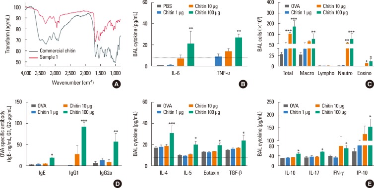
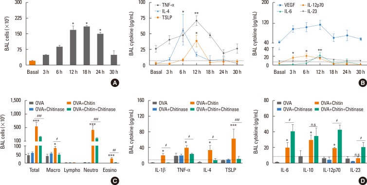
Purpose: Chitin is a potent adjuvant in the development of immune response to inhaled allergens in the airways. According to other studies, chitin is known as multi-faced adjuvants which can induce Th2 responses. Recently, we found that TNF-α is a key mediator in the development of Th2 cell response to inhaled allergens. Here, we evaluated the immunologic mechanisms in the development of airway hypersensitivity to inhaled allergens, enhanced by house dust mite (HDM)-derived chitin.

Methods: The role of TNF-α and TLRs was evaluated in an airway hypersensitivity mouse model induced by a sensitization with an allergen (ovalbumin, OVA) and HDM-derived chitin using mice with the null mutation of target genes.

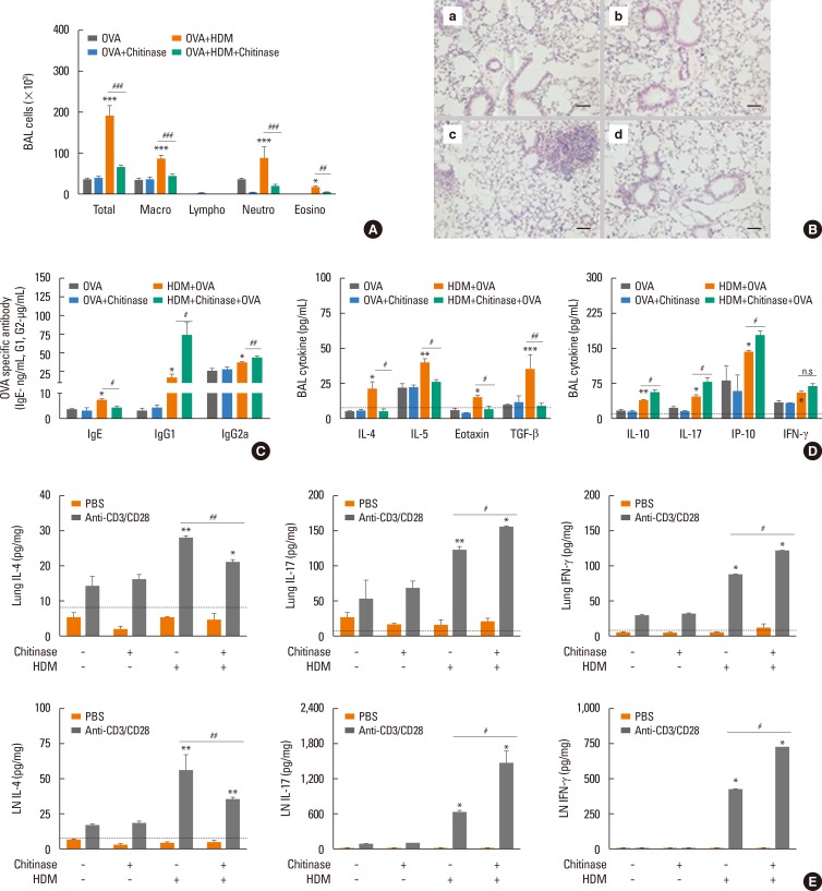
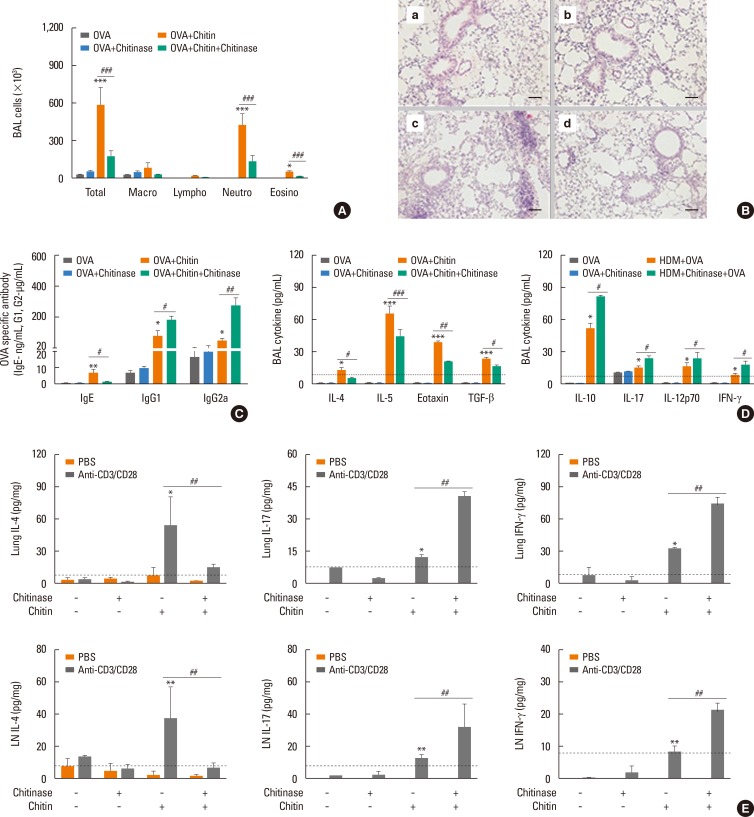
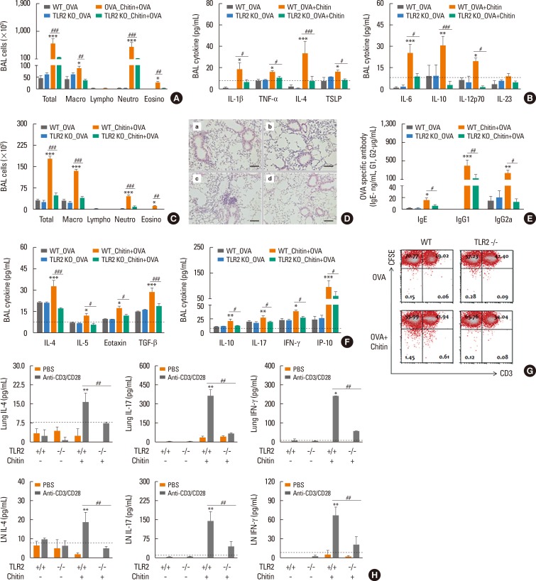
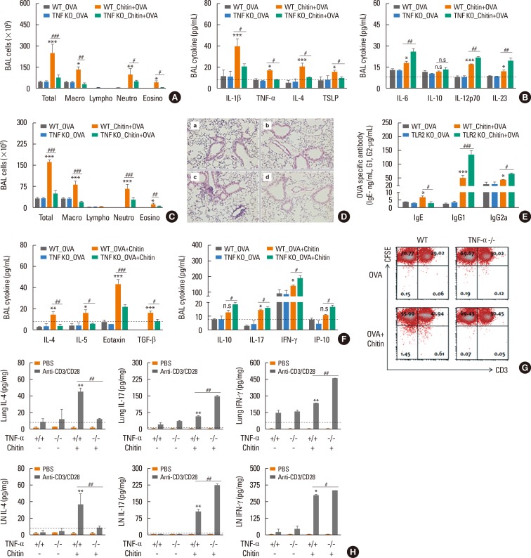
Results: The present study showed that airway sensitization with HDM-derived chitin plus OVA enhanced OVA-induced airway inflammation v. OVA alone. This phenotype was associated with the increased expression of Th1, Th2, and Th17 cytokines and also with the enhanced production of OVA-specific IgE, IgG1, and IgG2a. As for T cell responses, OVA-specific Th2 cell response, enhanced by chitin, was abolished by the treatment of chitinase, whereas Th1 and Th17 cell responses enhanced by this treatment. Moreover, the null mutation of the TNF-α gene revealed similar effects as the chitinase treatment. In contrast, all the OVA-specific T cell responses, enhanced by chitin, were blocked by the absence of TLR2, but not of TLR1, TLR4, or TLR6.

Conclusions: In conclusion, these data suggest that HDM-derived chitin may enhance airway hypersensitivity to inhaled allergens, via the TLR2-dependent pathway, and that chitin-induced TNF-α can be a key mediator in the development of Th2 cell response to inhaled allergens.

Keywords: Chitin; TNF-α; Th2 cell response; house dust mite.


Conflict of interest statement

There are no financial or other issues that might lead to conflict of interest.

Figures

Fig. 1
Fig. 1. Th2-dominant airway inflammation induced by HDM extracts plus OVA is down-regulated by chitinase treatment. For all panels, wild-type mice (C57BL/6 background) were sensitized with 75 µg of OVA+100 µg of HDM with or without chitinase, then challenged with OVA (50 µg) alone. The evaluations were performed 24 hours after last OVA challenge. *P<0.05; **P<0.01; ***P<0.001 relative to individual control groups, OVA or OVA+Chitinase, respectively; #P<0.05; ##P<0.01; ###P<0.001; n.s, not significant. Data are presented as the mean±SEM (n=5 mice per group) are from a single experiment representative of at least 3 independent experiments. (A) Bronchoalveolar lavage (BAL) cellularity after acute allergen challenge. (B) Lung histologic findings (a, OVA; b, OVA+chitinase; c, OVA+HDM; d, OVA+HDM+chitinase; magnification: 100×, scale bar: 200 µm). (C) The production of OVA-specific IgE, IgG1, and IgG2a in serum after acute OVA challenge. (D) Cytokine productions of IL-4, IL-5, eotaxin, TGF-β, IL-10, IL-17, IP-10, and IFN-γ in BAL fluid after last allergen challenge. For (E) panel, cells were isolated from lung-draining lymph nodes (LN) and lung tissues, and incubated with PBS or anti-CD3 and CD28 antibodies for 12 hours. Levels of each cytokine were evaluated in supernatant fraction. (E) Levels of IL-4, IL-10, IL-17, and IFN-γ from lung cells, and lung-draining LN cells.
Fig. 2
Fig. 2. Chitins extracted from HDM have an adjuvant effect to enhance adaptive immune response to inhaled allergens. (A) Fourier transform infrared (FTIR) spectroscopy of commercial crab chitin (commercial chitin) and HDM derived chitin (sample1). For (B) panel, HDM-derived chitin was sensitized to wild type (WT) mouse airways and then evaluated after 12 hours. For (C-E) panels, wild-type mice (C57BL/6 background) were sensitized with 75 µg of OVA, or 75 µg of OVA+1, 10, or 100 µg of HDM-derived chitin. Data are presented as the mean±SEM (n=5 mice per group) are from a single experiment representative of at least 2 independent experiments. *P<0.05; **P<0.01; ***P<0.001 relative to the OVA group; (B) Levels of IL-6, and TNF-α in BAL fluid after sensitization of each dose of HDM-derived chitin. For (C-E), mice were challenged with OVA (50 µg) alone and evaluated 24 hours after the last OVA challenge. (C) Bronchoalveolar lavage (BAL) cellularity after 24 hours of last OVA challenge. (D) Levels of OVA-specific IgE, IgG1, and IgG2a in serum after acute challenge. (E) Levels of IL-4, IL-5, eotaxin, TGF-β, IL-10, IL-17, IFN-γ, and IP-10 in BAL fluid after last acute OVA challenge.
Fig. 3
Fig. 3. Th2-type innate response, induced by HDM-derived chitin is abolished by chitinase treatment. Data are presented as the mean±SEM (n=5 mice per group) are from a single experiment representative of at least 3 independent experiments. *P<0.05; **P<0.01; ***P<0.001; n.s, not significant. relative to basal or the OVA group; For panel (A) and (B), 100 µg of HDM-derived chitin was sensitized once to wild type (WT) mouse airways and then evaluated at various time points. (A) Inflammatory cell infiltration of airway after HDM-derived chitin sensitization. (B) Levels of TNF-α, IL-4, TSLP, VEGF, IL-6, IL-12p70, and IL-23 in bronchoalveolar lavage (BAL) fluids. For (C-D), the evaluation was performed at 12 hours after HDM-derived chitin sensitization. (C) BAL cellularity after sensitization. (D) Levels of IL-1β, TNF-α, IL-4, TSLP, IL-6, IL-10, IL-12p70, and IL-23 in BAL fluid after sensitization.
Fig. 4
Fig. 4. OVA-induced Th2-cell response enhanced by HDM-derived chitin is abolished by chitinase treatment. For all panels, wild-type (WT) mice (C57BL/6 background) were sensitized with 75 µg of OVA, 75 µg of OVA+chitinase, 75 µg of OVA+100 µg of HDM-derived chitin, or 75 µg of OVA+100 µg of HDM-derived chitin+chitinase, then challenged with OVA (50 µg) alone, and evaluated 24 hours after last OVA challenge. Data are presented as the mean±SEM (n=5 mice per group) are from a single experiment representative of at least 3 independent experiments. *P<0.05; **P<0.01; ***P<0.001 relative to the OVA group; #P<0.05; ##P<0.01; ###P<0.001. (A) Bronchoalveolar lavage (BAL) cellularity after acute OVA challenge. (B) Lung histologic findings (a, OVA; b, OVA+chitinase; c, OVA+HDM-derived chitin; d, OVA+HDM-derived chitin+chitinase; magnification: 100×, scale bar: 200 µm). (C) Levels of OVA-specific IgE, IgG1, IgG2a in serum after acute challenge. (D) Levels of IL-4, IL-5, eotaxin, TGF-β, IL-10, IL-17, IL-12p70, and IFN-γ in BAL fluid after acute challenge. For panels (E), cells were isolated from lung-draining lymph nodes (LNs) and lung tissues, and incubated with PBS or CD3 and CD28 antibodies for 12 hours. Levels of each cytokine were evaluated in supernatant fraction. (E) Levels of IL-4, IL-10, IL-17, and IFN-γ from lung and lung-draining LN cells.
Fig. 5
Fig. 5. OVA-induced adaptive immune responses enhanced by HDM-derived chitin is abolished by the absence of TLR2. For all panels, TLR2-deficient mice and wild-type (WT) mice (C57BL/6 background) were sensitized with 75 µg of OVA, or 75 µg of OVA+100 µg of HDM derived chitin. *P<0.05; **P<0.01; ***P<0.001 relative to the wild-type OVA group; #P<0.05; ##P<0.01; ###P<0.001. Data are presented as the mean±SEM (n=5 mice per group) are from a single experiment representative of at least 2 independent experiments. For (A-C) panels, evaluations were performed after 12-hour sensitization. (A) Bronchoalveolar lavage (BAL) cellularity after sensitization. (B) Levels of IL-1β, TNF-α, IL-4, TSLP, IL-6, IL-10, IL-12p70, and IL-23 in BAL fluid after sensitization. For panels (C-H), mice were challenged with OVA (50 µg) alone, and evaluated 24 hours after last OVA challenge. (C) BAL cellularity after acute challenge. (D) Lung histologic findings (a, WT_OVA; b, TLR2 KO_OVA; c, WT_Chitin+OVA; d, TLR2 KO_OVA+chitin; magnification: 100×, scale bar: 200 µm). (E) Levels of OVA-specific IgE, IgG1, IgG2a in serum after acute challenge. (F) Levels of IL-4, IL-5, eotaxin, TGF-β, IL-10, IL-17, IFN-γ, and IP-10 in BAL fluid after acute challenge. For panels (G, H), cells were isolated from lung-draining lymph nodes (LNs) and lung tissues. (G) The levels of memory T-cell proliferation by stimulation of OVA in LN cells from each mouse. For (H), isolated cells were incubated with PBS or CD3 and CD28 antibodies for 12 hours. Levels of individual cytokines were evaluated in supernatant fraction. (H) Levels of IL-4, IL-10, IL-17, and IFN-γ from lung and lung-draining LN cells.
Fig. 6
Fig. 6. OVA-induced Th2-cell response, enhanced by HDM-derived chitin is abolished by the absence of TNF-α. Th2 responses induced by airway sensitization with ovalbumin (OVA) plus HDM-derived chitin is diminished in the absence of TNF-α, whereas the Th1- and Th17-cell responses are enhanced. For all panels, TNF-α-deficient (TNF KO) mice and wild-type (WT) mice (C57BL/6 background) were sensitized with 75 µg of OVA, or 75 µg of OVA+100 µg of HDM-derived chitin. Data are presented as the mean±SEM (n=5 mice per group) are from a single experiment representative of at least two independent experiments. *P<0.05; **P<0.01; ***P<0.001 relative to the wild-type OVA group; #P<0.05; ##P<0.01; ###P<0.001; n.s, not significant. For panel (A) and (B), the evaluation was performed 12 hours after sensitization. (A) Bronchoalveolar lavage (BAL) cellularity after sensitization. (B) Levels of IL-1β, TNF-α, IL-4, TSLP, IL-6, IL-10, IL-12p70, and IL-23 in BAL fluid after sensitization. For (C-H), mice were challenged with OVA (50 µg) alone and evaluated 24 hours after last OVA challenge. (C) BAL cellularity after acute challenge. (D) Lung histologic findings (a, WT_OVA; b, TNF KO_OVA; c, WT_OVA+chitin; d, TNF KO_OVA+chitin; magnification: 100×, scale bar: 200 µm). (E) Levels of OVA-specific IgE, IgG1, and IgG2a in serum after acute challenge. (F) Levels of IL-4, IL-5, eotaxin, TGF-β, IL-10, IL-17, IP-10, and IFN-γ in BAL fluid after acute challenge. For panels (G, H), cells were isolated from lung-draining lymph nodes (LNs) and lung tissues. (G) The levels of memory T-cell proliferation by stimulation of OVA in LN cells from each mouse. For (H), isolated cells were incubated with PBS or CD3 and CD28 antibodies for 12 hours. Levels of individual cytokine were evaluated in supernatant fraction. (H) Levels of IL-4, IL-10, IL-17, and IFN-γ from lung and lung-draining LN cells.

Similar articles

Cited by

References

    1. Dowse GK, Turner KJ, Stewart GA, Alpers MP, Woolcock AJ. The association between Dermatophagoides mites and the increasing prevalence of asthma in village communities within the Papua New Guinea highlands. J Allergy Clin Immunol. 1985;75:75–83. - PubMed
    1. Charpin D, Birnbaum J, Haddi E, Genard G, Lanteaume A, Toumi M, et al. Altitude and allergy to house-dust mites. A paradigm of the influence of environmental exposure on allergic sensitization. Am Rev Respir Dis. 1991;143:983–986. - PubMed
    1. Platts-Mills TA, Vervloet D, Thomas WR, Aalberse RC, Chapman MD. Indoor allergens and asthma: report of the Third International Workshop. J Allergy Clin Immunol. 1997;100:S2–S24. - PubMed
    1. Neville AC, Parry DA, Woodhead-Galloway J. The chitin crystallite in arthropod cuticle. J Cell Sci. 1976;21:73–82. - PubMed
    1. Fuhrman JA, Piessens WF. Chitin synthesis and sheath morphogenesis in Brugia malayi microfilariae. Mol Biochem Parasitol. 1985;17:93–104. - PubMed