. 2016 Apr 19:6:24346.
doi: 10.1038/srep24346.

Cellular uptake of exogenous calcineurin B is dependent on TLR4/MD2/CD14 complexes, and CnB is an endogenous ligand of TLR4

Affiliations

Cellular uptake of exogenous calcineurin B is dependent on TLR4/MD2/CD14 complexes, and CnB is an endogenous ligand of TLR4

Jinju Yang et al. Sci Rep. .

Abstract

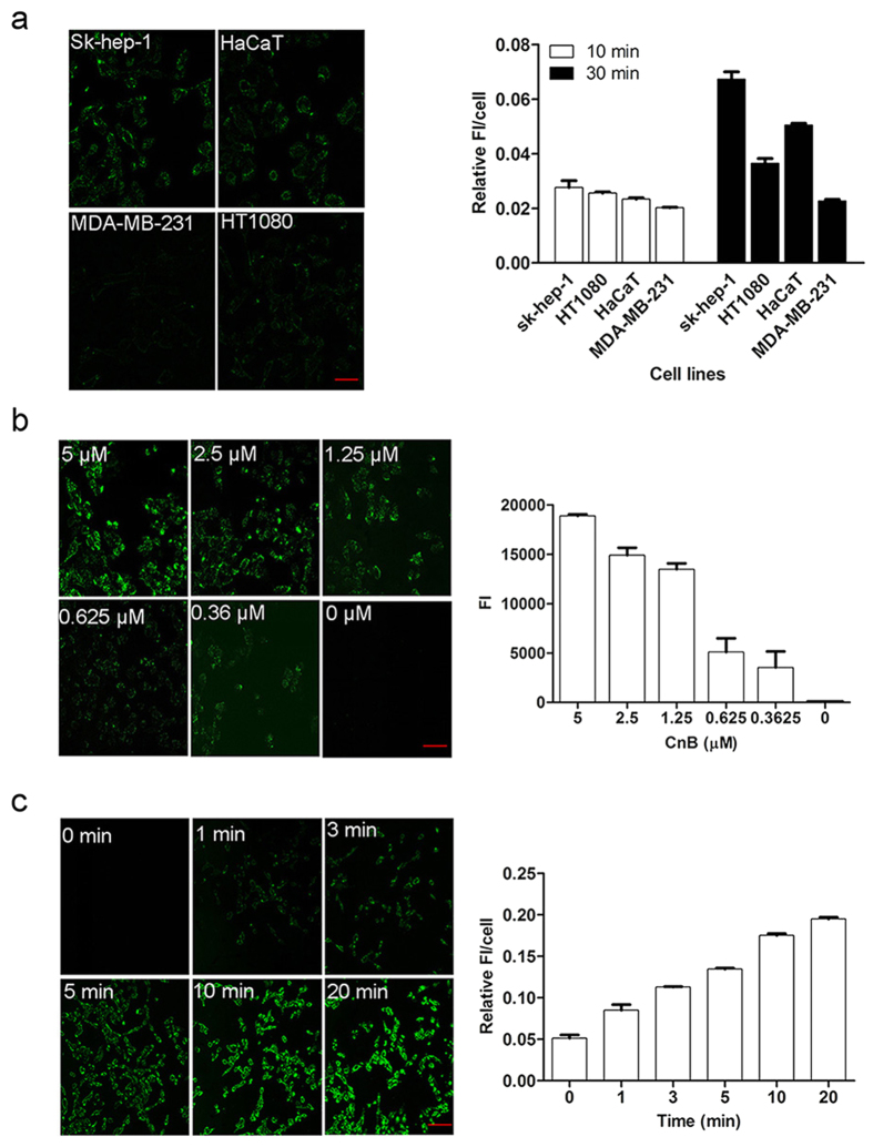
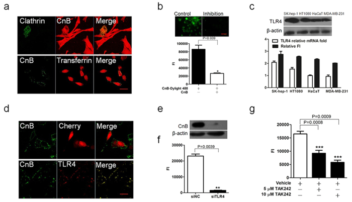
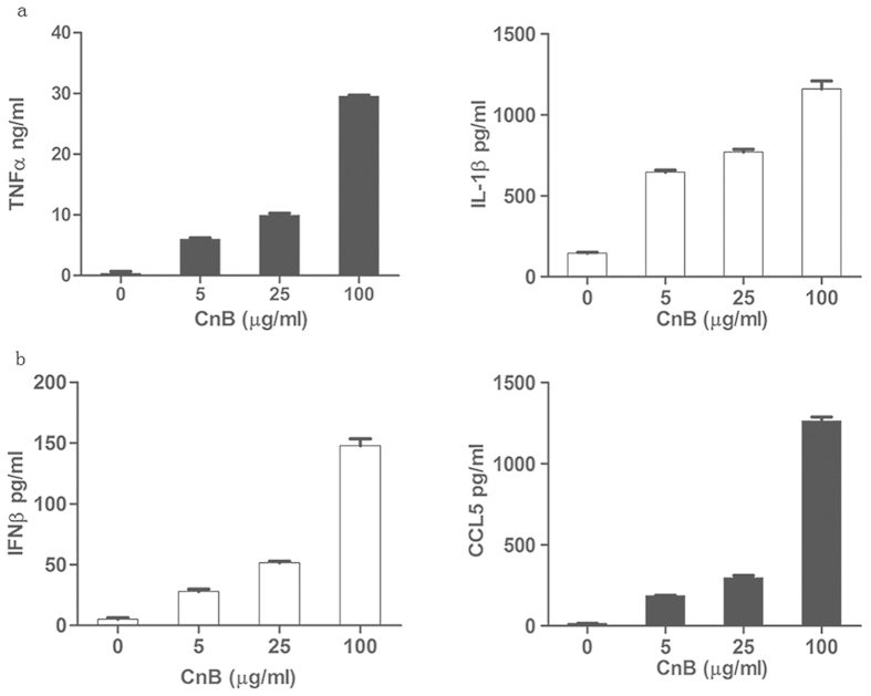
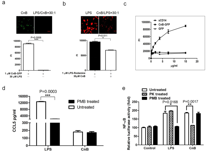
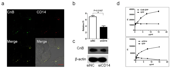
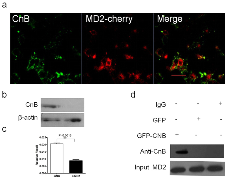
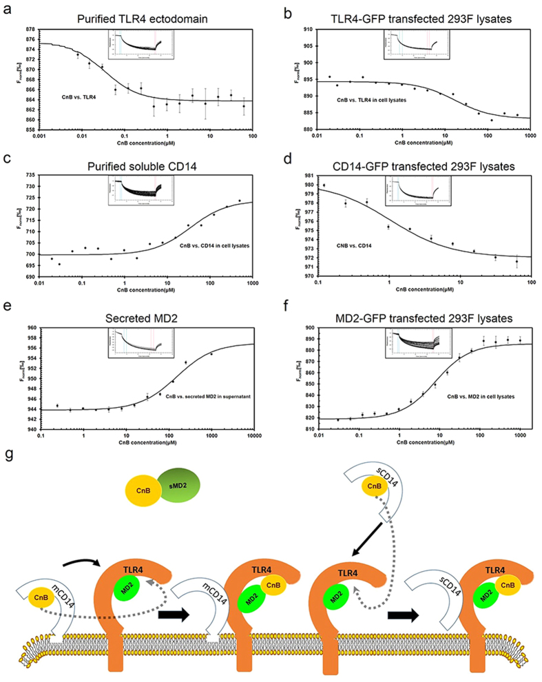
Our previous research showed that recombinant calcineurin B (rhCnB) stimulates cytokine secretion by immune cells, probably through TLR4. Exogenous CnB can be incorporated into many different tumour cells in vitro, but the mode of uptake and receptors required remain unknown. Here, we report that exogenous CnB is taken up by cells in a time- and concentration-dependent manner via clathrin-dependent receptor-mediated internalization. Our findings further confirm that uptake is mediated by the TLR4/MD2 complex together with the co-receptor CD14. The MST results revealed a high affinity between CnB and the TLR4 receptor complex. No binding was detected between CnB and LPS. CnB inhibited the uptake of LPS, and LPS also inhibited the uptake of CnB. These results indicate that the uptake of exogenous CnB did not occur through LPS and that CnB was not a chaperone of LPS. Thus, we conclude that TLR4 receptor complexes were required for the recognition and internalization of exogenous CnB. CnB could be a potential endogenous ligand of TLR4 and function as an agonist of TLR4. These properties of CnB support its potential for development as an anti-cancer drug.


Conflict of interest statement

The authors declare no competing financial interests.

Figures

Figure 1
Figure 1. Exogenous CnB was taken up by different cell lines in a concentration- and time-dependent manner.
(a) Exogenous CnB was incorporated into different cell lines. (b) Comparison of the uptake of different concentrations of CnB in SK-HEP-1 cells. (c) Comparison of CnB uptake at different time points. The cells were co-incubated with CnB-GFP, fixed and visualized using a Zeiss LSM700 confocal laser scanning microscope. The data were quantified using ImageJ software. FI represents the fluorescence intensity. The scale bar represents 50 μm, and the data represent mean ± s.e.m. from three independent experiments.
Figure 2
Figure 2. The uptake of exogenous CnB occurred via TLR4 receptor-mediated internalization.
(a) Co-localization of rhodamine-labelled CnB with clathrin-GFP (upper panel) or CnB-GFP with rhodamine-labelled transferrin (lower panel) in SK-HEP-1 cells. The clathrin-GFP transfected cells were co-incubated with 5 μM CnB-rhodamine, or SK-HEP-1 cells were co-incubated with 5 μM CnB-GFP mixed with 5 μM rhodamine-labelled transferrin for 30 min, and visualized using a confocal laser scanning microscope (×63, scale bar 10 μm). (b) Free CnB inhibits the uptake of the fluorescently labelled CnB. The cells were co-incubated with excess CnB and DyLight 488-labeled CnB or labelled CnB alone for 30 min and visualized using an inverted fluorescence microscope (upper panel, scale bar 50 μm, 20×). The fluorescence intensity was quantified using a microplate reader (lower panel). (c) Positive correlation between CnB uptake and TLR4 expression. 5 × 105 cells from different cell lines were co-incubated with 5 μM CnB for 10 min, subjected to Trizol treatment and RNA extraction. Extracted mRNA was used for qPCR analysis of TLR4. The qPCR results were analyzed and compared with CnB-GFP uptake (lower panel). 5 × 106 cells from different cell lines were co-incubated with 5 μM CnB for 10 min and lysed with RIPA buffer, the samples were used for detecting the protein level of TLR4 by western blot analysis (upper panel). (d) Co-localization of exogenous CnB-GFP and TLR4-cherry. The TLR4-cherry- or cherry-transfected Hek293 cells were co-incubated with 5 μM CnB-GFP for 30 min, and visualized using a confocal laser scanning microscope (63×, scale bar 20 μm). (e,f) Effect of TLR4 knock down on CnB uptake. The influence of TLR4 knock down on CnB uptake was analysed by western blot analysis (e) or FI (f), (scale bar 50 μm, 20×). (g) TAK242 inhibited CnB uptake. The SK-HEP-1 cells were pre-incubated with 10 μM or 5 μM TAK242 or vehicle for 3 h, followed by co-incubation with CnB-GFP for 30 min. The co-incubated cells were washed, acid-stripped and quantified by a microplate reader. Bars represent mean ± s.e.m. from three independent experiments. *P < 0.05, **P < 0.01, ***P < 0.005 (t-test, two-tailed).
Figure 3
Figure 3. CnB triggered cytokine secretion via the TLR4 signalling pathway in RAW264.7 macrophages, but not in SK-HEP-1 cells.
(a) CnB induced the secretion of pro-inflammatory cytokines secretion through the MyD88-dependent pathway in the macrophage cell line. (b) CnB induced CCL5 and IFNβ secretion through the TRIF-dependent pathway in the macrophage cell line. The RAW264.7 cells were treated with 5, 25, and 100 μg/ml CnB for 24 h, and the SK-HEP-1 cells were treated with 200, 400, and 800 μg/ml CnB for 48 h. The amounts of secreted cytokines were determined by ELISA. Data represent mean ± s.e.m. from three independent experiments.
Figure 4
Figure 4. Uptake of exogenous CnB did not occur via binding to LPS, and CnB induced cytokine secretion.
(a) The uptake of CnB could be inhibited by LPS. (b) The uptake of LPS could be inhibited by CnB. A 30-fold excess of LPS or CnB was mixed with CnB-GFP or rhodamine-labelled LPS and incubated with the SK-HEP-1 cells for 30 min. The results were visualized using an inverted fluorescence microscope (upper panel, scale bar 50 μm, 20×) and quantified using a microplate reader (lower panel). (c) CnB-GFP did not bind to LPS. To a black ELISA plate, 10 μg/ml LPS was immobilized and incubated with different concentrations of CnB-GFP, DyLight 488-labeled CD14 or GFP to evaluate the binding between CnB and LPS. (d) The CnB-induced cytokine production was not due to LPS contamination. The RAW264.7 cells were incubated with 1 μg/ml LPS, 100 μg/ml CnB, 1 μg/ml LPS in the presence of 100 μg/ml polymyxin B or 100 μg/ml CnB in the presence of 100 μg/ml polymyxin B for 24 h and the levels of the secreted cytokines in the supernatant were measured by ELISA. (e) NF-κB was activated by CnB in the TLR4-transfected Hek293 cells. The Hek293 cells were transfected with the TLR4-pcDNA3.1 plasmid and incubated for 24 h. The cells were plated in 24-well plates (1 × 105/well) and co-transfected with the pNF-κB-luc and pRL-null-Renilla-luc plasmids. Twenty-four hours post-transfection, the cells were co-incubated with 1 μg/ml LPS, 400 μg/ml CnB, 1 μg/ml LPS in the presence of 100 μg/ml polymyxin B, 400 μg/ml CnB in the presence of 100 μg/ml polymyxin B, 1 μg/ml proteinase K-treated LPS or 400 μg/ml proteinase K-treated CnB for 12 h. Luciferase activity was measured using a Dual-luciferase Reporter system. The data were normalized to the control. Data represent three independent experiments (mean ± s.e.m., n = 3). *P < 0.05, **P < 0.01, ***P < 0.005 (t-test, two-tailed).
Figure 5
Figure 5. The uptake of exogenous CnB is CD14-dependent.
(a) Co-localization of exogenous CnB and CD14. The CD14-cherry-transfected Hek293 cells were co-incubated with CnB-GFP for 30 min, fixed, and visualized using a confocal laser scanning microscope (63×, scale bar 10 μm). (b,c) Influence of CD14 knock down on CnB uptake. The influence of CD14 knock down on CnB uptake was analysed by western blot analysis (b) or FI (c). (d) The binding of CnB to sCD14 or sCD14 to CnB. To black ELISA plates, 10 μg/ml CnB or sCD14 was immobilized, and CnB-GFP or DyLight 488-labeled CD14 were incubated with the corresponding ELISA plates. Data represent mean ± s.e.m. from three independent experiments. *P < 0.05 (t-test, two-tailed).
Figure 6
Figure 6. Exogenous CnB uptake is MD2-dependent.
(a) Co-localization of CnB and MD2. The MD2-cherry-transfected Hek293 cells were co-incubated with CnB-GFP for 30 min, fixed, and visualized using a LSM700 confocal laser scanning microscope (63×, scale bar 10 μm). (b,c) Influence of MD2 knock down on CnB uptake. The influence of MD2 knock down on CnB uptake was analysed by western blot analysis (b) or FI (c). (d) Co-IP of CnB and MD2. The MD2-transfected HEK293 cells were lysed, and the lysates were co-incubated with CnB-GFP or GFP for 2 h at 4 °C. Next, rabbit anti-MD2 pAbs or rabbit IgG1 were added and incubated overnight. Protein A beads were used to capture the complex for 2 h, and the beads were then washed five times and boiled for 5 min. The interaction was detected by western blot analysis of anti-CnB antibody. Data represent mean ± s.e.m from two independent experiments. **P < 0.01 (t-test, two-tailed).
Figure 7
Figure 7. MST measurements of the interaction between CnB and the TLR4 receptor complexes.
(a) Measurement of rhCnB binding to the purified TLR4 ectodomain. (b) Measurement of CnB binding to the full-length TLR4 from transfected HEK293 lysates. (c) Measurement of rhCnB binding to purified soluble CD14. (d) Measurement of rhCnB binding to full-length membrane-anchored CD14 from transfected HEK293 lysates. (e) Measurement of the binding of rhCnB to secreted MD2 in the supernatant from transfected HEK293 cells. (f) Measurement of rhCnB binding to MD2 in the transfected Hek293 cell lysates. The purified TLR4 ectodomain or soluble CD14 was labelled with DyLight 488, and the concentration of labelled protein was adjusted to 20 nM. GFP-tagged TLR4, CD14 and MD2 constructs were transfected into HEK293 cells, incubated for 48 h, and lysed with RIPA buffer. Secreted MD2 was obtained from the supernatant of the MD2-transfected hek293 cells. The lysates were diluted according to fluorescence intensity. The recombinant CnB protein was dissolved to a 500 μM concentration using MST buffer and 16 1:1 dilution samples were prepared. The labelled proteins or GFP-tagged receptors lysates were added into each ligand dilution and mixed. After 10 min incubation, each solution was added to Standard Treated Capillaries for Thermophoresis. The data were analysed using NT. Analysis software. All data are representative of at least two independent experiments. (g) Model depicting the recognition of exogenous CnB. Membrane-anchored or soluble CD14 first recognized CnB and transported it to the TLR4/MD2 complex located on the plasma membrane, followed by internalization of the CnB/TLR4 receptor complexes and signalling through TLR4. Free MD2 also recognized and bound to CnB, although the affinity was lower than that of the MD2 on the plasma membrane.

Similar articles

Cited by

References

    1. Hemenway C. S. & Heitman J. Calcineurin. Structure, function, and inhibition. Cell Biochem Biophys. 30, 115–151 (1999). - PubMed
    1. Li W. & Handschumacher R. E. Identification of two calcineurin B-binding proteins: tubulin and heat shock protein 60. Biochim Biophys Acta. 1599, 72–81 (2002). - PubMed
    1. Saeki M. et al.. Calcineurin potentiates the activation of procaspase-3 by accelerating its proteolytic maturation. J Biol Chem. 282, 11786–11794 (2007). - PubMed
    1. Li N., Zhang Z., Zhang W. & Wei Q. Calcineurin B subunit interacts with proteasome subunit alpha type 7 and represses hypoxia-inducible factor-1α activity via the proteasome pathway. Biochem Biophys Res Commun. 405, 468–472 (2011). - PubMed
    1. Jing L. et al.. Calcineurin subunit B activates dendritic cells and acts as a cancer vaccine adjuvant. Int Immunol. 23, 327–334 (2011). - PubMed

Publication types

MeSH terms