Linking F-box protein 7 and parkin to neuronal degeneration in Parkinson's disease (PD)
- PMID: 27090516
- PMCID: PMC4835861
- DOI: 10.1186/s13041-016-0218-2
Linking F-box protein 7 and parkin to neuronal degeneration in Parkinson's disease (PD)
Abstract
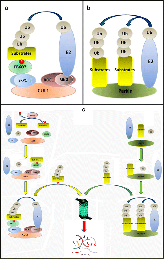
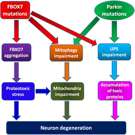
Mutations of F-box protein 7 (FBXO7) and Parkin, two proteins in ubiquitin-proteasome system (UPS), are both implicated in pathogenesis of dopamine (DA) neuron degeneration in Parkinson's disease (PD). Parkin is a HECT/RING hybrid ligase that physically receives ubiquitin on its catalytic centre and passes ubiquitin onto its substrates, whereas FBXO7 is an adaptor protein in Skp-Cullin-F-box (SCF) SCF(FBXO7) ubiquitin E3 ligase complex to recognize substrates and mediate substrates ubiquitination by SCF(FBXO7) E3 ligase. Here, we discuss the overlapping pathophysiologic mechanisms and clinical features linking Parkin and FBXO7 with autosomal recessive PD. Both proteins play an important role in neuroprotective mitophagy to clear away impaired mitochondria. Parkin can be recruited to impaired mitochondria whereas cellular stress can promote FBXO7 mitochondrial translocation. PD-linked FBXO7 can recruit Parkin into damaged mitochondria and facilitate its aggregation. WT FBXO7, but not PD-linked FBXO7 mutants can rescue DA neuron degeneration in Parkin null Drosophila. A better understanding of the common pathophysiologic mechanisms of these two proteins could unravel specific pathways for targeted therapy in PD.
Keywords: FBXO7; Mitochondria; Mitophagy; Parkin; Parkinson’s disease; Protein aggregation; Proteotoxicity; Ubiquitin proteasome system.
Figures


Similar articles
-
F-box protein 7 mutations promote protein aggregation in mitochondria and inhibit mitophagy.Hum Mol Genet. 2015 Nov 15;24(22):6314-30. doi: 10.1093/hmg/ddv340. Epub 2015 Aug 26. Hum Mol Genet. 2015. PMID: 26310625
-
Gsk3β and Tomm20 are substrates of the SCFFbxo7/PARK15 ubiquitin ligase associated with Parkinson's disease.Biochem J. 2016 Oct 15;473(20):3563-3580. doi: 10.1042/BCJ20160387. Epub 2016 Aug 8. Biochem J. 2016. PMID: 27503909 Free PMC article.
-
Pathophysiological mechanisms linking F-box only protein 7 (FBXO7) and Parkinson's disease (PD).Mutat Res Rev Mutat Res. 2018 Oct-Dec;778:72-78. doi: 10.1016/j.mrrev.2018.10.001. Epub 2018 Oct 17. Mutat Res Rev Mutat Res. 2018. PMID: 30454685 Review.
-
The Parkinson's disease-linked proteins Fbxo7 and Parkin interact to mediate mitophagy.Nat Neurosci. 2013 Sep;16(9):1257-65. doi: 10.1038/nn.3489. Epub 2013 Aug 11. Nat Neurosci. 2013. PMID: 23933751 Free PMC article.
-
Beyond ubiquitination: the atypical functions of Fbxo7 and other F-box proteins.Open Biol. 2013 Oct 9;3(10):130131. doi: 10.1098/rsob.130131. Open Biol. 2013. PMID: 24107298 Free PMC article. Review.
Cited by
-
Fbxo7 promotes Cdk6 activity to inhibit PFKP and glycolysis in T cells.J Cell Biol. 2022 Jul 4;221(7):e202203095. doi: 10.1083/jcb.202203095. Epub 2022 Jun 7. J Cell Biol. 2022. PMID: 35670764 Free PMC article.
-
Parkinson Disease from Mendelian Forms to Genetic Susceptibility: New Molecular Insights into the Neurodegeneration Process.Cell Mol Neurobiol. 2018 Aug;38(6):1153-1178. doi: 10.1007/s10571-018-0587-4. Epub 2018 Apr 26. Cell Mol Neurobiol. 2018. PMID: 29700661 Free PMC article. Review.
-
PARK Genes Link Mitochondrial Dysfunction and Alpha-Synuclein Pathology in Sporadic Parkinson's Disease.Front Cell Dev Biol. 2021 Jul 6;9:612476. doi: 10.3389/fcell.2021.612476. eCollection 2021. Front Cell Dev Biol. 2021. PMID: 34295884 Free PMC article. Review.
-
The Role of Oxidative Stress in Parkinson's Disease.Antioxidants (Basel). 2020 Jul 8;9(7):597. doi: 10.3390/antiox9070597. Antioxidants (Basel). 2020. PMID: 32650609 Free PMC article. Review.
-
Pluripotent Stem Cell Derived Neurons as In Vitro Models for Studying Autosomal Recessive Parkinson's Disease (ARPD): PLA2G6 and Other Gene Loci.Adv Exp Med Biol. 2021;1347:115-133. doi: 10.1007/5584_2021_643. Adv Exp Med Biol. 2021. PMID: 33990932 Free PMC article.
References
Publication types
MeSH terms
Substances
LinkOut - more resources
Full Text Sources
Other Literature Sources
Medical
Molecular Biology Databases

