. 2016 Jun 1;110(3):395-407.
doi: 10.1093/cvr/cvw080. Epub 2016 Apr 18.

Intracellular tortuosity underlies slow cAMP diffusion in adult ventricular myocytes

Affiliations

Intracellular tortuosity underlies slow cAMP diffusion in adult ventricular myocytes

Mark Richards et al. Cardiovasc Res. .

Abstract

Aims: 3',5'-Cyclic adenosine monophosphate (cAMP) signals in the heart are often confined to concentration microdomains shaped by cAMP diffusion and enzymatic degradation. While the importance of phosphodiesterases (degradative enzymes) in sculpting cAMP microdomains is well established in cardiomyocytes, less is known about cAMP diffusivity (DcAMP) and factors affecting it. Many earlier studies have reported fast diffusivity, which argues against sharply defined microdomains.

Methods and results: [cAMP] dynamics in the cytoplasm of adult rat ventricular myocytes were imaged using a fourth generation genetically encoded FRET-based sensor. The [cAMP]-response to the addition and removal of isoproterenol (β-adrenoceptor agonist) quantified the rates of cAMP synthesis and degradation. To obtain a read out of DcAMP, a stable [cAMP] gradient was generated using a microfluidic device which delivered agonist to one half of the myocyte only. After accounting for phosphodiesterase activity, DcAMP was calculated to be 32 µm(2)/s; an order of magnitude lower than in water. Diffusivity was independent of the amount of cAMP produced. Saturating cAMP-binding sites with the analogue 6-Bnz-cAMP did not accelerate DcAMP, arguing against a role of buffering in restricting cAMP mobility. cAMP diffused at a comparable rate to chemically unrelated but similar sized molecules, arguing for a common physical cause of restricted diffusivity. Lower mitochondrial density and order in neonatal cardiac myocytes allowed for faster diffusion, demonstrating the importance of mitochondria as physical barriers to cAMP mobility.

Conclusion: In adult cardiac myocytes, tortuosity due to physical barriers, notably mitochondria, restricts cAMP diffusion to levels that are more compatible with microdomain signalling.

Keywords: Diffusion reaction; FRET sensor; Mathematical modelling; Microfluidics; cAMP microdomains.


Figures

Figure 1
Figure 1
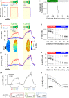
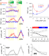
cAMP diffusivity measured using the H187 sensor is low in the cytoplasm of adult ventricular myocytes. (A) The experimental protocol is visualized with fluorescein added to one of two microstreams. (i) Time course of fluorescence emitted from extracellular fluorescein surrounding the proximal and distal regions of interest (ROI) of a myocyte. Manoeuvres 1 (both microstreams are released) and 2 (only fluorescein-containing microstream is released) are shaded yellow. (ii) Profile of fluorescence across the boundary between microstreams, showing the sharp (<10 µm) degree of microstream separation (n = 10/3). (B) Experiment on H187-expressing myocyte. 1 μM isoproterenol (ISO) included in one microstream (green) to active β-adrenoceptors; other microstream contained 10 µM propranolol, β-adrenoceptor antagonist (n = 11/4). (i) Time course of FRET ratio in proximal and distal ROIs; (ii) Longitudinal profile of FRET ratio, relative to boundary position, measured under resting conditions (light grey squares), during the last 10 s of Manoeuvre 1 (black circles) and during the last 10 s of Manoeuvre 2 (dark grey triangles). (C) Calibrated [cAMP] (i) time course and (ii) longitudinal profiles. (D) Mathematical model fit to experimental data. Best-fit DcAMP = 35 ± 3.4 µm2/s indicates slow cAMP diffusion inside adult cardiac cytoplasm. To illustrate the goodness-of-fit to data, longitudinal profiles were also simulated for a four-fold lower (dotted lines) and a four-fold higher (dashed lines) DcAMP. Inset: the partial differential equation describing cAMP dynamics is a linear combination of three processes (labelled a, b, and c). These can be parameterized sequentially by analysing different stages of the experiment. Dynamics in the third stage are determined by degradation only (c). With this information, it is then possible to characterize synthesis (b) by analysing the second stage. Having parameterized b and c, diffusion (a) can be obtained by analysing the first stage.
Figure 2
Figure 2
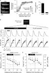
H187 does not interfere with the rate of cAMP diffusion. (A) Fluorescence recovery after photobleaching (FRAP) protocol for measuring H187 diffusivity in adult cardiac myocytes. Bleaching was performed in a 20 µm-wide region in the middle of the cell. Spatial profiles show fluorescence as a function of distance from the bleaching region at different time points during recovery. Continuous lines show best fit by diffusion model. H187 diffusivity (n = 17/3) was 15-fold slower than DcAMP measured in Figure 1; therefore, H187 cannot meaningfully accelerate cAMP diffusion. (B) Using SERCA activity as a bioassay of [cAMP] in adult cardiac myocytes that are not expressing H187. Cell paced at 2 Hz and imaged for [Ca2+] with Fluo3 fluorescence in line scan mode. Ca2+ transients (CaTs) measured in line scan sections on either side of boundary between ISO-containing and propranolol-containing microstreams. (B) Rate of CaT recovery as a function of [Ca2+] provides a measure of SERCA activity before (grey) and during (black) dual microperfusion (n = 9/2). Activation of flux indicates that cAMP had activated SERCA pumps locally. (C) (i) Peak recovery of CaT and (ii) systolic [Ca2+] measured before (grey) and during (black) dual microperfusion (n = 9/2). Right axis shows calibrated units, with Fluo3 Kd = 840 nM. The longitudinal profiles of SERCA activity (in the absence of H187 sensor) are comparable to longitudinal [cAMP] profiles reported with H187 (see Figure 1Dii), indicating that H187 does not slow cAMP diffusion.
Figure 3
Figure 3
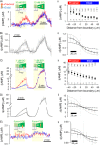
Cytoplasmic cAMP diffusivity is low, irrespective of the amount of cAMP produced or the type of β-receptor activated. (A) Experiment similar to that in Figure 1B repeated for lower (10 nM) dose of ISO (n = 15/4) to evoke a smaller proximal [cAMP]-rise. (B) Model fit to experimental data, with best-fit DcAMP = 32 ± 8.7 µm2/s confirming slow diffusion. (C) Experiment similar to that shown in Figure 1B repeated with phosphodiesterase inhibitor IBMX (100 µM) present in microstream containing 1 µM ISO (n = 12/4) to evoke a larger proximal [cAMP]-rise. (D) Mathematical model fit to experimental data, with best-fit DcAMP = 28 ± 3.0 µm2/s confirming slow diffusion. (D) Experiment similar to that in Figure 1B repeated using formoterol (100 nM) and CGP 20712A (1 µM) to selectively activate β2-receptors in the proximal end of the cell (n = 9/3). (i) The [cAMP]-rise was small, compared with the ISO response. A steady-state [cAMP] gradient can be resolved by time averaging over a 2-min interval during regional β2-adrenoceptor activation. (ii) Model fit to experimental data, with best-fit DcAMP = 24 ± 15.8 µm2/s confirming slow diffusivity. To illustrate the goodness-of-fit data, longitudinal profiles in Bii, Dii, and Eii were also simulated for a four-fold lower (dotted lines) and a four-fold higher (dashed lines) DcAMP.
Figure 4
Figure 4
Cytoplasmic cAMP diffusivity is not changed by cytoplasmic acidification. (A) Experiment similar to that shown in Figure 1B repeated on myocytes exposed to 80 mM acetate to acidify cytoplasm uniformly (n = 7/3). 5-(N,N)-dimethylamiloride (DMA; 30 µM) was included in both microstreams to inhibit pH regulation by Na+/H+ exchanger 1. (B) Model fit to experimental data, with best-fit DcAMP = 41 ± 12.2 µm2/s confirming slow diffusivity. (C) Experiment shown in Figure 4A repeated, but with acetate in the distal microstream only; DMA (30 µM) was included in both microstreams (n = 6/3). This protocol produces a pH-gradient overlying the gradient of β-adrenoceptor activation; higher pH in ISO-containing microstream favours greater cAMP production. (D) Model fit to experimental data with best-fit DcAMP = 32 ± 4.8 µm2/s confirming slow diffusivity. To illustrate the goodness-of-fit data, longitudinal profiles in Bii and Dii were also simulated for a four-fold lower (dotted lines) and a four-fold higher (dashed lines) DcAMP.
Figure 5
Figure 5
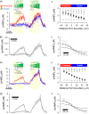
Cytoplasmic cAMP diffusivity is not affected by saturating cytoplasmic cAMP-binding sites with the analogue 6-Bnz-cAMP. (A) (i) Experiment similar to that shown in Figure 1B performed on myocyte AM-loaded with 20 µM (upper panel; n = 7/3; measured at half the normal acquisition frequency) or 5 µM (lower panel; n = 11/3) 6-Bnz-cAMP, a cAMP analogue with affinity for cAMP-binding sites. (ii) FRET ratio under resting conditions and at the proximal and distal end of the cell during dual microperfusion. The decreasing magnitude of FRET response to proximally presented ISO with higher doses of 6-Bnz-cAMP is due to a right shift in the [cAMP]-H187 calibration curve. (B) (i) [cAMP] time course in cells loaded with 5 µM 6-Bnz-cAMP. (ii) Longitudinal [cAMP] profiles, relative to boundary position, measured under resting conditions (light grey squares), during the last 10 s of dual microperfusion (black circles) and during the last 10 s of uniform exposure to agonist (dark grey triangles). (C) Model fit to (i) time course and (ii) longitudinal profile. Best-fit DcAMP = 29 ± 7.1 µm2/s is not different from measurements made in the absence of 6-Bnz-cAMP. To illustrate the goodness-of-fit data, longitudinal profiles were also simulated for a four-fold lower (dotted lines) and a four-fold higher (dashed lines) DcAMP.
Figure 6
Figure 6
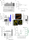
Cytoplasmic cAMP diffusivity in adult myocytes is comparable to that of similar sized molecules and is restricted by tortuosity. (A) (i) Summary of DcAMP measurements made by imposing [cAMP] gradients in adult myocytes by regional exposure to agonist. (ii) DcAMP is independent of the mean [cAMP]-rise evoked by regional exposure to agonist. (B) (i) Probing cytoplasmic tortuosity in terms of cytoplasmic calcein diffusion (measured by fluorescence recovery after photobleaching protocol) relative to calcein diffusivity in water (604 µm2/s). Measurements in adult cardiac myocytes under control conditions (n = 16/3), following hypotonic swelling (200 mOsm/kg solution; n = 14/3) or following detubulation with formamide (n = 14/3), and in HEK293 cells (n = 8) and HCT116 cells (n = 8). * denotes significant difference relative to adult cardiac myocyte (P < 0.05). (ii) Fluorescent staining of mitochondria (with MitoTracker Red), cytoplasm (with calcein), and nuclei (with Hoechst 33342) in adult or neonatal rat ventricular myocytes (excitation 555, 488, and 405 nm, respectively; emission collected at 580 ± 20, 520 ± 20, and 450 ± 20 nm). Mitochondrial distribution is more ordered and denser in adult myocytes compared with neonatal cells. (iii) Quantifying non-mitochondrial cell area. The mean intensity of MitoTracker Red fluorescence (Fmean) was measured in calcein-positive Hoechst 33342-negative pixels (i.e. non-nuclear cytoplasm). Non-nuclear cytoplasmic pixels with MitoTracker Red signals <0.5 × Fmean were defined as belonging to non-mitochondrial regions of the myocyte. The percentage of these pixels (relative to all non-nuclear cytoplasmic pixels) is lower in adult (n = 9/3) myocytes compared with neonatal (n = 12/3) myocytes, consistent with faster calcein diffusion in the latter. (C) Comparison of DcAMP (measured using dual microperfusion technique) with cytoplasmic diffusivity of the following fluorescent molecules (measured by FRAP): fluorescein (n = 8/3), MagFluo4 (n = 8/3), cAMP-fluorescein (n = 13/3) in control adult myocytes, cAMP-fluorescein (n = 12/3) in 5 µM 6-Bnz-cAMP-loaded adult myocytes, and cAMP-fluorescein diffusivity in neonatal myocytes (n = 6/4). Symbol α denotes significant difference (P < 0.05) relative to adult myocytes. (D) cAMP-fluorescein diffusivity (from C) and normalized calcein diffusivity (from Bi) plotted as a function of non-mitochondrial area fraction of myocyte (Biii). Star denotes diffusivity in water. Dashed line plots equation for diffusivity in tortuous medium, normalized to diffusivity in water: D/D0 = (2−2 × θ)/(2 + θ), where θ is the effective volume fraction of spherical impermeable inclusions.

Similar articles

Cited by

References

    1. Lomas O, Zaccolo M. Phosphodiesterases maintain signaling fidelity via compartmentalization of cyclic nucleotides. Physiology (Bethesda) 2014;29:141–149. - PMC - PubMed
    1. Lefkimmiatis K, Zaccolo M. cAMP signaling in subcellular compartments. Pharmacol Ther 2014;143:295–304. - PMC - PubMed
    1. Skalhegg BS, Tasken K. Specificity in the cAMP/PKA signaling pathway. Differential expression,regulation, and subcellular localization of subunits of PKA. Front Biosci 2000;5:D678–D693. - PubMed
    1. Smith FD, Scott JD. A-kinase-anchoring protein-Lbc connects stress signaling to cardiac hypertrophy. Mol Cell Biol 2013;33:2–3. - PMC - PubMed
    1. Kritzer MD, Li J, Dodge-Kafka K, Kapiloff MS. AKAPs: the architectural underpinnings of local cAMP signaling. J Mol Cell Cardiol 2012;52:351–358. - PMC - PubMed

Publication types

MeSH terms