

Matrine displayed antiviral activity in porcine alveolar macrophages co-infected by porcine reproductive and respiratory syndrome virus and porcine circovirus type 2
- PMID: 27080155
- PMCID: PMC4832146
- DOI: 10.1038/srep24401
Matrine displayed antiviral activity in porcine alveolar macrophages co-infected by porcine reproductive and respiratory syndrome virus and porcine circovirus type 2
Abstract
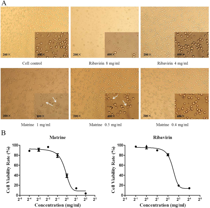
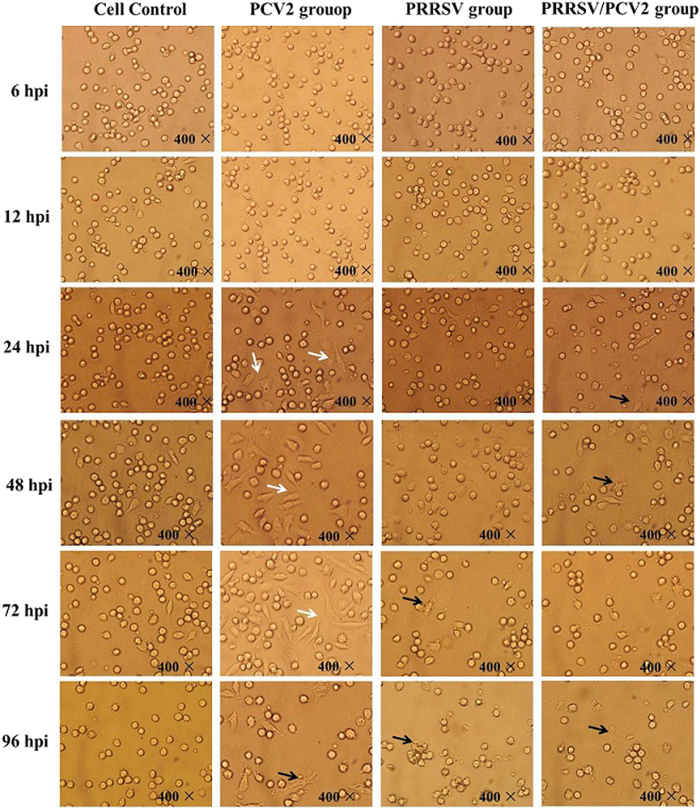
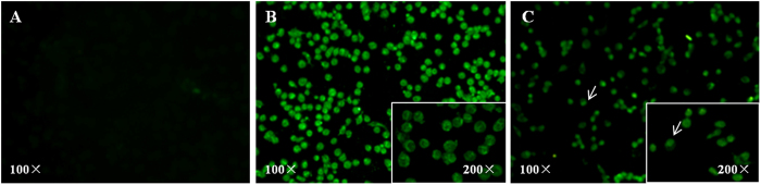
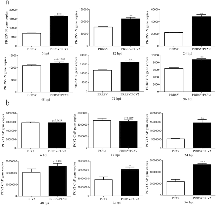
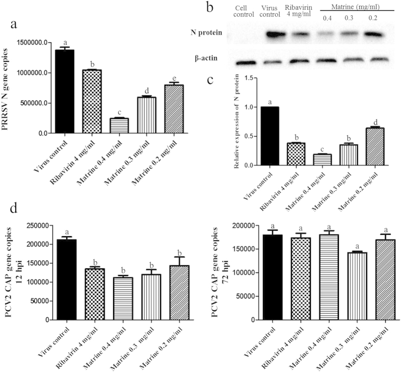
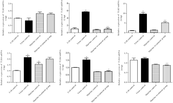
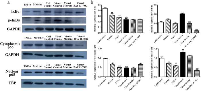
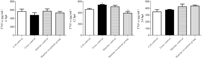
The co-infection of porcine reproductive respiratory syndrome virus (PRRSV) and porcine circovirus type 2 (PCV2) is quite common in clinical settings and no effective treatment to the co-infection is available. In this study, we established the porcine alveolar macrophages (PAM) cells model co-infected with PRRSV/PCV2 with modification in vitro, and investigated the antiviral activity of Matrine on this cell model and further evaluated the effect of Matrine on virus-induced TLR3,4/NF-κB/TNF-α pathway. The results demonstrated PAM cells inoculated with PRRSV followed by PCV2 2 h later enhanced PRRSV and PCV2 replications. Matrine treatment suppressed both PRRSV and PCV2 infection at 12 h post infection. Furthermore, PRRSV/PCV2 co- infection induced IκBα degradation and phosphorylation as well as the translocation of NF-κB from the cytoplasm to the nucleus indicating that PRRSV/PCV2 co-infection induced NF-κB activation. Matrine treatment significantly down-regulated the expression of TLR3, TLR4 and TNF-α although it, to some extent, suppressed p-IκBα expression, suggesting that TLR3,4/NF-κB/TNF-α pathway play an important role of Matrine in combating PRRSV/PCV2 co-infection. It is concluded that Matrine possesses activity against PRRSV/PCV2 co-infection in vitro and suppression of the TLR3,4/NF-κB/TNF-α pathway as an important underlying molecular mechanism. These findings warrant Matrine to be further explored for its antiviral activity in clinical settings.
Figures


Similar articles
-
Matrine exhibits antiviral activity in a PRRSV/PCV2 co-infected mouse model.Phytomedicine. 2020 Oct;77:153289. doi: 10.1016/j.phymed.2020.153289. Epub 2020 Jul 23. Phytomedicine. 2020. PMID: 32771536
-
The effect of infection order of porcine circovirus type 2 and porcine reproductive and respiratory syndrome virus on dually infected swine alveolar macrophages.BMC Vet Res. 2012 Sep 25;8:174. doi: 10.1186/1746-6148-8-174. BMC Vet Res. 2012. PMID: 23009687 Free PMC article.
-
Immune Molecules' mRNA Expression in Porcine Alveolar Macrophages Co-Infected with Porcine Reproductive and Respiratory Syndrome Virus and Porcine Circovirus Type 2.Viruses. 2023 Mar 17;15(3):777. doi: 10.3390/v15030777. Viruses. 2023. PMID: 36992486 Free PMC article.
-
Mechanism of PRRSV infection and antiviral role of polyphenols.Virulence. 2024 Dec;15(1):2417707. doi: 10.1080/21505594.2024.2417707. Epub 2024 Oct 21. Virulence. 2024. PMID: 39432383 Free PMC article. Review.
-
Matrine: Bioactivities and Structural Modifications.Curr Top Med Chem. 2016;16(28):3365-3378. doi: 10.2174/1568026616666160506131012. Curr Top Med Chem. 2016. PMID: 27150374 Review.
Cited by
-
Matrine Targets Intestinal Lactobacillus acidophilus to Inhibit Porcine Circovirus Type 2 Infection in Mice.Int J Mol Sci. 2023 Jul 25;24(15):11878. doi: 10.3390/ijms241511878. Int J Mol Sci. 2023. PMID: 37569261 Free PMC article.
-
Synthesis, characterization and in vitro biological evaluation of two matrine derivatives.Sci Rep. 2018 Oct 24;8(1):15686. doi: 10.1038/s41598-018-33908-8. Sci Rep. 2018. PMID: 30356148 Free PMC article.
-
Promising Therapeutic Candidate for Myocardial Ischemia/Reperfusion Injury: What Are the Possible Mechanisms and Roles of Phytochemicals?Front Cardiovasc Med. 2022 Feb 17;8:792592. doi: 10.3389/fcvm.2021.792592. eCollection 2021. Front Cardiovasc Med. 2022. PMID: 35252368 Free PMC article. Review.
-
Cepharanthine and Curcumin inhibited mitochondrial apoptosis induced by PCV2.BMC Vet Res. 2020 Sep 18;16(1):345. doi: 10.1186/s12917-020-02568-0. BMC Vet Res. 2020. PMID: 32948186 Free PMC article.
-
Matrine Exerts Pharmacological Effects Through Multiple Signaling Pathways: A Comprehensive Review.Drug Des Devel Ther. 2022 Mar 1;16:533-569. doi: 10.2147/DDDT.S349678. eCollection 2022. Drug Des Devel Ther. 2022. PMID: 35256842 Free PMC article. Review.
References
Publication types
MeSH terms
Substances
LinkOut - more resources
Full Text Sources
Other Literature Sources
Miscellaneous
