

What are the current and potential future roles for endoscopic ultrasound in the treatment of pancreatic cancer?
- PMID: 27076870
- PMCID: PMC4823670
- DOI: 10.4253/wjge.v8.i7.319
What are the current and potential future roles for endoscopic ultrasound in the treatment of pancreatic cancer?
Abstract
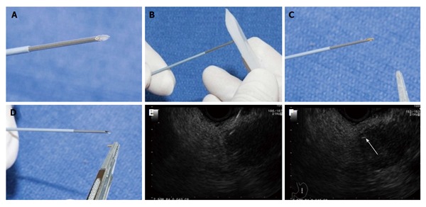
Pancreatic adenocarcinoma is the fourth leading cause of cancer-related death in the United States. Due to the aggressive tumor biology and late manifestations of the disease, long-term survival is extremely uncommon and the current 5-year survival rate is 7%. Over the last two decades, endoscopic ultrasound (EUS) has evolved from a diagnostic modality to a minimally invasive therapeutic alternative to radiologic procedures and surgery for pancreatic diseases. EUS-guided celiac plexus intervention is a useful adjunct to conventional analgesia for patients with pancreatic cancer. EUS-guided biliary drainage has emerged as a viable option in patients who have failed endoscopic retrograde cholangiopancreatography. Recently, the use of lumen-apposing metal stent to create gastrojejunal anastomosis under EUS and fluoroscopic guidance in patients with malignant gastric outlet obstruction has been reported. On the other hand, anti-tumor therapies delivered by EUS, such as the injection of anti-tumor agents, brachytherapy and ablations are still in the experimental stage without clear survival benefit. In this article, we provide updates on well-established EUS-guided interventions as well as novel techniques relevant to pancreatic cancer.
Keywords: Endoscopic ultrasound; Endoscopic ultrasound-guided ablation; Endoscopic ultrasound-guided anti-tumor therapy; Endoscopic ultrasound-guided biliary drainage; Endoscopic ultrasound-guided celiac plexus neurolysis and block; Endoscopic ultrasound-guided fiducial placement; Endoscopic ultrasound-guided gastrojejunal anastomosis; Palliation; Pancreatic cancer.
Figures
Similar articles
-
Interventional Therapy for Pancreatic Cancer.Gastrointest Tumors. 2016 Oct;3(2):81-89. doi: 10.1159/000446800. Epub 2016 Sep 6. Gastrointest Tumors. 2016. PMID: 27904860 Free PMC article. Review.
-
Endoscopic ultrasonography-guided tumor ablation.Dig Endosc. 2017 May;29(4):486-494. doi: 10.1111/den.12833. Epub 2017 Mar 16. Dig Endosc. 2017. PMID: 28171697 Review.
-
Endoscopic Ultrasound-Guided Treatment beyond Drainage: Hemostasis, Anastomosis, and Others.Clin Endosc. 2014 Sep;47(5):432-9. doi: 10.5946/ce.2014.47.5.432. Epub 2014 Sep 30. Clin Endosc. 2014. PMID: 25325004 Free PMC article. Review.
-
Current role of endoscopic ultrasound in the diagnosis and management of pancreatic cancer.World J Gastrointest Endosc. 2022 Jan 16;14(1):35-48. doi: 10.4253/wjge.v14.i1.35. World J Gastrointest Endosc. 2022. PMID: 35116098 Free PMC article. Review.
-
Endoscopic ultrasound guided interventional procedures.World J Gastrointest Endosc. 2015 Jun 10;7(6):628-42. doi: 10.4253/wjge.v7.i6.628. World J Gastrointest Endosc. 2015. PMID: 26078831 Free PMC article. Review.
Cited by
-
Endoscopic ultrasound role in pancreatic adenocarcinoma treatment: A review focusing on technical success, safety and efficacy.World J Gastroenterol. 2022 Jan 21;28(3):332-347. doi: 10.3748/wjg.v28.i3.332. World J Gastroenterol. 2022. PMID: 35110953 Free PMC article. Review.
-
Oxytocin and oxytocin receptor alterations, decreased survival, and increased chemoresistance in patients with pancreatic cancer.Hepatobiliary Pancreat Dis Int. 2020 Apr;19(2):175-180. doi: 10.1016/j.hbpd.2019.12.002. Epub 2019 Dec 14. Hepatobiliary Pancreat Dis Int. 2020. PMID: 31919036 Free PMC article.
-
Initial experience with endoscopic ultrasound-guided gallbladder drainage.Wideochir Inne Tech Maloinwazyjne. 2019 Apr;14(2):195-202. doi: 10.5114/wiitm.2018.79528. Epub 2018 Nov 14. Wideochir Inne Tech Maloinwazyjne. 2019. PMID: 31118983 Free PMC article.
-
The future of ERCP.Endosc Int Open. 2017 Apr;5(4):E272-E274. doi: 10.1055/s-0043-101697. Endosc Int Open. 2017. PMID: 28382325 Free PMC article. No abstract available.
References
-
- Jemal A, Siegel R, Xu J, Ward E. Cancer statistics, 2010. CA Cancer J Clin. 2010;60:277–300. - PubMed
-
- Yeo TP, Hruban RH, Leach SD, Wilentz RE, Sohn TA, Kern SE, Iacobuzio-Donahue CA, Maitra A, Goggins M, Canto MI, et al. Pancreatic cancer. Curr Probl Cancer. 2002;26:176–275. - PubMed
-
- Vonlaufen A, Joshi S, Qu C, Phillips PA, Xu Z, Parker NR, Toi CS, Pirola RC, Wilson JS, Goldstein D, et al. Pancreatic stellate cells: partners in crime with pancreatic cancer cells. Cancer Res. 2008;68:2085–2093. - PubMed
Publication types
LinkOut - more resources
Full Text Sources
Other Literature Sources
