. 2016 Apr;57(4):1749-56.
doi: 10.1167/iovs.16-19183.

A Central Role for Sympathetic Nerves in Herpes Stromal Keratitis in Mice

Affiliations

A Central Role for Sympathetic Nerves in Herpes Stromal Keratitis in Mice

Hongmin Yun et al. Invest Ophthalmol Vis Sci. 2016 Apr.

Abstract

Purpose: Herpes simplex virus type 1 (HSV-1) is a neurotrophic virus that can cause herpes stromal keratitis (HSK), a severe corneal inflammation that can lead to corneal scarring and blindness. This study identified neurologic changes that occur in HSV-1-infected corneas and related them to HSV-1-induced immunopathology.

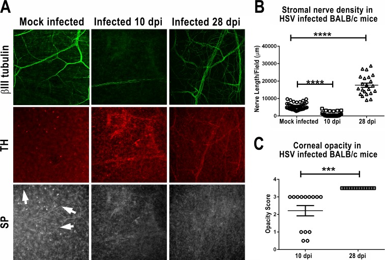
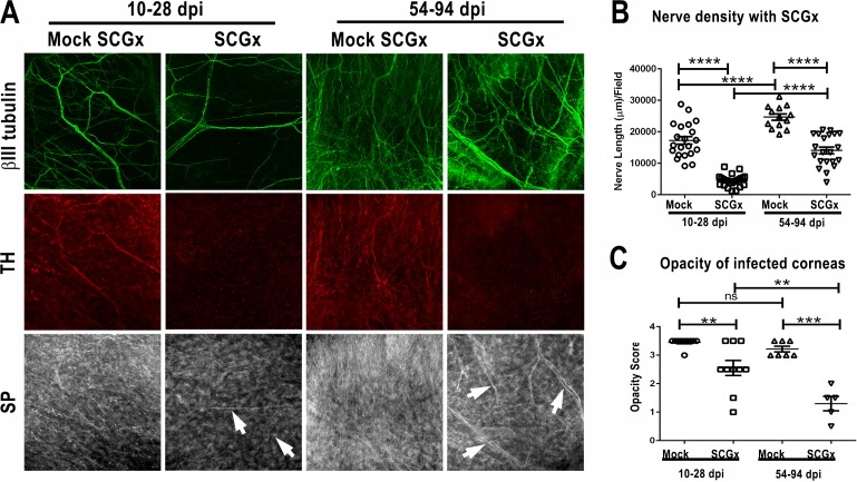
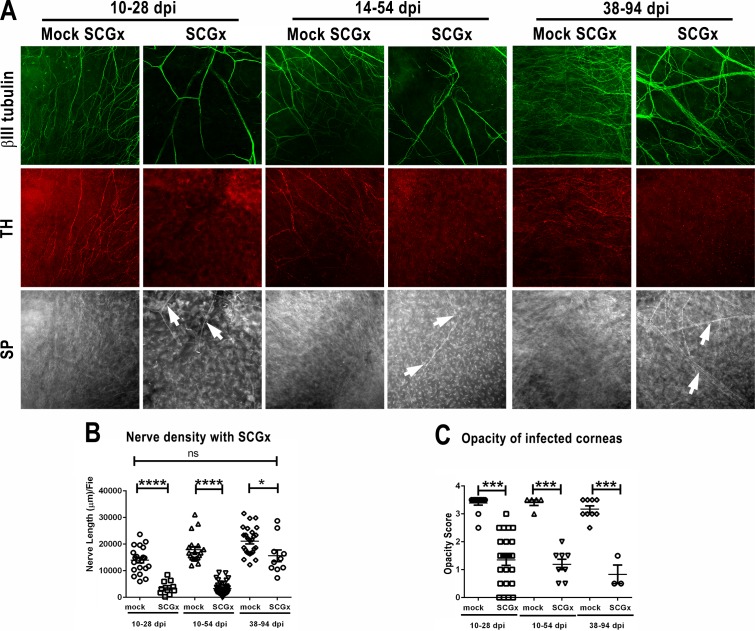
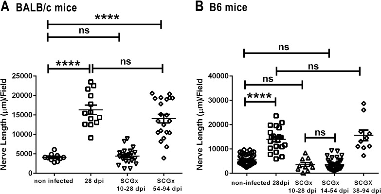
Methods: Corneas of BALB/c and C57BL/6 mice were infected with HSV-1 strains that induce HSK. Changes in sensory nerves were identified by immunofluorescence staining of sensory and sympathetic nerves for substance P (SP) and tyrosine hydroxylase (TH), respectively, and confocal microscopic examination. Some mice received superior cervical ganglionectomy (SCGx) to eliminate sympathetic nerves from the cornea.

Results: Normal corneas exclusively expressed sensory nerves that entered the stroma as large nerve stalks, branched to form a plexus at the epithelial/stromal interface, and extended termini into the epithelium. These nerves completely retracted from the infected cornea and were replaced by sympathetic nerves that sprouted extensively to hyperinnervate the corneal stroma but failed to form a plexus or extend termini into the epithelium. The hyperinnervating nerves expressed the sympathetic nerve marker TH and their invasion was blocked by performing SCGx. Moreover, the corneal opacity and neovascularization that normally characterizes HSK in this mouse model were largely abrogated by SCGx. Sensory nerves reinnervated infected corneas following SCGx, reformed a nerve plexus, and extended termini into the epithelium resulting in recovery of corneal sensitivity.

Conclusions: Sympathetic nerves have a central role in HSK in mice, preventing reinnervation by sensory nerves and promoting severe and persistent corneal inflammation.


Figures

Figure 1
Figure 1
BALB/c mice developed sympathetic nerve hyperinnervation associated with severe HSK at 28 dpi. BALB/c mice were mock infected or infected with 1 × 10⁵ pfu of HSV-1 KOS strain. At 10 or 28 dpi, the corneal opacity was recorded, and then the mice were killed, infected corneas were excised and fixed, and whole mounts were stained for the neuronal marker βIII tubulin (green), the sympathetic nerve marker TH (red), and the sensory nerve marker SP (gray). Confocal images were acquired and analyzed as described in Methods. (A) Changes in nerve innervation of the stroma at 10 and 28 dpi. The corneal stroma of mock infected mice show a low density of nerves that express SP but not TH. In contrast, corneas obtained at 10 dpi exhibit an almost complete lack of corneal nerves, whereas corneas obtained at 28 dpi show a corneal stroma that is hyperinnervated by nerve fibers that express the sympathetic marker TH but not the sensory marker SP. (B) Stromal nerve density measured by cumulative length of nerve fiber at 10 or 28 dpi. (C) Corneal opacity in HSV-infected mice recorded prior to death at 10 or 28 dpi. No opacity was observed in mock infected corneas (data not shown). ***P < 0.001, ****P < 0.0001.
Figure 2
Figure 2
B6 mice developed sympathetic nerve hyperinnervation associated with severe HSK at 28 dpi. B6 mice were mock infected or infected with 1 × 10⁵ pfu of the HSV-1 RE strain. At 10 or 28 dpi, the corneal opacity was recorded, and then the mice were killed, infected corneas were excised and fixed, and whole mounts were stained for the neuronal marker βIII tubulin (green), the sympathetic nerve marker TH (red), and the sensory nerve marker SP (gray). Confocal images were acquired and analyzed. (A) Changes in nerve innervation of the stroma at 10 and 28 dpi. Mock infected mice show a low density of nerves that express SP, but not TH, whereas corneas obtained at 10 dpi exhibit an almost complete lack of corneal nerves, yet corneas obtained at 28 dpi show a corneal stroma hyperinnervated by nerve fibers that express the sympathetic marker TH but not the sensory marker SP. (B) Stromal nerve density measured by cumulative length of nerve fiber at 10 or 28 dpi. (C) Corneal opacity in HSV-infected mice recorded prior to death at 10 or 28 dpi. Mock infected corneas remained clear (data not shown). **P < 0.01, ****P < 0.0001.
Figure 3
Figure 3
Superior cervical ganglionectomy eliminates sympathetic nerves, allows regrowth of sensory nerves, and prevents development of severe persistent HSK in BALB/c mice. BALB/c mice were infected with 1 × 10⁵ pfu of the HSV-1 KOS strain. Groups of infected mice received mock SCGx or SCGx at 10 dpi and were followed to 28 dpi, whereas other groups received mock SCGx or SCGx at 54 dpi and were followed to 94 dpi. Opacity scores were recorded on the day mice were killed. Whole mounted corneas were stained for βIII tubulin (green), TH (red), and SP (gray). (A) Superior cervical ganglionectomy administered at 10 dpi effectively eliminated the hyperinnervation at 28 dpi and permitted regrowth of a low density of SP-positive sensory nerves. Administering SCGx after hyperinnervation when sympathetic nerves had established (54 dpi) did not eliminate the hyperinnervating fibers, but the nerves did not express the sympathetic nerve marker TH. Note the hyperinnervation of the corneal stroma of mice that received mock SCGx at 10 or 54 dpi, with nerve fibers that expressed the sympathetic nerve marker TH but not the sensory nerve marker SP. (B) Nerve density in corneal stromas of mock or SCGx-treated mice at 28 and 94 dpi was quantitated as the cumulative length of nerve fibers in 500 × 500-μm areas of the corneal stroma. (C) Opacity of infected corneas was recorded just prior to death at 28 and 94 dpi. **P < 0.01, ***P < 0.001, ****P < 0.0001.
Figure 4
Figure 4
Superior cervical ganglionectomy eliminates sympathetic nerves, allows regrowth of sensory nerves, and prevents development of severe persistent HSK in B6 mice. B6 mice were infected with 1 × 10⁵ pfu of the HSV-1 RE strain. Groups of infected mice received mock SCGx or SCGx at 10, 14, or 38 dpi and were followed to 28, 54, and 94 dpi, respectively. Opacity scores were recorded on the day the mice was killed. Corneas were excised and fixed, and whole mounts were stained for βIII tubulin (green), TH (red), and SP (gray). Confocal images were acquired and analyzed as described in the Methods. (A) Superior cervical ganglionectomy before hyperinnervation was established (10 and 14 dpi) effectively eliminated the hyperinnervation with TH-positive sympathetic nerve fibers and permitted regrowth of a low density of SP-positive sensory nerves. Performing SCGx after hyperinnervation with sympathetic nerves was established (38 dpi) did not eliminate the hyperinnervating fibers, but the nerves failed to express the sympathetic nerve marker TH. Note the hyperinnervation of the corneal stroma of mice that received mock SCGx at 10, 14, and 54 dpi, with nerve fibers that expressed the sympathetic nerve marker TH but not the sensory nerve marker SP. (B) Nerve density in corneal stromas of mock or SCGx treated mice at 28, 54, and 94 dpi quantitated as the cumulative length of nerve fibers in 500 × 500-μm areas of the corneal stroma. (C) Opacity of infected corneas was recorded just prior to death at 28, 54, and 94 dpi. **P< 0.05, ***P < 0.001, ****P < 0.0001.
Figure 5
Figure 5
Early SCGx allows regeneration of normal density sensory nerves in BALB/c and B6 mice with HSK. (A) BALB/c and (B) B6 mice were mock infected or infected with 1 × 10⁵ pfu of the HSV-1 KOS or RE strain, respectively. BALB/c mice received mock SCGx at 10 dpi, and corneas were excised at 28 dpi, or mice received SCGx at 10 or 54 dpi and corneas were excised at 28 and 94 dpi, respectively. B6 mice received mock SCGX at 10 dpi, and corneas were excised at 28 dpi, or received SCGx at 10, 14, or 38 dpi and corneas were excised at 28, 54, and 94 dpi, respectively. Corneal whole mounts were stained for βIII tubulin. Nerve density in various areas of the corneal stroma was imaged with confocal microscopy and quantified with FIJI. ****P < 0.0001.

Similar articles

Cited by

References

    1. Liesegang TJ. Herpes simplex virus epidemiology and ocular importance. Cornea. 2001; 20: 1–13. - PubMed
    1. Rowe AM,, St Leger AJ,, Jeon S,, Dhaliwal DK,, Knickelbein JE,, Hendricks RL. Herpes keratitis. Prog Retin Eye Res. 2012; 32: 88–101. - PMC - PubMed
    1. Tang Q,, Hendricks RL. IFN-gamma regulates PECAM-1 expression and neutrophil infiltration into herpes simplex virus-infected mouse corneas. J Exp Med. 1996; 184: 1435–1447. - PMC - PubMed
    1. Tang Q,, Chen W,, Hendricks RL. Proinflammatory functions of IL-2 in herpes simplex virus corneal infection. J Immunol. 1997; 158: 1275–1283. - PubMed
    1. Suryawanshi A,, Veiga-Parga T,, Rajasagi NK,, et al. Role of IL-17 and Th17 cells in herpes simplex virus-induced corneal immunopathology. J Immunol. 2011; 187: 1919–1930. - PMC - PubMed

Publication types

MeSH terms

LinkOut - more resources