

Tracking cells implanted into cynomolgus monkeys (Macaca fascicularis) using MRI
- PMID: 27062993
- PMCID: PMC4976245
- DOI: 10.1538/expanim.15-0125
Tracking cells implanted into cynomolgus monkeys (Macaca fascicularis) using MRI
Abstract
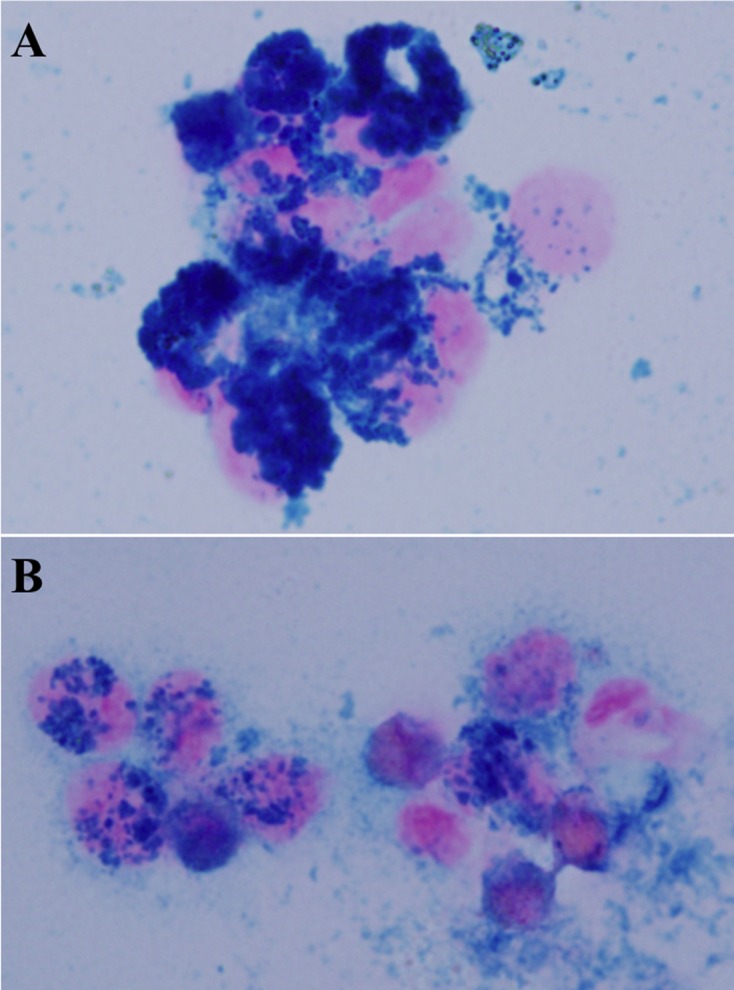
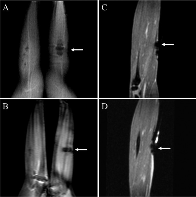
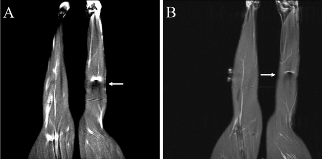
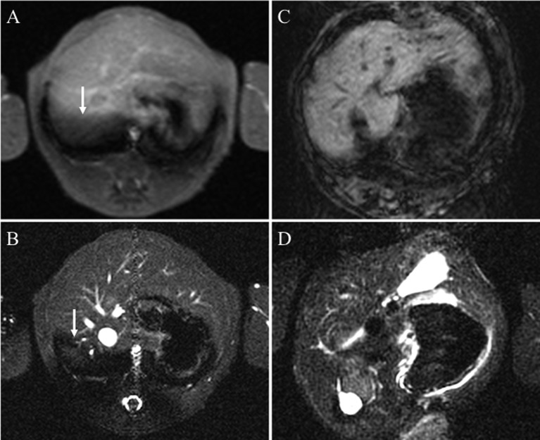
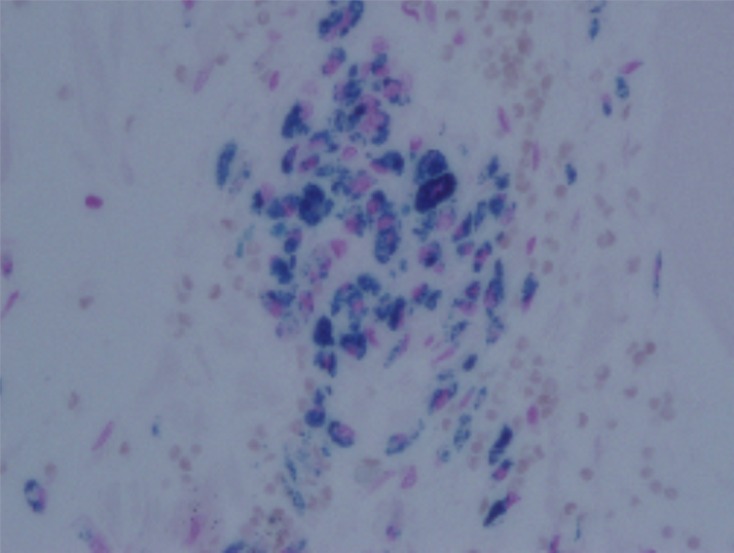
Regenerative therapy with stem cell transplantation is used to treat various diseases such as coronary syndrome and Buerger's disease. For instance, stem-cell transplantation into the infarcted myocardium is an innovative and promising strategy for treating heart failure due to ischemic heart disease. Basic studies using small animals have shown that transplanted cells improve blood flow in the infarcted region. Magnetic resonance imaging (MRI) can noninvasively identify and track transplanted cells labeled with superparamagnetic iron oxide (SPIO). Although clinical regenerative therapies have been clinically applied to patients, the fate of implanted cells remains unknown. In addition, follow-up studies have shown that some adverse events can occur after recovery. Therefore, the present study evaluated the ability of MRI using a 3T scanner to track implanted peripheral blood mononuclear cells labeled with SPIO on days 0 and 7 after intramuscular (i.m.) and intravenous (i.v.) injection into a cynomolgus monkey. Labeled cells were visualized at the liver and triceps surae muscle on MR images using T1- and T2-weighted sequences and histologically localized by Prussian blue staining. The transplanted cells were tracked without abnormal clinical manifestations throughout this study. Hence, MRI of cynomolgus monkey transplanted SPIO-labeled cells is a safe and efficient method of tracking labeled cells that could help to determine the mechanisms involved in regenerative therapy.
Figures
Similar articles
-
Labeling of cynomolgus monkey bone marrow-derived mesenchymal stem cells for cell tracking by multimodality imaging.Sci China Life Sci. 2011 Nov;54(11):981-7. doi: 10.1007/s11427-011-4239-x. Epub 2011 Dec 16. Sci China Life Sci. 2011. PMID: 22173303
-
Whole body MRI and fluorescent microscopy for detection of stem cells labeled with superparamagnetic iron oxide (SPIO) nanoparticles and DiI following intramuscular and systemic delivery.Methods Mol Biol. 2013;1052:177-93. doi: 10.1007/7651_2013_13. Methods Mol Biol. 2013. PMID: 23733536
-
Magnetic resonance monitoring of superparamagnetic iron oxide (SPIO)-labeled stem cells transplanted into the inner ear.Neurosci Res. 2015 Jun;95:21-6. doi: 10.1016/j.neures.2015.01.010. Epub 2015 Jan 30. Neurosci Res. 2015. PMID: 25645157
-
In vivo tracking of stem cells in brain and spinal cord injury.Prog Brain Res. 2007;161:367-83. doi: 10.1016/S0079-6123(06)61026-1. Prog Brain Res. 2007. PMID: 17618991 Review.
-
Considerations for the clinical use of contrast agents for cellular MRI in regenerative medicine.Contrast Media Mol Imaging. 2013 Nov-Dec;8(6):439-55. doi: 10.1002/cmmi.1547. Contrast Media Mol Imaging. 2013. PMID: 24375900 Review.
Cited by
-
Molecular Imaging of Stem Cell Transplantation for Liver Diseases: Monitoring, Clinical Translation, and Theranostics.Stem Cells Int. 2016;2016:4058656. doi: 10.1155/2016/4058656. Epub 2016 Dec 14. Stem Cells Int. 2016. PMID: 28070195 Free PMC article. Review.
References
-
- Ageyama N., Hanazono Y., Shibata H., Ohto K., Ono F., Nagashima T., Ueda Y., Donahue R.E., Hasegawa M., Ozawa K., Yoshikawa Y., Terao K.2002. Safe and efficient methods of autologous hematopoietic stem cell transplantation for biomedical research in cynomolgus monkeys. Comp. Med. 52: 445–451. - PubMed
-
- Amado L.C., Saliaris A.P., Schuleri K.H., St John M., Xie J.S., Cattaneo S., Durand D.J., Fitton T., Kuang J.Q., Stewart G., Lehrke S., Baumgartner W.W., Martin B.J., Heldman A.W., Hare J.M.2005. Cardiac repair with intramyocardial injection of allogeneic mesenchymal stem cells after myocardial infarction. Proc. Natl. Acad. Sci. USA 102: 11474–11479. doi: 10.1073/pnas.0504388102 - DOI - PMC - PubMed
-
- Arbab A.S., Bashaw L.A., Miller B.R., Jordan E.K., Lewis B.K., Kalish H., Frank J.A.2003. Characterization of biophysical and metabolic properties of cells labeled with superparamagnetic iron oxide nanoparticles and transfection agent for cellular MR imaging. Radiology 229: 838–846. doi: 10.1148/radiol.2293021215 - DOI - PubMed
MeSH terms
Substances
LinkOut - more resources
Full Text Sources
Other Literature Sources
Medical