. 2016 Apr 8;10(4):e0004609.
doi: 10.1371/journal.pntd.0004609. eCollection 2016 Apr.

Neutrophils Contribute to the Protection Conferred by ArtinM against Intracellular Pathogens: A Study on Leishmania major

Affiliations

Neutrophils Contribute to the Protection Conferred by ArtinM against Intracellular Pathogens: A Study on Leishmania major

Rafael Ricci-Azevedo et al. PLoS Negl Trop Dis. .

Abstract

ArtinM, a D-mannose binding lectin from Artocarpus heterophyllus, has immunomodulatory activities through its interaction with N-glycans of immune cells, culminating with the establishment of T helper type 1 (Th1) immunity. This interaction protects mice against intracellular pathogens, including Leishmania major and Leishmania amazonensis. ArtinM induces neutrophils activation, which is known to account for both resistance to pathogens and host tissue injury. Although exacerbated inflammation was not observed in ArtinM-treated animals, assessment of neutrophil responses to ArtinM is required to envisage its possible application to design a novel immunomodulatory agent based on carbohydrate recognition. Herein, we focus on the mechanisms through which neutrophils contribute to ArtinM-induced protection against Leishmania, without exacerbating inflammation. For this purpose, human neutrophils treated with ArtinM and infected with Leishmania major were analyzed together with untreated and uninfected controls, based on their ability to eliminate the parasite, release cytokines, degranulate, produce reactive oxygen species (ROS), form neutrophil extracellular traps (NETs) and change life span. We demonstrate that ArtinM-stimulated neutrophils enhanced L. major clearance and at least duplicated tumor necrosis factor (TNF) and interleukin-1beta (IL-1β) release; otherwise, transforming growth factor-beta (TGF-β) production was reduced by half. Furthermore, ROS production and cell degranulation were augmented. The life span of ArtinM-stimulated neutrophils decreased and they did not form NETs when infected with L. major. We postulate that the enhanced leishmanicidal ability of ArtinM-stimulated neutrophils is due to augmented release of inflammatory cytokines, ROS production, and cell degranulation, whereas host tissue integrity is favored by their shortened life span and the absence of NET formation. Our results reinforce the idea that ArtinM may be considered an appropriate molecular template for the construction of an efficient anti-infective agent.


Conflict of interest statement

The authors have declared that no competing interests exist.

Figures
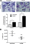
Fig 1
Fig 1. ArtinM enhances L. major uptake and killing by human neutrophils.
Human neutrophils were treated or were not treated with ArtinM, and infected with L. major promastigotes (MOI 3:1). A–L. major uptake. After incubation for 3 and 20 h, the cells were centrifuged on slides and stained for evaluation by light microscopy. Arrows indicate internalized parasites. In the graphic, data are expressed as the mean of neutrophils with intracellular parasites ± SD. * p<0.05; ** p<0.01 in comparison to untreated cells at the same period. One way ANOVA followed by Bonferroni's post-test. B–L. major killing. After 3 h of infection the cultures were washed to discard uninternalized parasites, fed with Schneider`s medium and cultured for additional 48 h. Motile promastigotes were counted, and the data are expressed as mean of viable parasites ± SD. ** p<0.002: compared to untreated, Student t test. Each assay was carried out in triplicate. The shown data are representative from three different experiments.
Fig 2
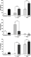
Fig 2. ArtinM stimulates the production of inflammatory cytokines by L. major-infected or uninfected neutrophils.
Human neutrophils were treated with or without ArtinM or PMA, and either infected or not infected with L. major promastigotes (MOI 3:1). At 20 h post-infection, the supernatant of these cells was collected for cytokine measurement by ELISA. A–TNF, B–TGFβ-1 and C–IL-1β concentrations are expressed as mean ± SD. * p<0.05; **** p<0.001 in comparison to untreated cells, or as indicated. One way ANOVA followed by Bonferroni's post-test. ND stands for “not detected”. ELISA assays were carried out in triplicate. The shown data are representative from three different experiments.
Fig 3
Fig 3. ArtinM stimulates the degranulation of L. major-infected or uninfected neutrophils.
A–Intracellular levels of myeloperoxidase. Human neutrophils were incubated for 20 h with ArtinM or medium only (untreated), and infected or not infected with L. major promastigotes (MOI 3:1); cells were permeabilized and reacted with anti-MPOPE. Cells were analyzed by flow cytometry and data were expressed as normalized mean ± SD of the median fluorescence intensity. * p<0.02, **p<0,01. Student's t test. B–Elastase releasing. Human neutrophils were stimulated with ArtinM or medium only (untreated); Cell supernatants were monitored for 30 min for enzymatic activity, using the substrate N-succinyl-Ala-Ala-Val-p-nitroanilide. The NE activity was expressed as the area under curve (AUC) ± SD. ** p<0.05 in comparison to untreated cells. Student's t test. C–Elastase leishmanicidal activity. Supernatants from neutrophils treated for 1 h or not treated with ArtinM were incubated for 30 min (or not incubated) with anti-MPO or anti-NE antibodies (both 1:500). Next, supernatants were added to L. major culture (1x10⁵), in Schneider’s medium. After 24 h, live parasites were quantified by reaction with Alamar Blue. Data are expressed as the number of viable parasites ± SD. * p<0.05 in comparison to the supernatant from untreated cells. One way ANOVA, followed by Bonferroni's post-test. Each assay was carried out in duplicate. The shown data are representative from three different experiments.
Fig 4
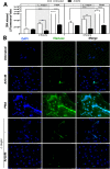
Fig 4. ArtinM inhibits NET formation induced by L. major.
Human neutrophils were incubated with ArtinM, PMA, ArtinM+PMA, or medium (untreated), and were either infected or not infected with L. major promastigotes (3:1). A–DNA quantitation. DNA released at 2 and 4 h after treatment/infection was quantified in the cell supernatants by fluorescence detection (Ex./Em. 480/520 nm) after reaction with SYTOX green. Data are expressed as a mean of fluorescence intensity ± SD. **p<0.01; ***p<0.001; **** p<0.0001. Two way ANOVA followed by Bonferroni's post-test. DNA quantitation assays were performed in triplicate and, the data shown are representative of four independent experiments. B–NET immunofluorescence detection. Neutrophils were plated onto poly-l-lysine coated slides, treated or untreated with PMA or ArtinM, and either infected or not infected with L. major promastigotes (MOI 3:1). After 6 h, the cells were stained with DAPI (DNA blue stain) and with anti-NE antibody (green). Merged images confirmed NETs by colocalization of staining. NET immunofluorescence detection assays were performed in duplicate and the data shown are representative of three independent experiments.
Fig 5
Fig 5. ArtinM augments ROS production by L. major-infected neutrophils.
Human neutrophils were treated with PMA, fMLP, ArtinM or medium (untreated). ROS production was quantified by reaction with luminol (A) and lucigenin (B), producing chemiluminescent photons (CPS). Data on PMA stimulation or L. major promastigotes infected neutrophils (3:1), which were treated or not treated with ArtinM, are shown in panel C. The amounts of ROS released are expressed as mean AUCxCPS ± SD. ** p<0.01; *** p<0.001; **** p<0.0001. One way ANOVA followed by Bonferroni's post-test. Each assay was carried out in triplicate. The data shown are representative of three independent experiments.
Fig 6
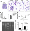
Fig 6. ArtinM treatment postpones apoptosis of uninfected neutrophils.
Human neutrophils were incubated (indicated period) with medium (untreated), lysis buffer, ArtinM or IL-8. A–Neutrophil morphology. Neutrophils were cytocentrifuged and stained for evaluation by light microscopy. Arrows indicate neutrophils with nuclear condensation. B–Phosphatidylserine exposure. Neutrophils were labeled with Annexin V-FITC and analyzed by flow cytometry. Data are expressed as mean of percentage of AnnexinV+ neutrophils ± SD. *** p<0.001 in comparison with the untreated cells, two way ANOVA followed by Bonferroni's post-test. ND stands for “not detected”. CJC-1 monomer detection. Neutrophils were incubated with JC-1 probe and green fluorescence detection (Ex/Em = 485/528) was performed at 3 and 20 h after treatment. Data are expressed as mean of fluorescence intensity ± SD. *** p<0.01 comparing with untreated curve, two way ANOVA followed by Bonferroni's post-test. D and E—Electrophoretic detection of DNA fragmentation. Neutrophils were incubated for 24 and 48 h with ArtinM or medium (untreated). Their genomic DNA was analyzed by gel electrophoresis. Images are shown on panel D. Panel E represents the band area in pixels2, regarding the electrophoretic detection of DNA fragmentation (ImageJ Software). Each assay was carried out in triplicate. The shown data are representative from three different experiments.
Fig 7
Fig 7. ArtinM induces early apoptosis of L. major infected neutrophils.
Human neutrophils were incubated with medium (untreated), lysis buffer, ArtinM, or IL-8, and infected or not with L. major promastigotes (MOI 3:1). At 3 h post infection, cells were labeled with Annexin V-FITC and analyzed by flow cytometry. Data are expressed as mean of percentage of AnnexinV+ neutrophils ± SD. ** p<0.01; *** p<0.001 in comparison to untreated cells, or as indicated. Two way ANOVA followed by Bonferroni's post-test. Each assay was carried out in triplicate. The shown data are representative from three different experiments.
Fig 8
Fig 8. ArtinM increases the leishmanicidal capacity of human neutrophils: a model.
Once infected with L. major, non-stimulated neutrophils produce high levels of TGF-β, and low levels of TNF, and IL-1β, which are associated with decreased cell degranulation and postponed cell death. In contrast, ArtinM-treated neutrophils become activated, produce high levels of TNF, and IL-1β, while TGF-b is reduced, and enhance degranulation. Collectively, these ArtinM induced responses augments the leishmanicidal capacity of neutrophils.

Similar articles

Cited by

References

    1. Andre FE, Booy R, Bock HL, Clemens J, Datta SK, et al. (2008) Vaccination greatly reduces disease, disability, death and inequity worldwide. Bull World Health Organ 86: 140–146. 10.2471/BLT.07.040089 - DOI - PMC - PubMed
    1. Levine MM, Robins-Browne R (2009) Vaccines, global health and social equity. Immunol Cell Biol 87: 274–278. 10.1038/icb.2009.15 - DOI - PubMed
    1. Robbins JB, Schneerson R, Szu SC (1995) Perspective: hypothesis: serum IgG antibody is sufficient to confer protection against infectious diseases by inactivating the inoculum. J Infect Dis 171: 1387–1398. - PubMed
    1. Griffiths KL, Khader SA (2014) Novel vaccine approaches for protection against intracellular pathogens. Curr Opin Immunol 28: 58–63. 10.1016/j.coi.2014.02.003 - DOI - PMC - PubMed
    1. Mifsud EJ, Tan ACL, Jackson DC (2014) TLR agonists as modulators of the innate immune response and their potential as agents against infectious disease. Front Immunol 5 10.3389/fimmu.2014.00079 - DOI - PMC - PubMed

Publication types

Grants and funding

This study was supported by grants from São Paulo Research Foundation (grants FAPESP #2009/60642-3, #2012/13419-7, and #2013/04088-0), and Financiadora de Estudos e Projetos (grant FINEP# 01100455900). The funders had no role in study design, data collection and analysis, decision to publish, or preparation of the manuscript.

LinkOut - more resources