. 2016 Apr 15;30(8):931-45.
doi: 10.1101/gad.277665.116. Epub 2016 Apr 7.

PolySUMOylation by Siz2 and Mms21 triggers relocation of DNA breaks to nuclear pores through the Slx5/Slx8 STUbL

Affiliations

PolySUMOylation by Siz2 and Mms21 triggers relocation of DNA breaks to nuclear pores through the Slx5/Slx8 STUbL

Chihiro Horigome et al. Genes Dev. .

Abstract

High-resolution imaging shows that persistent DNA damage in budding yeast localizes in distinct perinuclear foci for repair. The signals that trigger DNA double-strand break (DSB) relocation or determine their destination are unknown. We show here that DSB relocation to the nuclear envelope depends on SUMOylation mediated by the E3 ligases Siz2 and Mms21. In G1, a polySUMOylation signal deposited coordinately by Mms21 and Siz2 recruits the SUMO targeted ubiquitin ligase Slx5/Slx8 to persistent breaks. Both Slx5 and Slx8 are necessary for damage relocation to nuclear pores. When targeted to an undamaged locus, however, Slx5 alone can mediate relocation in G1-phase cells, bypassing the requirement for polySUMOylation. In contrast, in S-phase cells, monoSUMOylation mediated by the Rtt107-stabilized SMC5/6-Mms21 E3 complex drives DSBs to the SUN domain protein Mps3 in a manner independent of Slx5. Slx5/Slx8 and binding to pores favor repair by ectopic break-induced replication and imprecise end-joining.

Keywords: DNA damage; Mms21; SUMO; Siz2; Slx5; nuclear organization; nuclear pores.


Figures

Figure 1.
Figure 1.
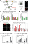
SUMO E3 ligases are required for the DSB anchoring to the nuclear periphery. (A) Shown is chromosome III (Chr III) in GA-6844 bearing deleted homologous donor loci (hmlΔ/hmrΔ) and a lacO array inserted 4.4 kb from the HO cut site at MAT. GFP-LacI and CFP-Nup49 label DSB and pores, respectively. Relocation to Mps3 was previously shown to occur in S or G2 phase, coincident with extensive resection. (B) Locus position was scored relative to the nuclear diameter in the locus’ plane of focus using a spinning disc confocal image stack. Distance over diameter ratios are binned into three equal zones. (C) Position of cleaved MAT position relative to CFP-Nup49 in siz1Δ siz2Δ (GA-7968) and mms21ΔC (JC3654) after 120 min on galactose. The mutants compromise relocation in both G1- and S-phase cells. (#) Significantly nonrandom based on cell number and confidence values from a proportional test comparing random and experimental distributions; (*) significantly different distribution between wild type and the mutant; (red dotted line) 33% or random distribution. Cleavage efficiency, nuclei counted, and statistical significance for all imaging experiments are summarized in Supplemental Table S1. (D) Scoring of MAT colocalization with the pore cluster in nup133ΔN (GA-7314) after cut induction. Scoring criteria are shown at the left, and results comparing wild type (GA-7314) and siz2Δ (GA-7970) are at the right. A gray-shaded zone between dotted lines represents empirically (top) and computationally (bottom) determined limits of stochastic colocalization (Horigome et al. 2014, 2015). (E) Pore-ChIP (chromatin immunoprecipitation) was performed with Mab414 monoclonal (Abcam) with wild-type and siz2Δ isogenic derivatives of JKM179 (Lee et al. 1998). HO cleavage was induced for the indicated times on galactose. Quantitative PCR (qPCR) at 1.6 kb from the HO cut site was performed in triplicate on two biological replicates. See the Supplemental Material for normalization techniques. (F) Chromosome III in the MATa strain was used for ChIP, with the positions of primer/probe sets shown. ChIP of MAT colocalization with HA-tagged Mps3 is shown for wild-type (GA-8306) and siz2Δ (GA-8541) cells at the indicated times after cut induction. Data from two independent experiments quantified in triplicate are represented as mean ± SEM. (G) The position of the cleaved MAT relative to CFP-Nup49 in smt3-3KR (GA-9072) after 120 min on galactose, as in C. (n.s.) Not significantly different from random.
Figure 2.
Figure 2.
The STUbL Slx5/8 promotes DSB relocation to the nuclear pore. (A) ChIP for HA-tagged Slx5 monitored MAT locus binding of Slx5 at the indicated time after cut induction on galactose in wild-type (JC3020), siz2Δ (JC3668), smt3-3KR (JC3214), and a nontagged strain (JC727). Data from three independent experiments are represented as mean ± SEM. (B) MAT position relative to CFP-Nup49 in wild-type (GA-6844), slx5Δ (GA-7097), and slx8Δ (GA-7098) after 120 min on galactose. Symbols and scoring are as in Figure 1, C and G. (C) Scoring of MAT colocalization with the pore cluster in nup133ΔN (GA-7314) after cut induction as in Figure 1D. Strains scored at the indicated times on galactose were wild type (GA-7314) and slx5Δ (GA-7969). (Pink/red shaded region) Colocalization. (D) ChIP for HA-tagged Mps3 monitored MAT locus association with Mps3 at the indicated times after cut induction in wild-type (GA-8306) and slx5Δ (GA-8539) cells. Data from three independent experiments are represented as mean ± SEM.
Figure 3.
Figure 3.
Targeted polySUMO and Slx5 promote chromatin relocation to the nuclear pore, bypassing Siz2 and Slx8 activities. (A) Scheme of the LexA fusion protein targeting to PES4::lacO-LexA for the zoning assay (GA-1461) and to LYS2::lacO-LexA in a strain bearing nup133ΔN (GA-8194) for nuclear pore colocalization of the intact targeted locus. (B,C) The position of lacO/LexA-tagged PES4 was visualized by GFP-LacI and scored in cells classified as G1 or S phase. Strains carried GFP-Nup49 (GA-1461) and the indicated LexA fusion proteins (polySUMO = 4×Smt3) (see the Supplemental Material) or LexA alone expressed from pAT4 derivatives. LexA fusion was expressed in wild-type (wt) (GA-1461), siz2Δ (GA-4447), slx5Δ (GA-4448), or slx8Δ (GA-4449) strains. Position was scored as in Figure 1C. (D) Pore cluster colocalization for LexA-tagged LYS2 in a strain bearing nup133ΔN (GA-8194) transformed with the indicated LexA fusion. Colocalization (pink to red) is as described in Figure 1D.
Figure 4.
Figure 4.
The targeted monoSUMO construct shifts chromatin to the nuclear periphery in S phase but not to nuclear pores. (A) The indicated LexA fusion proteins are expressed in the wild-type (wt) strain (GA-1461) and siz2Δ (GA-4447). The position of lacO/LexA-tagged ARS607 was visualized by GFP-LacI and scored as in Figure 1C. (B) Pore cluster colocalization for LexA-tagged LYS2 in a strain bearing nup133ΔN (GA-8194) transformed with the indicated LexA fusions. Colocalization (pink to red) was scored as in Figure 1D. (C) In a wild-type strain (GA-1461) expressing LexA-smt3-3KR and either an empty vector or the Mps3N′ construct, PES4 position was scored as in A. (D) Summary of the chromatin positioning roles of monoSUMO and polySUMO chains based on the targeting assays.
Figure 5.
Figure 5.
The SMC5/6 complex, the recruiter Rtt107, and the efficient interaction of Nse5 with Slx5/Slx8 facilitate DSB anchoring to Mps3. (A) Depiction of Nse5 interactions with SUMO (Smt3), Slx5, and Nse6 and the effects of the nse5L247A mutation (Supplemental Fig. S5; see the text). (B) The position of the cleaved MAT locus relative to CFP-Nup49 in wild-type (GA-6844), nse5-L247A (JC3161), smc6-9 (JC3131), and rtt107Δ (GA-7092) cells after 120 min on galactose at 30°C is shown. Binning into G1 and S as well as the symbols are as in Figure 1C. (C) ChIP for Nup84-13MYC monitored the MAT locus after 240 min on galactose in wild-type (GA-4133), nse5-L247A (JC3154), and smc6-9 (JC3150) cells grown at 30°C. Data from four experiments are represented as mean ± SEM. PCR probes were at the indicated distances from the HO cut site. (D) ChIP against 3HA-Mps3 (anti-HA) at the indicated times on galactose. Enrichment of MAT (0.6 kb from the cut site) over uncut SMC2 was quantified by qPCR in wild type (JC3167) and nse5-L247A (JC3114). For the smc6-9 temperature-sensitive alleles (JC3115), strains were grown at 25°C and then transferred for 1 h to 37°C to inactivate the smc6-9 allele before HO induction at 35°C. Data from three independent experiments are represented as mean ± SEM. (E) ChIP for HA-tagged Slx5 monitored MAT locus association at the indicated time after cut induction in wild-type (JC3020), nse5-L247A (JC3621), smc6-9 (JC3198), and nontagged (JC727) cells at 30°C. Data from three independent experiments are represented as mean ± SEM.
Figure 6.
Figure 6.
Slx8 and pore proteins favor repair by BIR and imprecise end-joining. (A) Cell survival after HO cut induction. Cleavage at the MAT locus by the HO endonuclease was induced by galactose for the indicated times. Cells were washed and plated on YPAD plates, and CFUs were scored after 2 d at 30°C. The rates of the viable cells in wild-type (GA-6844), siz2Δ (GA-6858), nse5-L247A (JC3161), slx5Δ (GA-7097), siz2Δ slx5Δ (GA-9206), and nse5-L247A slx5Δ (GA-9355) strains were normalized to cell count before cut induction. (B) BIR assay. A recipient cassette composed of a 3′ truncated LYS2 gene (ly), a 36-base-pair cut site for the HO endonuclease, and a kanR marker was incorporated into chromosome V (ChrV). A donor cassette composed of a TRP1 marker and a 5′ truncation of LYS2 (ys2) was inserted 60 kb from the telomere of chromosome I (ChrI). The two mutant lys2 fragments share 2.1 kb of homology. The HO endonuclease was expressed under the control of a galactose promoter. Lys+ cells lacking kanamycin resistance were scored for wild-type (GA-8994), nup60Δ (GA-9185), slx8Δ (GA-9186), and pol32Δ (GA-9090) cells. Error bars indicate standard deviation. Significance was determined by Student's t-test. (C) Precise NHEJ and imprecise NHEJ were performed and analyzed as described in the Materials and Methods on GA-8860 (wild type) and GA-8471 (nup84Δ).
Figure 7.
Figure 7.
Model for the role of SUMO and the Slx5 STUbL subunit in break relocation. Siz2 is damage-associated and deposits SUMO on various repair substrates that are monoSUMOylated by Mms21. Slx5/Slx8 is itself SUMOylated and recruited to the DSB in a polySUMOylation-dependent manner. Siz2, polySUMOylation, and its recognition by Slx5/Slx8 are required for DSB relocation to pores in both G1 and S. Nup84 components are recognized by Slx5. In contrast, monoSUMOylation can shift chromatin to Mps3 in an SMC5/6-dependent but Slx5/8-independent manner. Rtt107 phosphorylation by Mec1 recruits the SMC5/6 complex to DSBs. A component of SMC5/6, Nse5, interacts with Slx5. The SUMO E3 ligase Mms21 is recruited with the SMC5/6 complex. Ubiquitination of SUMOylated target proteins by the Slx5/Slx8 STUbL most likely results in subsequent degradation by the proteasome at the NE to enable repair pathways such as BIR.

Similar articles

Cited by

References

    1. Agmon N, Liefshitz B, Zimmer C, Fabre E, Kupiec M. 2013. Effect of nuclear architecture on the efficiency of double-strand break repair. Nat Cell Biol 15: 694–699. - PubMed
    1. Albuquerque CP, Wang G, Lee NS, Kolodner RD, Putnam CD, Zhou H. 2013. Distinct SUMO ligases cooperate with Esc2 and Slx5 to suppress duplication-mediated genome rearrangements. PLoS Genet 9: e1003670. - PMC - PubMed
    1. Azam M, Lee JY, Abraham V, Chanoux R, Schoenly KA, Johnson FB. 2006. Evidence that the S. cerevisiae Sgs1 protein facilitates recombinational repair of telomeres during senescence. Nucleic Acids Res 34: 506–516. - PMC - PubMed
    1. Bennett CB, Lewis LK, Karthikeyan G, Lobachev KS, Jin YH, Sterling JF, Snipe JR, Resnick MA. 2001. Genes required for ionizing radiation resistance in yeast. Nat Genet 29: 426–434. - PubMed
    1. Bermudez-Lopez M, Pocino-Merino I, Sánchez H, Bueno A, Guasch C, Almedawar S, Bru-Virgili S, Garí E, Wyman C, Reverter D, et al. 2015. ATPase-dependent control of the Mms21 SUMO ligase during DNA repair. PLoS Biol 13: e1002089. - PMC - PubMed

Publication types

MeSH terms

Substances

LinkOut - more resources