. 2016 Jun 1;27(11):1834-44.
doi: 10.1091/mbc.E15-03-0123. Epub 2016 Apr 6.

Rab43 regulates the sorting of a subset of membrane protein cargo through the medial Golgi

Affiliations

Rab43 regulates the sorting of a subset of membrane protein cargo through the medial Golgi

John V Cox et al. Mol Biol Cell. .

Abstract

To evaluate the role of cytoplasmic domains of membrane-spanning proteins in directing trafficking through the secretory pathway, we generated fluorescently tagged VSV G tsO45 with either the native G tail (G) or a cytoplasmic tail derived from the chicken AE1-4 anion exchanger (G(AE)). We previously showed that these two proteins progressed through the Golgi with distinct kinetics. To investigate the basis for the differential sorting of G and G(AE), we analyzed the role of several Golgi-associated small GTP-binding proteins and found that Rab43 differentially regulated their transport through the Golgi. We show that the expression of GFP-Rab43 arrested the anterograde transport of G(AE) in a Rab43-positive medial Golgi compartment. GFP-Rab43 expression also inhibited the acquisition of endoH-resistant sugars and the surface delivery of G(AE), as well as the surface delivery of the AE1-4 anion exchanger. In contrast, GFP-Rab43 expression did not affect the glycosylation or surface delivery of G. Unexpectedly, down-regulation of endogenous Rab43 using small interfering RNA resulted in an increase in the accumulation of G(AE) on the cell surface while having minimal effect on the surface levels of G. Our data demonstrate that Rab43 regulates the sorting of a subset of membrane-spanning cargo as they progress through the medial Golgi.


Figures

FIGURE 1:
FIGURE 1:
Colocalization of endogenous Rab43 with markers of the early secretory pathway. (A) PH5CH8 human hepatocytes were fixed, permeabilized, and stained with a mouse monoclonal antibody specific for Rab43 (green) and rabbit polyclonal antibodies specific for GM130, giantin, or mannosidase II (Mann. II). After incubation with the appropriate secondary antibodies, the cells were imaged by confocal microscopy. (B) Percentage colocalization of endogenous Rab43 with these various markers. (C) In similar analyses, COS7 cells were transfected with GFP-Rab43 and the percentage colocalization of this fusion with GM130, giantin, or mannosidase II (Mann. II) quantified by confocal microscopy. Average colocalization (±SD) from 25 cells from two independent experiments. Scale bars, 10 μm.
FIGURE 2:
FIGURE 2:
Quantification of GAE and G colocalization with various markers of the early secretory pathway in PH5CH8 cells. PH5CH8 hepatocytes expressing GAE-EGFP or G-EGFP were grown overnight at restrictive temperature and then shifted to permissive temperature for various times ranging from 0 to 30 min. At each time point, the cells were fixed and stained with antibodies specific for (A) GM130, (B) giantin, (C) mannosidase II, or (D) Rab43 and analyzed by confocal microscopy. The data points reflect percentage colocalization of GAE or G with the various early secretory pathway markers. The colocalization values shown represent the average value obtained from Z-stacks of at least 25 different cells from two independent experiments. Additional analyses revealed that the observed differences in the colocalization of G and GAE with giantin at the 15-min time point (p < 0.0001), with mannosidase II at the 20-min time point (p = 0.0002), and with endogenous Rab43 at the 25-min time point (p = 0.0006) were all statistically significant using the Student’s two-tailed t test.
FIGURE 3:
FIGURE 3:
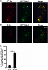
Differential effect of GFP-Rab43 on GAE and G sorting. COS7 cells transfected with GFP-Rab43 were subsequently infected with replication-defective adenoviruses encoding (A) GAE -Ds or (B) G-Ds and grown at the restrictive temperature for 24 h. The cells were then shifted to permissive temperature for 30 min, fixed, and imaged by confocal microscopy. The merged images show that GAE accumulated in a GFP-Rab43-positive compartment, whereas G did not. (C) Percentage colocalization of GAE and G with GFP-Rab43. The values represent the average (±SD) from at least 20 cells from two independent experiments. **p < 0.0001 (Student’s two-tailed t test). Scale bars, 5 μm.
FIGURE 4:
FIGURE 4:
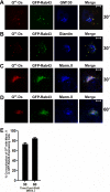
GAE accumulates in a mannosidase II– and GFP-Rab43–containing compartment in GFP-Rab43–expressing cells. COS7 cells transfected with GFP-Rab43 were subsequently infected with adenovirus expressing GAE-Ds and grown at the restrictive temperature for 24 h. The cells were then shifted to permissive temperature for 30 min (A–C) or 60 min (D) and fixed. The cells were then permeabilized, stained with antibodies specific for GM130 (A), giantin (B), or mannosidase II (C, D), and incubated with Alexa Fluor 647–conjugated secondary antibody. The cells were then imaged by confocal microscopy. Scale bars, 5 μm. (E) Percentage colocalization of GAE-Ds with mannosidase II after a 30- or 60-min temperature shift. Average values from 20 different cells from two independent experiments.
FIGURE 5:
FIGURE 5:
Effect of GAE expression on the localization of GFP-Rab43. COS7 cells transfected with GFP-Rab43 were subsequently infected with adenoviral vectors expressing GAE-Ds or G-Ds. The cells were grown at the restrictive temperature for 24 h and then shifted to permissive temperature for 60 min before fixation (A) or the cells were fixed before being shifted to permissive temperature (B). The cells were then permeabilized and stained with antibodies specific for GM130, giantin, or mannosidase II and imaged by confocal microscopy. At least 15 cells from two independent experiments were used to calculate the percentage colocalization of GFP-Rab43 with these markers of the early secretory pathway in cells expressing GAE-Ds or G-Ds. **p < 0.0001 (Student’s two-tailed t test).
FIGURE 6:
FIGURE 6:
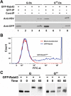
Coprecipitation of GAE with GFP-Rab43. (A) COS7 cells expressing GFP-Rab43 were infected with adenovirus encoding GAE-Ds or G-Ds and grown at restrictive temperature for 24 h. The cells were then shifted to permissive temperature for 60 min and harvested. After being washed in PBS, the cells were lysed by sonication and centrifuged at 5000 × g for 10 min. The supernatants were then subjected to immunoprecipitation analysis using rabbit anti-GFP (GFP) antibodies (lanes 1 and 5), and the precipitates were immunoblotted with an anti-VSV antibody that recognizes the identical ectodomains of GAE and G and anti-GFP antibodies. Control GFP immunoprecipitates were prepared from cells that did not express GFP-Rab43 (lanes 2 and 6). Additional control immunoprecipitates were prepared using protein A agarose beads coated with normal rabbit serum (lanes 3 and 7). Lysates are included for comparison (lanes 4 and 8). (B) COS7 cells were transfected with GFP-Rab43, and 24 h posttransfection, cells expressing low levels of GFP-Rab43 were isolated using a fluorescence-activated cell sorter. (C) The GFP-Rab43low cells or unsorted mock-transfected cells were replated and infected with adenovirus encoding GAE or G. The cells were grown overnight at restrictive temperature and lysed (0 time) or shifted to permissive temperature for 60 min before lysis. In each instance, the lysates were incubated with endoglycosidase H and subjected to immunoblotting analysis with the anti-VSV antibody, which recognizes the identical ectodomains of GAE and G. GFP-Rab43 expression significantly blocked the acquisition of complex N-linked sugars by GAE but had minimal effect on the acquisition of complex N-linked sugars by G.
FIGURE 7:
FIGURE 7:
Differential effect of GFP-Rab43 on the surface accumulation of GAE and G. COS7 cells transfected with GFP-Rab43 were subsequently infected with adenoviral vectors expressing (A) GAE-Ds or (B) G-Ds and grown at the restrictive temperature for 24 h. The cells were then shifted to permissive temperature for 60 min and fixed. Surface GAE or G was stained in nonpermeabilized cells using the I1 monoclonal antibody, which recognizes an epitope in the ectodomains of both GAE and G, and an Alexa Fluor 647 secondary antibody. The cells were then imaged by confocal microscopy. The periphery of a cell coexpressing GFP-Rab43 and GAE-Ds is outlined in A. (C) Quantification of the surface levels of GAE-Ds and G-Ds in GFP-Rab43– expressing cells shifted to permissive temperature for times ranging from 0 to 120 min (averaged from 12–16 cells in two independent experiments). The difference in the surface delivery of GAE-Ds at the 120-min time point in the presence and absence of GFP-Rab43 is statistically significant, with p = 0.0003. Scale bars, 5 μm.
FIGURE 8:
FIGURE 8:
AE1-4 and GAE accumulate in the same compartment in GFP-Rab43–expressing cells. (A) COS7 cells were cotransfected with AE1-4, which has an extracellular V5 epitope tag, and GFP-Rab43 and subsequently infected with an adenoviral vector encoding GAE-Ds. The cells were grown overnight at restrictive temperature and then shifted to permissive temperature for 30 min, at which time the cells were fixed, permeabilized, and stained with anti-V5 antibodies. After incubation with an Alexa Fluor 647–conjugated secondary antibody, the cells were imaged by confocal microscopy. The majority of AE1-4 accumulated in a compartment that was positive for both GAE and GFP-Rab43. (B) COS7 cells were cotransfected with AE1-4 containing an extracellular V5 epitope tag and GFP-Rab43. At 24 h posttransfection, the cells were fixed, and nonpermeabilized cells were incubated with mouse anti-V5 antibodies to detect AE 1-4 on the cell surface. After extensive washing, the cells were permeabilized and incubated with rabbit antibodies specific for the cytoplasmic tail of AE1 and then with goat anti-mouse secondary antibody conjugated to Alexa Fluor 647 and goat anti-rabbit secondary antibody conjugated to Alexa Fluor 594. The cells were then imaged by confocal microscopy. AE1-4 accumulated on the surface of cells that did not express GFP-Rab43. However, in cells expressing GFP-Rab43 (indicated by arrows), AE1-4 was retained in an intracellular GFP-Rab43–containing compartment. (C) Quantification of the level of surface AE1-4 in Rab43-positive and negative cells. Values reflect the average value obtained from the analysis of at least 25 cells from two independent experiments. **p < 0.0001 (Student’s two-tailed t test). Scale bars, 5 μm.
FIGURE 9:
FIGURE 9:
Effect of Rab43 knockdown on GAE and G trafficking. (A) PH5CH8 cells were transfected with Rab43-specific SMART pool siRNAs (12.5 nM each) or with 50 nM scrambled control siRNA, and 4 d posttransfection, the cells were harvested. Lysates were analyzed by immunoblotting analysis with Rab43-specific rabbit polyclonal antibodies and actin monoclonal antibodies. (B) PH5CH8 cells transfected with the Rab43-specific SMART pool siRNAs or with the scrambled control siRNA were infected with adenovirus encoding GAE-EGFP or G-EGFP at 72 h posttransfection. After growth overnight at restrictive temperature, the cells were shifted to permissive temperature for 2 h. The cells were then shifted to 4°C and incubated with the I1 monoclonal antibody. After washing, cells were detergent lysed, and immune complexes were precipitated. The surface immunoprecipitate and lysate from each condition were analyzed by immunoblotting analysis with anti-VSV antibodies. Control experiments in which nontransfected cells were maintained at the restrictive temperature of 39.8°C are also shown. (C) PH5CH8 cells transfected with the Rab43-specific SMART pool siRNAs or with the scrambled control siRNA were infected with adenovirus encoding GAE-EGFP or G-EGFP at 72 h posttransfection. After growth overnight at restrictive temperature, the cells were shifted to permissive temperature for 0 or 2 h and fixed. Surface levels of GAE and G in nonpermeabilized cells were monitored by staining with the I1 monoclonal antibody, and surface-bound antibodies were detected using donkey anti-mouse immunoglobulin G conjugated to Alexa Fluor 594. The cells were imaged on a Zeiss AxioImager.M2 microscope, and the levels of surface GAE and G were quantified. The values presented represent the surface fluorescence normalized to total G and GAE levels at each time point.

Similar articles

Cited by

References

    1. Adair-Kirk TL, Cox KH, Cox JV. Intracellular trafficking of variant chicken kidney AE1 anion exchangers: role of alternative NH(2) termini in polarized sorting and Golgi recycling. J Cell Biol. 1999;147:1237–1248. - PMC - PubMed
    1. Balch WE, Kahn RA, Schwaninger R. ADP-ribosylation factor is required for vesicular trafficking between the endoplasmic reticulum and the cis-Golgi compartment. J Biol Chem. 1992;267:13053–13061. - PubMed
    1. Barr FA. Rab GTPase function in Golgi trafficking. Semin Cell Dev Biol. 2009;20:780–783. - PubMed
    1. Caster AH, Sztul E, Kahn RA. A role for cargo in Arf-dependent adaptor recruitment. J Biol Chem. 2013;288:14788–14804. - PMC - PubMed
    1. Chavrier P, Vingron M, Sander C, Simons K, Zerial M. Molecular cloning of YPT1/SEC4-related cDNAs from an epithelial cell line. Mol Cell Biol. 1990;10:6578–6585. - PMC - PubMed

Publication types

LinkOut - more resources