. 2016 Apr 12;15(2):264-73.
doi: 10.1016/j.celrep.2016.03.017. Epub 2016 Mar 31.

Manipulating TLR Signaling Increases the Anti-tumor T Cell Response Induced by Viral Cancer Therapies

Affiliations

Manipulating TLR Signaling Increases the Anti-tumor T Cell Response Induced by Viral Cancer Therapies

Juan J Rojas et al. Cell Rep. .

Abstract

The immune response plays a key role in enhancing the therapeutic activity of oncolytic virotherapies. However, to date, investigators have relied on inherent interactions between the virus and the immune system, often coupled to the expression of a single cytokine transgene. Recently, the importance of TLR activation in mediating adaptive immunity has been demonstrated. We therefore sought to influence the type and level of immune response raised after oncolytic vaccinia therapy through manipulation of TLR signaling. Vaccinia naturally activates TLR2, associated with an antibody response, whereas a CTL response is associated with TLR3-TRIF-signaling pathways. We manipulated TLR signaling by vaccinia through deglycosylation of the viral particle to block TLR2 activation and expression of a TRIF transgene. The resulting vector displayed greatly reduced production of anti-viral neutralizing antibody as well as an increased anti-tumor CTL response. Delivery in both naive and pre-treated mice was enhanced and immunotherapeutic activity dramatically improved.


Figures

Figure 1
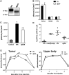
Figure 1. Deglycosylation of Vaccinia Virus Envelope
(A) Immunoblot showing deglycosylation of vaccinia virus envelope protein B5R. Purified WR (wild-type strain WR) and dgWR (deglycosylated) viruses were disrupted and blotted using an anti-B5R antibody. Decrease in protein weight corresponds to deglycosylation of the B5R protein. (B) Deglycosylation of virus envelope has no effect on vaccinia virus infectivity. Different mouse tumor cell lines were infected with TK– (strain WR with a deletion of the viral thymidine kinase gene) or its deglycosylated version at an MOI of 1, and viral luciferase expression was measured 3 hr after infection by bioluminescence imaging. Mean values + SD of three independent experiments are plotted. (C) Deglycosylation reduces TLR2 activation in vitro. 293 cells expressing mouse TLR2 were transfected with pNiFty (TLR-signaling reporter plasmid). Twenty-four hours after transfection, cells were infected at an MOI of 1 with WR or dgWR, and TLR2 activation was quantified 24 hr after infection by bioluminescence imaging. Means + SD of two independent experiments (performed in quadruplicate) are depicted. (D) STAT3 phosphorylation is depleted in splenic lymphocytes of mice injected with deglycosylated vaccinia. C57/BL6 mice were injected intravenously with 1 × 10⁷ plaque-forming units (PFUs) of WR or dgWR, and 1 hr after injection, spleens were excised, dissociated, and prepared for intracellular analysis. Percentage of pSTAT1pSTAT3+ lymphocytes was determined by flow cytometry. PBS and PAM(3)CSK(4) were used as controls. Values of individual mice and means ± SEM of the different treatments are plotted. (E) Deglycosylation of vaccinia virus (TK–) envelope selectively increases viral gene expression from tumors in vivo. Balb/c mice harboring subcutaneous tumors of Renca cells (mouse renal adenocarcinoma) were randomized and injected with a single intravenous dose of 1 × 10⁸ PFUs per mouse of TK– or dgTK– (both expressing firefly luciferase). Kinetics of viral gene expression from within the tumor (left) or for the upper body (lungs, spleen, and liver signal; right) was monitored by bioluminescence imaging of viral luciferase expression. Mean values of 12 or 13 animals + SD are plotted. *p < 0.05 compared with PBS or control; #p < 0.05 compared with TK– or WR group; ϕp < 0.05 compared with PAM(3)CSK(4) group.
Figure 2
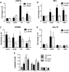
Figure 2. Oncolytic Vaccinia Virus Expressing the Mouse TRIF Protein Increases Activation of TLR-Responding Pathways and the Induction of Necroptosis
(A and B) Activation of NF-κB and IRF3 pathways after infection with TK-TRIF and TK-DAI. ELISA assays were utilized to determine concentrations of pNF-κB (A) and IRF3 (B) in cytoplasmic and nuclear extracts, respectively, of Renca, MC38, 4T1, or MEF cells infected with TK–, TK-TRIF, or TK-DAI at an MOI of 1. Analyses were performed 24 hr after infection. Data were obtained in quadruplicate from two independent experiments and are plotted as fold change versus TK– + SD. Dashed lines indicate TK– activation level. (C and D) mTRIF expression increases release of danger-associated molecular patterns (DAMPs). ELISA assays were utilized for determining the release of HMGB1 (C) and Hsp-70 (D) after infection with TK–, TK-TRIF, and TK-DAI viruses at an MOI of 1. Quantification was performed 24 hr after infection, and data are depicted as fold change versus TK– + SD, dashed lines indicating TK– release levels. (E) Percentage of apoptotic cells after infection with TK-TRIF and TK-DAI. A panel of mouse tumor cell lines was infected with the indicated viruses using an MOI of 1. At 48 hr after infection, percentage of necrotic and apoptotic cells were determined by flow cytometry by phosphatidylinositol (PI) and annexin V staining. Two independent experiments were performed, and means + SD are plotted.
Figure 3
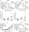
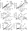
Figure 3. In Vivo Effects of TRIF Expression
(A) In vivo intratumoral concentration of cytokines and chemokines. Balb/c mice with established Renca subcutaneous tumors were randomized and injected with a single intravenous dose of 1 × 10⁸ PFUs per mouse of TK– or TK-TRIF. Four days after injection, tumors were harvested and concentrations of selected cytokines and chemokines were determined in tumor lysates by Luminex or ELISA assays. Fold change versus TK– from four to five mice per group + SD is plotted. Dashed line indicates TK– concentrations. *p < 0.05 compared with TK– group; #p < 0.05 compared with TK-DAI group. (B) Anti-tumor activity of TK-TRIF and TK-DAI. Renca or MC38 cells were implanted in Balb/c or C57/Bl6 mice, respectively, and mice were injected with PBS or 1 × 10⁸ PFUs of TK–, TK-TRIF, or TK-DAI through the tail vein. Tumor volumes were measured at indicated time points. n = 12–15 mice/group + SE. *p < 0.05 compared with PBS group; #p < 0.05 compared with TK– group; ϕ p < 0.05 compared with TK-DAI group.
Figure 4
Figure 4. In Vivo Immunotherapeutic Effects of TRIF Expression
(A) Viral gene expression in vivo. Renca or MC38 cells were implanted in Balb/c or C57/Bl6 mice, respectively, and mice were injected with PBS or 1 × 10⁸ PFUs of TK–, TK-TRIF, or TK-DAI through the tail vein. Viral luciferase expression from within the tumors was measured at indicated time points. n = 12–15 mice/group + SE. *, significant p < 0.05 compared with PBS group; #, significant p < 0.05 compared with TK– group; ϕ, significant p < 0.05 compared with TK-DAI group. (B) Altered T cell repertoire in the tumor after TK– or TK-TRIF injection. Balb/c mice with subcutaneous Renca tumors were treated with a single intravenous injection of PBS or 1 × 10⁸ PFUs/mouse of indicated viruses, and tumors were harvested at day 7 post-virus injection and evaluated for lymphocyte populations by flow cytometry. Percentages of (left) CD3+CD4+ and (right) CD3+CD8+ populations are plotted. (C) TK-TRIF improves the anti-tumor efficacy compared to TK-GMCSF in a mammary semi-orthotopic model. 4T1 cells were implanted in the mammary fat pad of Balb/c mice, and once the tumor was established, mice were injected with PBS or 1 × 10⁸ PFUs of TK–, TK-TRIF, or TK-GMCSF through the tail vein. (Left) Tumor volumes and (right) viral luciferase expression from within the tumors were measured at indicated time points. n = 12–14 mice/group + SE.
Figure 5
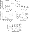
Figure 5. Combination of Envelope Deglycosylation and Mouse TRIF Expression Boosts Anti-tumor Cellular Responses and Enhances Safety
(A) Cellular immune response to vaccinia virus evaluated by IFN-γ ELISpot assay. At day 7, post-virus administration spleens were harvested from mice injected intravenously with 1 × 10⁸ PFUs of indicated viruses or PBS (Balb/c mice bearing Renca tumors) and evaluated for the amount of CTLs recognizing vaccinia virus. Values of individual mice and means ± SEM are depicted. (B) Serum-neutralizing antibody titers. A neutralizing assay was performed to determine circulating anti-vaccinia antibody levels for mice injected with 1 × 10⁸ PFUs of TK–, dgTK–, TK-TRIF, or dgTK-TRIF. Nabs titers were determined by the highest dilution of serum that resulted in at least 50% inhibition of cell-killing capacity. Values of individual mice and means ± SEM are plotted. (C) CTL response was measured as in (A), except ELIPSOT was performed against Renca cell lysate. (D) Viral gene expression in vivo after dgTK-TRIF administration. Renca tumors were implanted in Balb/c mice, and mice were injected with PBS or 1 × 10⁸ PFUs of TK–, TK-TRIF, or dgTK-TRIF through the tail vein. Viral luciferase expression from within the tumors was measured at indicated time points. n = 12–14 mice/group + SE. (E) Body weight change after intravenous viral administration. Balb/c mice were injected intravenously with 1 × 10⁸ PFUs per mouse of TK–, TK-TRIF, or dgTK-TRIF. PBS administration was used in the control group. TK-injected mice presented more than 10% reduction in body weight at day 6 after virus injection, whereas TK-TRIF- and dgTK-TRIF-injected mice presented a similar weight profile as those injected with PBS.
Figure 6
Figure 6. In Vivo Anti-tumor Activity in Different Models
(A) Balb/c-bearing Renca (left) or C57/BL6-bearing MC38 (right) tumors were treated with a single intravenous dose of indicated viruses (1 × 10⁸ PFUs/mouse). Tumor growth was followed by caliper measurements. Means of 12–15 mice per group + SE are depicted. (B) dgTK-TRIF improves anti-tumor activity compared to TK-mGMCSF. Immunocompetent mice harboring Renca, MC38, Pan02, or 4T1 tumors were injected intravenously with a dose of 1 × 10⁸ PFUs/mouse of TK-mGMCSF or dgTK-TRIF. Relative tumor volume after virus administration is plotted (n = 12–15 mice/group + SE). *p < 0.05 compared with PBS group; ψp < 0.05 compared with TK-GMCSF group.
Figure 7
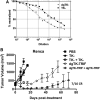
Figure 7. dgTK-TRIF Displays Enhanced Systemic Delivery and Therapeutic Activity during Repeat Cycles of Treatment
(A) Deglycosylated virus is resistant to anti-vaccinia neutralizing antibody. 1 × 10³ PFUs of TK– and deglycosylated TK– were mixed with limiting dilutions of VIG (vaccinia immunoglobulin G [IgG]) for 30 min before addition to a fresh cell layer and luciferase reading taken 24 hr later (uninfected cells and virus with no VIG were used to define 0% and 100% neutralization). (B) Enhanced therapeutic activity of repeated delivery of deglycosylated virus. Mice (BALB/c-bearing Renca tumors) were treated i.v. with 1 × 10⁸ PFUs of indicated virus. After 7 days, mice were treated with a second dose of the same viruses. Tumors were measured over time (n = 12–15 per group). dgTK-TRIF displayed significantly enhanced therapeutic activity (p < 0.05) relative to all other treatment groups from day 10 onward.

Similar articles

Cited by

References

    1. Andtbacka RHI, Collichio FA, Amatruda T, Senzer NN, Chesney J, Delman KA, Spitler LE, Puzanov I, Doleman S, Ye Y, et al. OPTiM: A randomized phase III trial of talimogene laherparepvec (T-VEC) versus subcutaneous (SC) granulocyte-macrophage colony-stimulating factor (GM-CSF) for the treatment (tx) of unresected stage IIIB/C and IV melanoma. J. Clin. Oncol. 2013;31:LBA9008.
    1. Chakrabarti S, Sisler JR, Moss B. Compact, synthetic, vaccinia virus early/late promoter for protein expression. Biotechniques. 1997;23:1094–1097. - PubMed
    1. Chen H, Sampath P, Hou W, Thorne SH. Regulating cytokine function enhances safety and activity of genetic cancer therapies. Mol. Ther. 2013;21:167–174. - PMC - PubMed
    1. Essajee S, Kaufman HL. Poxvirus vaccines for cancer and HIV therapy. Expert Opin. Biol. Ther. 2004;4:575–588. - PubMed
    1. Ganly I, Kirn D, Eckhardt G, Rodriguez GI, Soutar DS, Otto R, Robertson AG, Park O, Gulley ML, Heise C, et al. A phase I study of Onyx-015, an E1B attenuated adenovirus, administered intratumorally to patients with recurrent head and neck cancer. Clin. Cancer Res. 2000;6:798–806. - PubMed

Publication types

Substances