. 2016 Apr 11;213(1):49-63.
doi: 10.1083/jcb.201510098. Epub 2016 Apr 4.

Doa1 targets ubiquitinated substrates for mitochondria-associated degradation

Affiliations

Doa1 targets ubiquitinated substrates for mitochondria-associated degradation

Xi Wu et al. J Cell Biol. .

Abstract

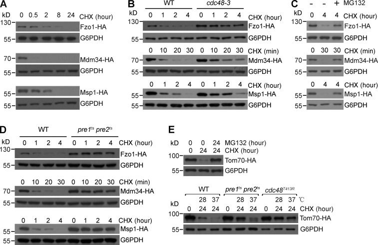
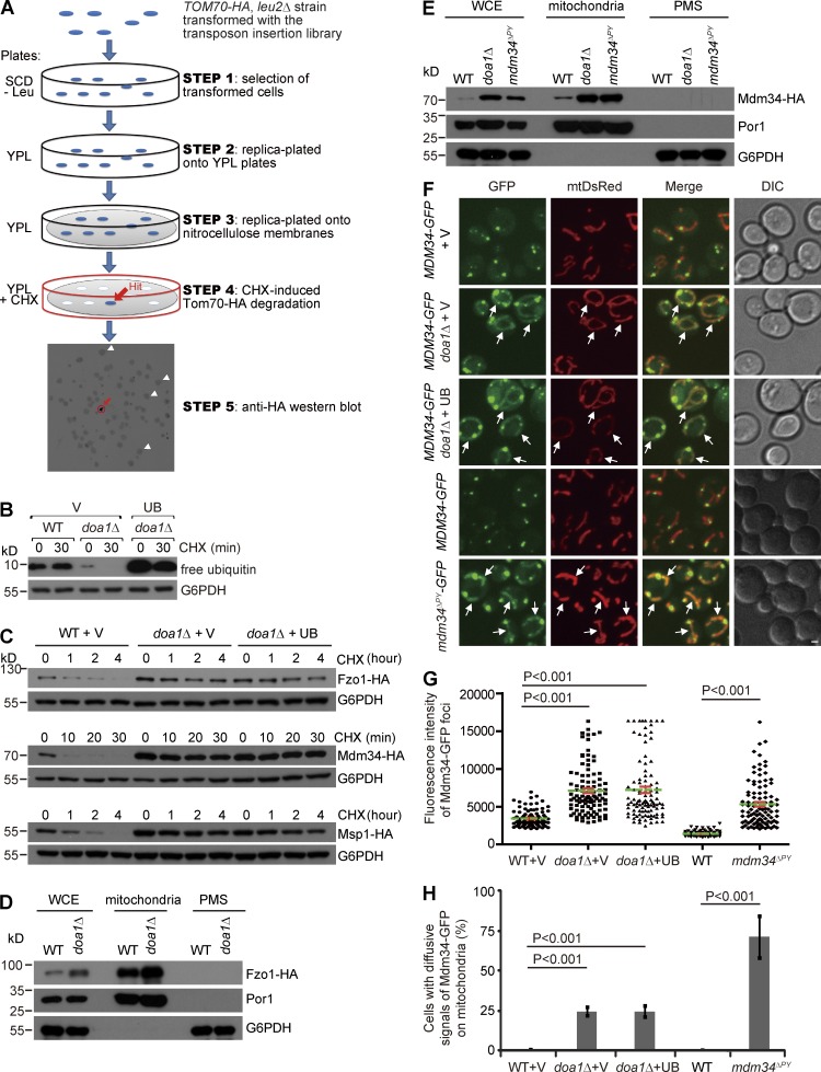
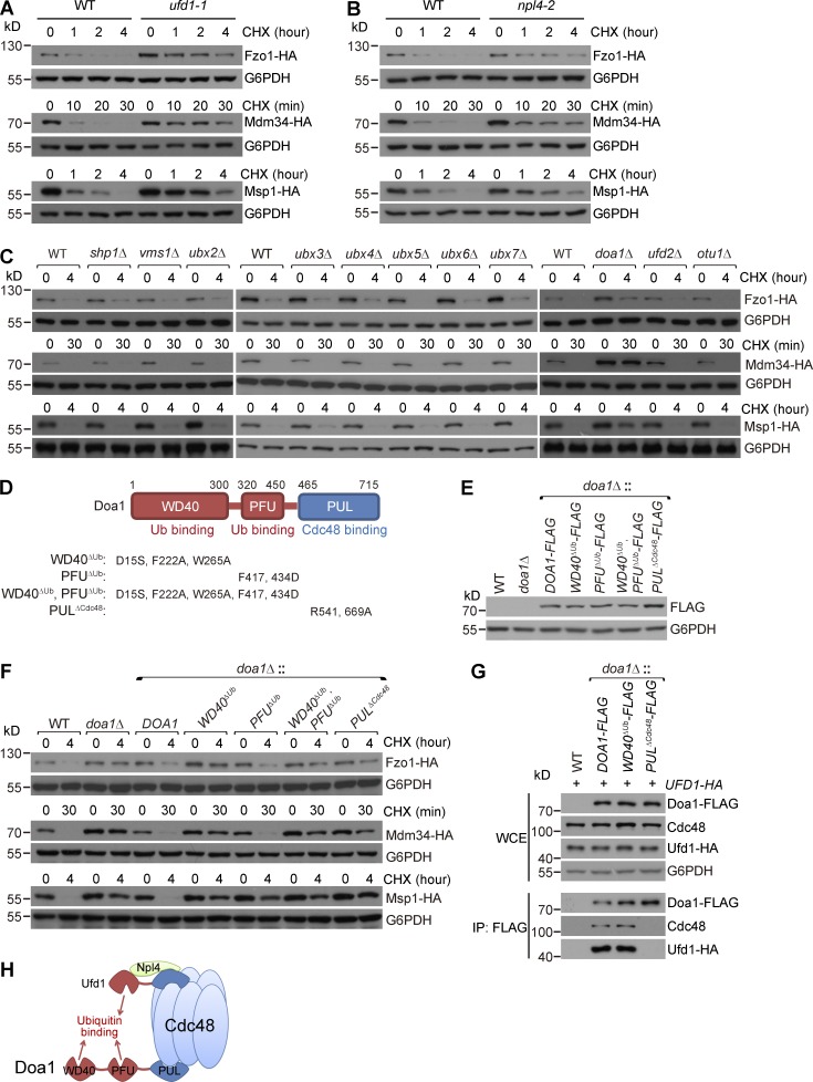
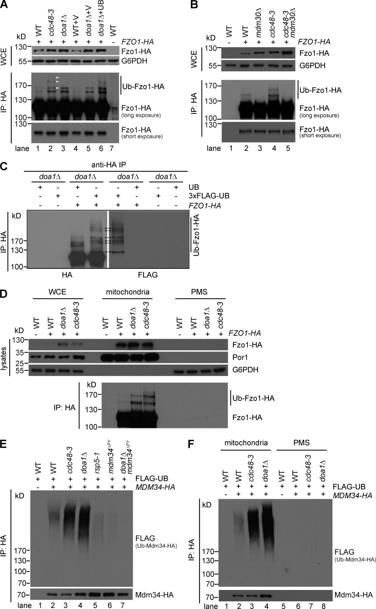
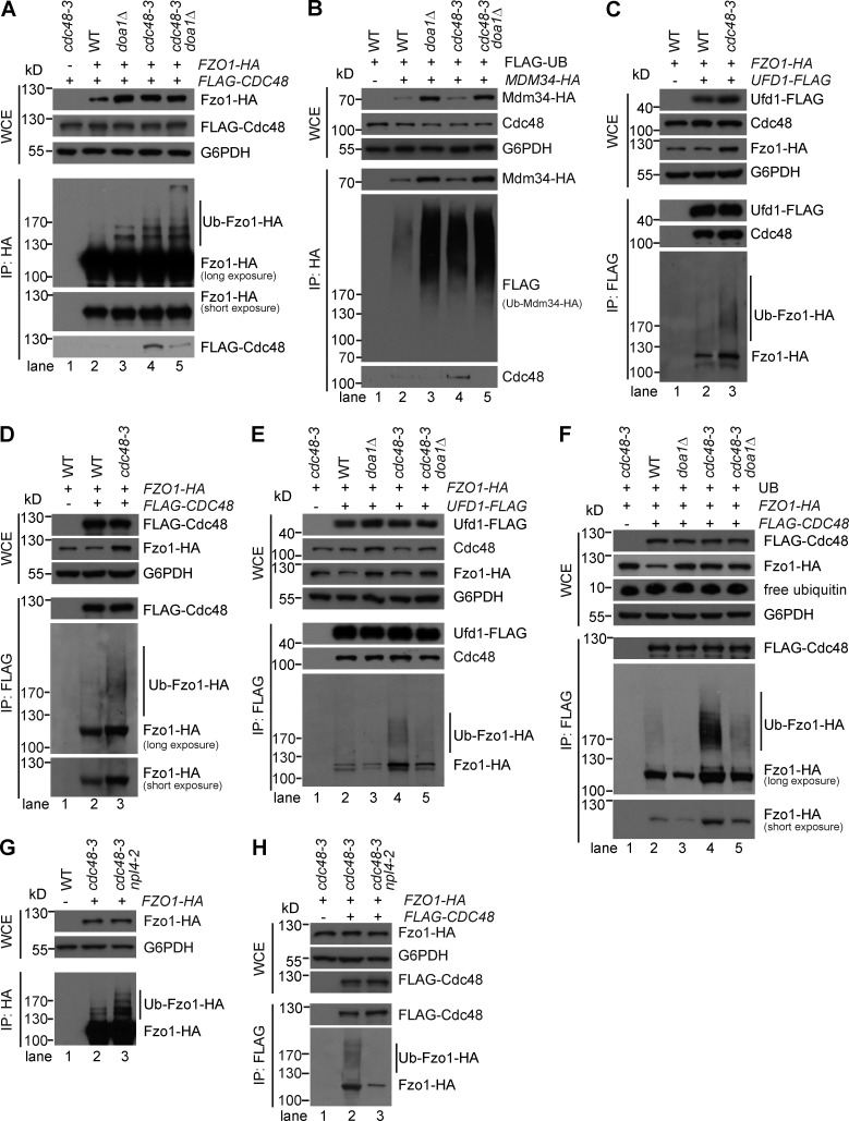
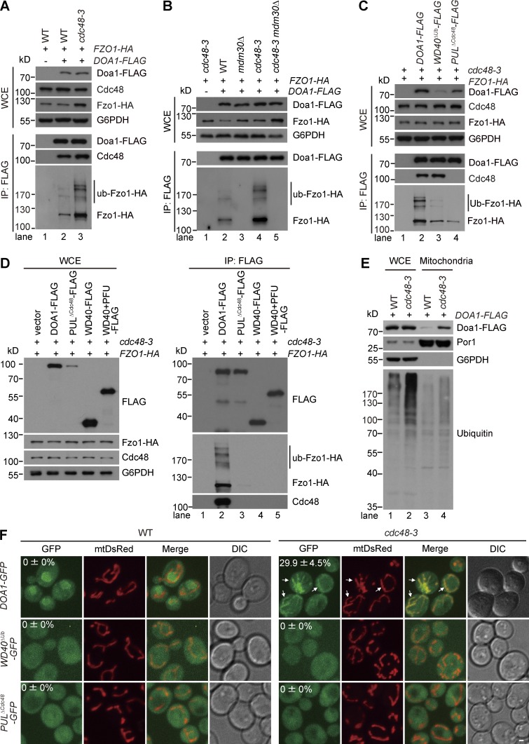
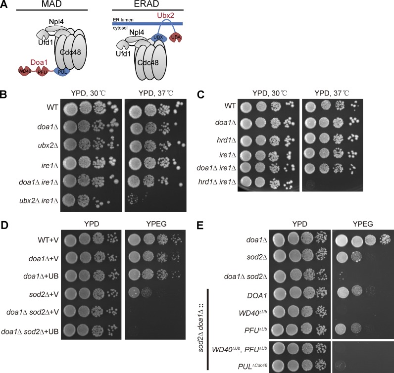
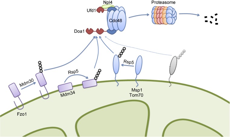
Mitochondria-associated degradation (MAD) mediated by the Cdc48 complex and proteasome degrades ubiquitinated mitochondrial outer-membrane proteins. MAD is critical for mitochondrial proteostasis, but it remains poorly characterized. We identified several mitochondrial Cdc48 substrates and developed a genetic screen assay to uncover regulators of the Cdc48-dependent MAD pathway. Surprisingly, we identified Doa1, a substrate-processing factor of Cdc48 that inhibits the degradation of some Cdc48 substrates, as a critical mediator of the turnover of mitochondrial Cdc48 substrates. Deletion ofDOA1causes the accumulation and mislocalization of substrates on mitochondria. Profiling of Cdc48 cofactors shows that Doa1 and Cdc48(-Ufd1-Npl4)form a functional complex mediating MAD. Biochemically, Doa1 interacts with ubiquitinated substrates and facilitates substrate recruitment to the Cdc48(-Ufd1-Npl4)complex. Functionally, Doa1 is critical for cell survival under mitochondrial oxidative stress, but not ER stress, conditions. Collectively, our results demonstrate the essential role of the Doa1-Cdc48(-Ufd1-Npl4)complex in mitochondrial proteostasis and suggest that Doa1 plays dual roles on the Cdc48 complex.


Figures

Figure 1.
Figure 1.
Identification of mitochondrial Cdc48 substrates. If not otherwise indicated, all the tagged proteins depicted in this and other figures were expressed from the endogenous chromosomal loci. (A) The FZO1-HA, MDM34-HA, and MSP1-HA strains were grown to log phase and then treated with cycloheximide (CHX) and collected at the indicated time points. Anti-G6PDH blots are shown as loading controls. (B) The FZO1-HA, MDM34-HA, and MSP1-HA strains in wild-type (WT) or cdc48-3 background were grown to log phase at 25°C and then treated with CHX at 28°C (semirestrictive temperature). (C) The FZO1-HA, MDM34-HA, and MSP1-HA strains were grown to log phase and then treated with CHX alone or CHX + MG132 as indicated. (D) The FZO1-HA, MDM34-HA, and MSP1-HA strains in WT or pre1ts pre2ts background were treated as in B. (E) The TOM70-HA CEN.PK strain was grown to log phase and then treated with CHX and MG132 as indicated (top). The TOM70-HA CEN.PK strains in WT, pre1ts pre2ts, or cdc48T413R background were grown to log phase at 25°C and then treated with CHX at 28°C or at 37°C (restrictive temperature; bottom).
Figure 2.
Figure 2.
Doa1 mediates the turnover of mitochondrial Cdc48 substrates, and deletion of DOA1 causes substrate accumulation and mislocalization on mitochondria. (A) Schematic illustration of the colony screen assay (see Materials and methods for details). Blue indicates Tom70-HA signal. In the representative Western blot image, white arrowheads point to colonies with normal degradation of Tom70-HA, and the red arrow points to a colony defective in Tom70-HA degradation. (B) The WT and doa1Δ strains transformed with a high-copy plasmid expressing ubiquitin (UB) or the empty vector (V) were grown to log phase before CHX treatment. (C) The FZO1-HA, MDM34-HA, and MSP1-HA strains in WT or doa1Δ background were transformed with V or UB plasmids and grown to log phase before CHX treatment. (D) Subcellular fractionation of the FZO1-HA and FZO1-HA doa1Δ strains. Lysates from the whole-cell extracts (WCEs), mitochondria-enriched fraction, and postmitochondria supernatant (PMS) were analyzed by Western blot. G6PDH and Por1 are the markers for cytoplasm and mitochondria, respectively. (E) Subcellular fractionation of the MDM34-HA, MDM34-HA doa1Δ, and mdm34ΔPY-HA strains were analyzed as in D. (F) The indicated WT or mutant strains chromosomally expressing Mdm34-GFP and mtDsRed (labeling mitochondria) were grown in glucose media to log phase before imaging. Z projections and differential interference contrast images are shown. Bar, 1 µm. White arrows point to diffusive mitochondrial signals of Mdm34-GFP. (G) Quantitative analysis of the foci intensity of Mdm34-GFP. Data values represent means (green line) ± SEM (red line). n = 100 for each strain. Similar results were obtained in two additional, independent experiments. Data were analyzed by one-way analysis of variance followed by Tukey post tests. (H) Quantitative analysis of the percentage of cells with diffusive mitochondrial signals of Mdm34-GFP. Data values represent means ± SEM from three independent experiments, with at least 200 cells counted in each experiment. Data were analyzed by one-way analysis of variance followed by Tukey post tests. Note that ubiquitin overexpression did not rescue the increased foci intensity or the mislocalization phenotype of Mdm34-GFP in doa1Δ cells.
Figure 3.
Figure 3.
The Doa1Cdc48-Ufd1-Npl4 complex mediates the degradation of mitochondrial Cdc48 substrates. (A and B) The FZO1-HA, MDM34-HA, and MSP1-HA strains in WT, ufd1-1, or npl4-2 background were grown to log phase at 25°C and then treated with CHX at 28°C. (C) The FZO1-HA, MDM34-HA, and MSP1-HA strains in WT or the indicated deletion mutant background were grown to log phase and then treated with CHX. (D) Domain architecture of Doa1 and the mutations that disrupt the function of the indicated domains. (E) The coding sequence of DOA1 was deleted and then replaced with WT or mutated DOA1 sequences with a C-terminal FLAG tag. Their expression levels were analyzed by anti-FLAG Western blot. (F) The FZO1-HA, MDM34-HA, and MSP1-HA strains in WT or the indicated mutant background were grown to log phase and then treated with CHX. The WD40 and PUL domains of Doa1 were required for the degradation of all the three substrates, whereas the PFU domain was only required for Msp1-HA degradation. (G) The UFD1-HA strains in WT, DOA1-FLAG, doa1WD40ΔUb-FLAG, or doa1PULΔCdc48-FLAG background were subject to anti-FLAG IP. WCE and immunoprecipitates were analyzed by Western blot. (H) Illustration of the Doa1–Cdc48-Ufd1-Npl4 complex. Doa1 and the Ufd1-Npl4 heterodimer bind to the C and N terminus of the Cdc48 hexamer, respectively.
Figure 4.
Figure 4.
Deletion of DOA1 causes the accumulation of ubiquitinated Fzo1-HA and Mdm34-HA in the mitochondria-enriched fraction. (A and B) The FZO1-HA strains in WT or the indicated mutant background were grown to log phase at 28°C before being subject to anti-HA IP. WCE and immunoprecipitates were analyzed by Western blot. The anti-HA antibody was used to probe the nonubiquitinated and ubiquitinated Fzo1-HA (Ub-Fzo1-HA). (C) The doa1Δ and FZO1-HA doa1Δ strains were transformed with high-copy ubiquitin-expressing plasmids (UB or 3xFLAG-UB as indicated). The anti-HA immunoprecipitates were analyzed by anti-HA and anti-FLAG antibodies. In comparison with cells transformed with UB, cells harboring 3xFLAG-UB slowed the migration of Ub-Fzo1-HA. The corresponding bands on both blots are highlighted by arrows. (D) Lysates from the mitochondria-enriched and PMS fractions of the indicated strains (grown at 28°C) were subject to anti-HA IP. Lysates from different fractions and immunoprecipitates were analyzed by Western blot. (E) The MDM34-HA strains in WT or the indicated mutant background were transformed with a high-copy ubiquitin-expressing plasmid (FLAG-UB) and grown to log phase at 28°C. The anti-HA immunoprecipitates were analyzed by anti-HA and anti-FLAG antibodies. Ub-Mdm34-HA: ubiquitinated Mdm34-HA. (F) Lysates from the mitochondria-enriched and PMS fractions of the indicated strains (grown at 28°C) were subject to anti-HA IP. The immunoprecipitates were analyzed as in E.
Figure 5.
Figure 5.
Doa1 is required for the recruitment of ubiquitinated Fzo1-HA and Mdm34-HA to the Cdc48-Ufd1-Npl4 complex. (A–H) The indicated strains were grown to log phase at 28°C before being subject to anti-HA (A, B, and G) or anti-FLAG (C–F and H) IP. WCE and immunoprecipitates were analyzed by Western blot. Strains in B and F were transformed with a high-copy plasmid expressing FLAG-ubiquitin (FLAG-UB) and ubiquitin (UB), respectively.
Figure 6.
Figure 6.
Doa1 interacts with Ub-Fzol-HA and accumulates on mitochondria in cdc48-3 cells. (A–C) The indicated strains were grown to log phase at 28°C before being subject to anti-FLAG IP. WCE and immunoprecipitates were analyzed by Western blot. (D) The FZO1-HA cdc48-3 strain was transformed with low-copy plasmids expressing FLAG-tagged, WT, or mutant forms of Doa1 and analyzed as in A. (E) Lysates from the WCE and the mitochondria-enriched fraction of the indicated strains (grown at 28°C) were analyzed by Western blot. (F) The DOA1-GFP, doa1WD40ΔUb-GFP, and doa1PULΔCdc48-GFP strains in WT or cdc48-3 background were grown to log phase at 25°C, then switched to 28°C for 5 h before imaging. All the strains chromosomally expressed mtDsRed. Z projections and differential interference contrast images are shown. Bar, 1 µm. White arrows point to cells with mitochondrial localization of Doa1-GFP. The percentages of cells with mitochondrial signals of Doa1-GFP were shown on the top of the GFP images. Data values represent means ± SEM from three independent experiments, with at least 600 cells counted in each experiment.
Figure 7.
Figure 7.
Doa1 is critical for cell survival under mitochondrial oxidative stress, but not ER stress, conditions. (A) Illustration of the Cdc48 complexes for MAD and ERAD, respectively. (B) The indicated strains in W303 strain background were grown in glucose media to log phase and then spotted on glucose (YPD) plates in a 10-fold serial dilution and then incubated for 3 d at 30°C or 37°C. (C) The indicated strains in BY4741 strain background were analyzed as in B. (D) The indicated strains were grown in glucose media to log phase and then spotted on YPD or ethanol and glycerol (YPEG) plates in a 10-fold serial dilution, and then incubated for 2–4 d at 30°C. (E) The indicated strains were analyzed as in D. WT or mutated DOA1 sequences were integrated into the chromosomal locus of DOA1 in sod2Δ doa1Δ cells. The growth test result of the last two strains was cut out from the full image shown in Fig. S5 H.
Figure 8.
Figure 8.
Cartoon illustration of the MAD pathway mediated by the Doa1–Cdc48-Ufd1-Npl4 complex. MOM proteins with different topologies are ubiquitinated by ubiquitin E3 ligases, such as Mdm30 and Rsp5. Doa1 in the Doa1-Cdc48-Ufd1-Npl4 complex is the primary factor to recognize and recruit ubiquitinated MOM proteins to the Ufd1–Npl4 heterodimer and then to Cdc48. By hydrolyzing ATP, Cdc48 dislodges ubiquitinated MOM proteins from membrane and presents them to proteasome for degradation. The dashed lines indicate potential additional mitochondrial substrates of Doa1.

Comment in

  • Doa1 is a MAD adaptor for Cdc48.
    Zhang T, Ye Y. Zhang T, et al. J Cell Biol. 2016 Apr 11;213(1):7-9. doi: 10.1083/jcb.201603078. Epub 2016 Apr 4. J Cell Biol. 2016. PMID: 27044894 Free PMC article.

Similar articles

Cited by

References

    1. Alexandru G., Graumann J., Smith G.T., Kolawa N.J., Fang R., and Deshaies R.J.. 2008. UBXD7 binds multiple ubiquitin ligases and implicates p97 in HIF1alpha turnover. Cell. 134:804–816. 10.1016/j.cell.2008.06.048 - DOI - PMC - PubMed
    1. Anton F., Fres J.M., Schauss A., Pinson B., Praefcke G.J., Langer T., and Escobar-Henriques M.. 2011. Ugo1 and Mdm30 act sequentially during Fzo1-mediated mitochondrial outer membrane fusion. J. Cell Sci. 124:1126–1135. 10.1242/jcs.073080 - DOI - PubMed
    1. Arlt H., Tauer R., Feldmann H., Neupert W., and Langer T.. 1996. The YTA10-12 complex, an AAA protease with chaperone-like activity in the inner membrane of mitochondria. Cell. 85:875–885. 10.1016/S0092-8674(00)81271-4 - DOI - PubMed
    1. Baek G.H., Cheng H., Choe V., Bao X., Shao J., Luo S., and Rao H.. 2013. Cdc48: a swiss army knife of cell biology. J. Amino Acids. 2013:183421 10.1155/2013/183421 - DOI - PMC - PubMed
    1. Bartolome F., Wu H.-C., Burchell V.S., Preza E., Wray S., Mahoney C.J., Fox N.C., Calvo A., Canosa A., Moglia C., et al. . 2013. Pathogenic VCP mutations induce mitochondrial uncoupling and reduced ATP levels. Neuron. 78:57–64. 10.1016/j.neuron.2013.02.028 - DOI - PMC - PubMed

Publication types

MeSH terms