The pro-apoptotic effects of TIPE2 on AA rat fibroblast-like synoviocytes via regulation of the DR5-caspase-NF-κB pathway in vitro
- PMID: 27013892
- PMCID: PMC4778775
- DOI: 10.2147/OTT.S92907
The pro-apoptotic effects of TIPE2 on AA rat fibroblast-like synoviocytes via regulation of the DR5-caspase-NF-κB pathway in vitro
Abstract
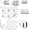
TIPE2, also known as TNFAIP8L2, a member of the tumor necrosis factor-alpha-induced protein-8 (TNFAIP8) family, is known as an inhibitor in inflammation and cancer, and its overexpression induces cell death. We examined the role of TIPE2 with respect to adjuvant arthritis (AA)-associated pathogenesis by analyzing the TIPE2 regulation of death receptor (DR5)-mediated apoptosis in vitro. The results showed that TIPE2 was detected in normal fibroblast-like synoviocytes (FLSs), but scarcely observed in AA-FLSs. Therefore, recombinant MIGR1/TIPE2(+/+) and control MIGR1 lentivirus vectors were transfected to AA-FLSs, which were denoted as TIPE2(+/+)-FLSs and MIGR1-FLSs, respectively. Our results showed that TIPE2(+/+)-FLSs were highly susceptible to ZF1-mediated apoptosis, and ZF1 was our own purification of an anti-DR5 single chain variable fragment antibody. Under the presence of TIPE2, the expression of DR5 was significantly increased compared with that of the MIGR1-FLS group. In contrast, the level of phosphorylated nuclear factor-kappa B (pNF-κB) was lower in the TIPE2(+/+)-FLS group treated with ZF1, whereas the activity of caspase was higher. Moreover, the rate of apoptosis in the TIPE2(+/+)-FLS group, which was pretreated with caspase inhibitor Z-VAD-FMK, was significantly decreased. In contrast, the apoptosis occurrence in the MIGR1-FLS group increased significantly with the pretreatment of the NF-κB inhibitor Bay. These results indicated that TIPE2 increased the apoptosis of AA-FLSs by enhancing DR5 expression levels, thereby promoting the activation of caspase and inhibiting the activation of NF-κB in AA-FLSs. TIPE2 might potentially act as a therapeutic target for rheumatoid arthritis.
Keywords: DR5; TIPE2; adjuvant arthritis; fibroblast-like synoviocytes; rheumatoid arthritis.
Figures


Similar articles
-
Regulatory Roles of Tumor Necrosis Factor-α-Induced Protein 8 Like-Protein 2 in Inflammation, Immunity and Cancers: A Review.Cancer Manag Res. 2020 Dec 14;12:12735-12746. doi: 10.2147/CMAR.S283877. eCollection 2020. Cancer Manag Res. 2020. PMID: 33364825 Free PMC article. Review.
-
CYLD suppression enhances the pro-inflammatory effects and hyperproliferation of rheumatoid arthritis fibroblast-like synoviocytes by enhancing NF-κB activation.Arthritis Res Ther. 2018 Oct 3;20(1):219. doi: 10.1186/s13075-018-1722-9. Arthritis Res Ther. 2018. PMID: 30285829 Free PMC article.
-
Effects of sorafenib on fibroblast-like synoviocyte apoptosis in rats with adjuvant arthritis.Int Immunopharmacol. 2020 Jun;83:106418. doi: 10.1016/j.intimp.2020.106418. Epub 2020 Apr 30. Int Immunopharmacol. 2020. PMID: 32199349
-
Recombinant human endostatin inhibits TNF-alpha-induced receptor activator of NF-κB ligand expression in fibroblast-like synoviocytes in mice with adjuvant arthritis.Cell Biol Int. 2016 Dec;40(12):1340-1348. doi: 10.1002/cbin.10689. Epub 2016 Oct 26. Cell Biol Int. 2016. PMID: 27730697
-
TIPE2 as a potential therapeutic target in chronic viral hepatitis.Expert Opin Ther Targets. 2019 Jun;23(6):485-493. doi: 10.1080/14728222.2019.1608948. Epub 2019 Apr 22. Expert Opin Ther Targets. 2019. PMID: 30995133 Review.
Cited by
-
TIPE Family of Proteins and Its Implications in Different Chronic Diseases.Int J Mol Sci. 2018 Sep 29;19(10):2974. doi: 10.3390/ijms19102974. Int J Mol Sci. 2018. PMID: 30274259 Free PMC article. Review.
-
Loss of TIPE2 Has Opposing Effects on the Pathogenesis of Autoimmune Diseases.Front Immunol. 2019 Sep 24;10:2284. doi: 10.3389/fimmu.2019.02284. eCollection 2019. Front Immunol. 2019. PMID: 31616442 Free PMC article.
-
Apoptosis, Autophagy, NETosis, Necroptosis, and Pyroptosis Mediated Programmed Cell Death as Targets for Innovative Therapy in Rheumatoid Arthritis.Front Immunol. 2021 Dec 24;12:809806. doi: 10.3389/fimmu.2021.809806. eCollection 2021. Front Immunol. 2021. PMID: 35003139 Free PMC article. Review.
-
Regulatory Roles of Tumor Necrosis Factor-α-Induced Protein 8 Like-Protein 2 in Inflammation, Immunity and Cancers: A Review.Cancer Manag Res. 2020 Dec 14;12:12735-12746. doi: 10.2147/CMAR.S283877. eCollection 2020. Cancer Manag Res. 2020. PMID: 33364825 Free PMC article. Review.
References
LinkOut - more resources
Full Text Sources
Other Literature Sources

