Review
. 2016 Apr;16(4):201-18.
doi: 10.1038/nrc.2016.25.

Targeting metastasis

Affiliations
Review

Targeting metastasis

Patricia S Steeg. Nat Rev Cancer. 2016 Apr.

Abstract

Tumour metastasis, the movement of tumour cells from a primary site to progressively colonize distant organs, is a major contributor to the deaths of cancer patients. Therapeutic goals are the prevention of an initial metastasis in high-risk patients, shrinkage of established lesions and prevention of additional metastases in patients with limited disease. Instead of being autonomous, tumour cells engage in bidirectional interactions with metastatic microenvironments to alter antitumour immunity, the extracellular milieu, genomic stability, survival signalling, chemotherapeutic resistance and proliferative cycles. Can targeting of these interactions significantly improve patient outcomes? In this Review preclinical research, combination therapies and clinical trial designs are re-examined.


Conflict of interest statement

Competing interests statement

The authors declare competing interests: see Web version for details.

Figures

Figure 1 |
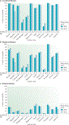
Figure 1 |. Few improvements in 5-year survival for cancer patients initially diagnosed with metastatic disease.
The percentage of patients surviving for 5 years is plotted based on their initial disease staging of localized (organ confined), regional (invasion to lymph nodes) or distant (metastases detected by imaging) using the US National Cancer Institute Surveillance, Epidemiology and End Results (SEER) registries,. Data covering 1995–2000 and 2004–2010 were reported in 2005 and 2015, respectively, to determine where improvements were attained. With few exceptions, 5-year survival after a diagnosis of localized disease was excellent; where it was low in 2005, gains were observed in 2015. Regional disease survival rates fluctuated by cancer type, but the majority saw increased survival in the later reporting period. Patients with metastatic disease at diagnosis had lower overall 5-year survival rates, with fewer than 20% of patients surviving after 5 years for half of the cancer sites. The increase in survival between the 2005 and 2015 reporting periods was under 3% in three of the four cancer types for which increased survival was seen. For each type, stage categories may not total 100% because of insufficient information for all cases. Beneath each plot is the incidence of each stage at diagnosis for the reporting period. *Localized and regional data were combined.
Figure 2
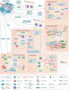
Figure 2. Functional interactions between tumour cells and the metastatic microenvironment in colonization.
a | Premetastatic niche. Primary tumour cells upregulate vascular endothelial growth factor (VEGF), causing VEGF receptor-positive (VEGFR+) haematopoietic bone marrow cells (also called myeloid-derived suppressor cells (MDSCs)) to migrate to the lung, upregulating fibronectin deposition in the extracellular matrix (ECM) by resident fibroblasts and producing inflammatory cytokines. Disseminated tumour cells then home to these locations for preferential colonization,. A premetastatic niche is also formed by tumour secretion of VEGF, tumour necrosis factor (TNF) or transforming growth factor-β (TGFβ), stimulating lung tissue to produce S100A8 and S100A9 chemokines, which serve as chemoattractants for alveolar and peritoneal macrophages and tumour cells,. Primary tumour hypoxic conditions favour formation of a premetastatic niche by producing lysyl oxidase (LOX) to alter the microenvironment, carbonic anhydrase (CAIX) to mobilize MDSCs, and suppression of natural killer (NK) cell activation. Exosomes produced by the primary tumour educate MDSCs and alter the premetastatic microenvironment ECM and metabolism directly,,. b | ECM and fibroblasts. Colonizing tumour cells functionally interact with altered levels of hyaluronic acid, fibronectin, tenascin C and collagens in the ECM. The ECM is remodelled by various proteases produced by tumour cells and the activated microenvironment, with downstream effects on adhesion and tumour viability. Integrins, receptors for ECM components, mediate many interactions between tumour cells and the altered ECM to effect colonization. Fibroblasts in the microenvironment are activated by tumour cells or their secreted factors; comigration of primary tumour fibroblasts with tumour cells to metastatic sites also occurs in model systems. Activated fibroblasts contribute to multiple aspects of colonization, including angiogenesis, inflammation, immunity and tumour growth potential. Fibrosis is an out-of-control activation of myofibroblasts to produce higher amounts of ECM. When fibrosis is induced by drugs or radiation treatment, experimental metastases are elevated. c | Innate immunity. Bone marrow-derived myeloid cells, which are macrophage-like, are stimulated to migrate to sites of metastasis by TGFβ, which promotes metastatic colonization by diminishing arginase, reactive oxygen species (ROS) and interferon-γ (IFNγ) production, leading to decreased T cell-dependent antitumour immunity. Other tumour-derived factors,, ECM components and hypoxia mobilize myeloid cells. Their activity is regulated by growth factors, toll-like receptor (TLR) and peroxisome proliferator-activated receptor-γ (PPARγ) signalling; a chemokine (C-X3-C motif) ligand 1 (CX3CL1) loop promotes their viability. Myeloid cells also transdifferentiate into metastasis effector cells. Macrophages from host tissue and circulation also facilitate colonization through chemokine cascades. Reciprocal signalling between macrophages and tumour cells enhances the viability of both cell populations,. NK cells also join the site of colonization and provide innate immune functions. d | T cell-mediated immunity. Influx and activation of tumour infiltrating lymphocytes (TILs) is mediated by chemokines such as C-X-C motif chemokine ligand 16 (CXCL16). Programmed cell death protein 1 (PD1) immune checkpoint expression is increased on CD8+ TILs in the metastatic microenvironment. In turn, CD8+ TILs secrete IFNγ to upregulate PD1 ligand 1 (PDL1) on metastatic tumour cells,. The PD1–PDL1 pathway inactivates the cytotoxic T-lymphocyte (CTL) effector arm. Tumour cells halt immune responses in several ways. They produce interleukins (ILs), leading to the production of granulocyte-colony stimulating factor (G-CSF), mobilization of neutrophils and inactivation of CTLs. Tumour chemokine networks can recruit T regulatory (Treg) cells, shutting down NK cell activity. A balance of tumour zinc finger E-box binding homeobox 1 (ZEB1) and microRNA miR-200 regulates PDL1 expression. e | Vascular system. Angiogenesis is stimulated by hypoxia. Endothelial cells proliferate, migrate and encircle to form capillaries. The process is facilitated by MDSCs, which are ‘educated’ by tumour-derived exosomes. Other pathways such as angiopoietin (ANGPT) signalling, stabilize vessels. Residual hypoxia stimulates tumour invasion,–. Other sources of blood supply include co-option of the existing vasculature and vasculogenic mimicry, which is the formation of blood-conducting tubes by tumour cells–,. CCL, chemokine (C-C motif) ligand; CCR, chemokine (C-C motif) receptor; MIF, macrophage migration inhibitory factor; SAA, serum amyloid A.
Figure 3 |
Figure 3 |. A wealth of mechanistic translational targets in metastasis to the brain.
In addition to the basic pathways of colonization, bidirectional interactions between tumour cells and the microenvironments of specific tissues foster metastatic colonization. In the brain, tumour cells breach the blood–brain barrier to extravasate using tissue-nonspecific and tissue-specific adhesion molecules and proteases,. Tumour cells then adhere to the vascular basement membrane via β1 integrins. Activated astrocytes congregate around the developing metastasis and are stimulated by tumour-derived cytokines. The astrocytes produce growth factors that are stimulatory for the tumour cells,, and elevate tumour cell expression of receptors for cytokines in the microenvironment. Systemic hormones such as oestrogen can further activate astrocytes to stimulate colonization. Activated microglia, resident macrophage-like cells, also surround a developing metastasis. Their activation is determined by a balance of tumour cell stimulatory and inhibitory factors. In turn, they activate tumour cells via the WNT pathway. The brain is infiltrated by myeloid-derived suppressor cells (MDSCs) in a premetastatic phase, fuelled by cyclooxygenase 2 (COX2) and T cells. Tumours shut down T cell responses using ATP binding cassette transporter 1 (TAP1; also known as ABCB2). The brain microenvironment mounts a protective reaction by the secretion of reactive oxygen species (ROS). Overexpression of DNA double strand break repair genes by tumour cells attenuates the ROS-induced DNA damage and overexpression of serpins inactivates death signals from the microenvironment. Neuronal cell death is a consequence of metastasis formation, but is attenuated by tumour-derived pigment-epithelial-derived factor (PEDF). It is likely that many of the mediators of brain metastatic colonization are active in other anatomical locations as well, via a different set of host microenvironmental cells, suggesting that any therapeutics that are developed may be more broadly applicable. CTL, cytotoxic T lymphocyte; IL-6, interleukin 6; JAG1, Jagged 1; PDGF, platelet-derived growth factor.
Figure 4 |
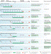
Figure 4 |. Meaningful incorporation of preclinical metastasis models into drug development.
Potential experimental designs for an antimetastatic investigational agent are mapped along a timeline. Tumour cell injection can either be orthotopic, into the tissue of origin (white needles) or haematogenous for experimental metastasis (red needles). The investigational agent (purple arrowheads) can be delivered for all or part of the assay; optimally it should reflect the oral or intravenous dosing to be used in the clinic. Standard of care (SOC) therapy (green line), at a clinically achievable dose, can be added before, concurrent with or after the investigational agent and can use agents approved in the adjuvant or metastatic setting. a | Standard drug development uses primary tumour growth as an end point, which completely neglects the metastatic process. b–e | Model systems with different degrees of applicability to adjuvant setting trials preventing metastatic colonization. f–h | Model systems with different applicabilities to metastatic setting trials. End points include the number and size of metastases by histology and imaging, pharmacodynamic markers of drug activity or tumour biology, and survival.

Similar articles

Cited by

References

    1. Brosnan JA & Iacobuzio-Donahue CA A new branch on the tree: next-generation sequencing in the study of cancer evolution. Semin. Cell Dev. Biol 23, 237–242 (2012). - PMC - PubMed
    1. Paget S The distribution of secondary growths in cancer of the breast. Lancet 1, 99–101 (1889). - PubMed
    2. The origin of the seed and soil hypothesis of metastasis.

    1. Hensel JA, Flaig TW & Theodorescu D Clinical opportunities and challenges in targeting tumour dormancy. Nat. Rev. Clin. Oncol 10, 41–51 (2013). - PubMed
    1. Husemann Y et al. Systemic spread is an early step in breast cancer. Cancer Cell 13, 58–68 (2008). - PubMed
    1. Bojovic B & Crowe DL Dysfunctional telomeres promote genomic instability and metastasis in the absence of telomerase activity in oncogene induced mammary cancer. Mol. Carcinogen 52, 103–117 (2013). - PubMed

MeSH terms