. 2016 May 15;310(10):L940-54.
doi: 10.1152/ajplung.00346.2015. Epub 2016 Mar 18.

Elevated expression of NEU1 sialidase in idiopathic pulmonary fibrosis provokes pulmonary collagen deposition, lymphocytosis, and fibrosis

Affiliations

Elevated expression of NEU1 sialidase in idiopathic pulmonary fibrosis provokes pulmonary collagen deposition, lymphocytosis, and fibrosis

Irina G Luzina et al. Am J Physiol Lung Cell Mol Physiol. .

Abstract

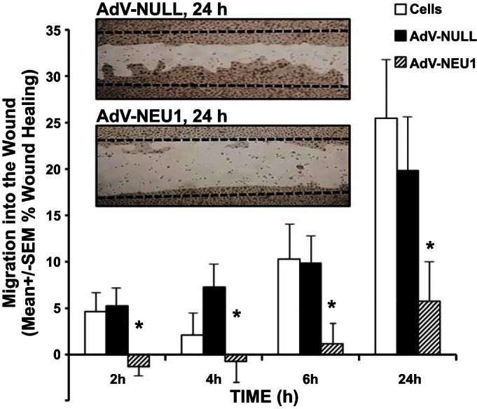
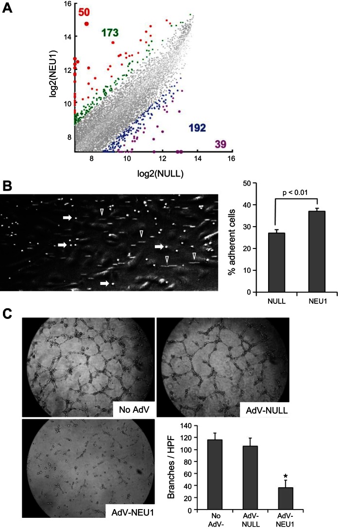
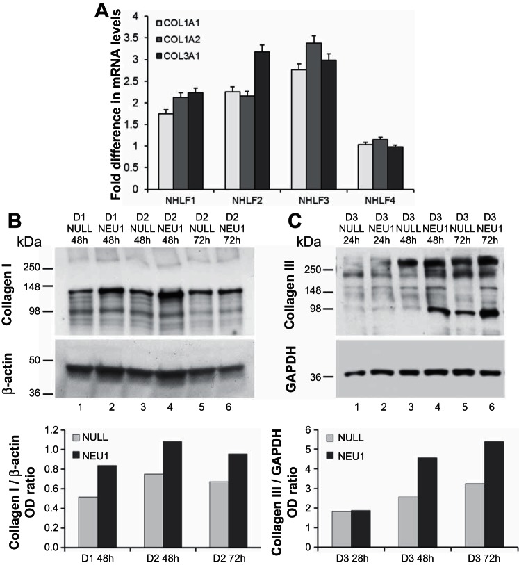
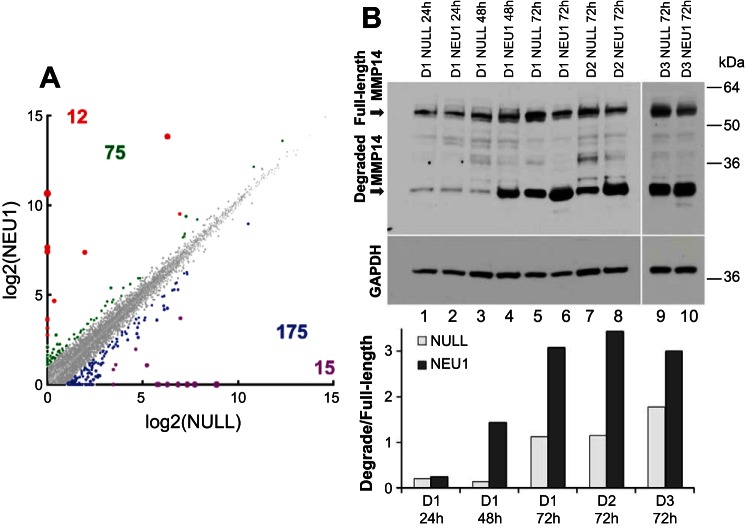
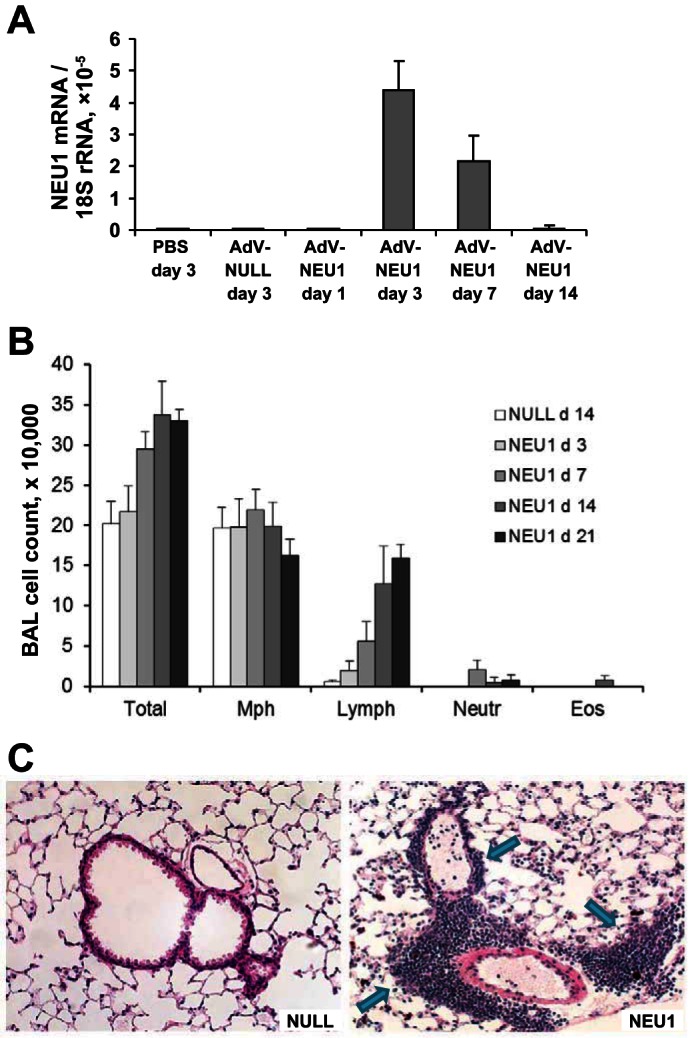
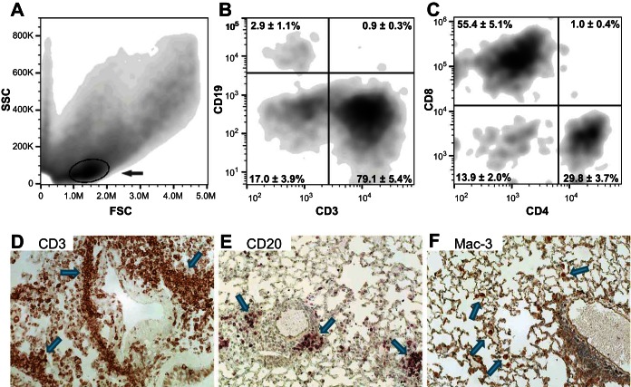
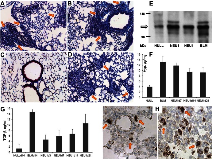
Idiopathic pulmonary fibrosis (IPF) poses challenges to understanding its underlying cellular and molecular mechanisms and the development of better therapies. Previous studies suggest a pathophysiological role for neuraminidase 1 (NEU1), an enzyme that removes terminal sialic acid from glycoproteins. We observed increased NEU1 expression in epithelial and endothelial cells, as well as fibroblasts, in the lungs of patients with IPF compared with healthy control lungs. Recombinant adenovirus-mediated gene delivery of NEU1 to cultured primary human cells elicited profound changes in cellular phenotypes. Small airway epithelial cell migration was impaired in wounding assays, whereas, in pulmonary microvascular endothelial cells, NEU1 overexpression strongly impacted global gene expression, increased T cell adhesion to endothelial monolayers, and disrupted endothelial capillary-like tube formation. NEU1 overexpression in fibroblasts provoked increased levels of collagen types I and III, substantial changes in global gene expression, and accelerated degradation of matrix metalloproteinase-14. Intratracheal instillation of NEU1 encoding, but not control adenovirus, induced lymphocyte accumulation in bronchoalveolar lavage samples and lung tissues and elevations of pulmonary transforming growth factor-β and collagen. The lymphocytes were predominantly T cells, with CD8(+) cells exceeding CD4(+) cells by nearly twofold. These combined data indicate that elevated NEU1 expression alters functional activities of distinct lung cell types in vitro and recapitulates lymphocytic infiltration and collagen accumulation in vivo, consistent with mechanisms implicated in lung fibrosis.

Keywords: fibroblasts; fibrosis; inflammation.


Figures

Fig. 1.
Fig. 1.
Photomicrographs of lung sections from healthy control (Ctrl) volunteers and IPF patients immunohistochemically stained for NEU1 (blue). A: a nonimmune immunoglobulin control was used in the same protocol. Counterstaining with eosin-Y alcoholic solution appears in light pink. A–C: overview of the sections with a ×10 objective, whereas the remaining panels (D–I) provide a more detailed view with a ×40 objective. Note that bronchial epithelium (D–F) shows more intense NEU1 positivity in the brush border in sections from patients with IPF (arrows in E and F) compared with a healthy control (arrowhead in D). Similarly, endothelial cells (G–I) appear to stain more intensely in patients with IPF (arrows in H and I) compared with a healthy control (arrowhead in G).
Fig. 2.
Fig. 2.
Expression of NEU1 in human lung fibroblasts from patients with IPF. A: photomicrograph of a fibroblastic focus area in a lung section from a patient with IPF after immunohistochemical staining for NEU1 (objective ×40). Arrows indicate selected stromal NEU1-positive cells, likely fibroblasts, staining in blue. B: reverse transcriptase-quantitative PCR for NEU1, -2, -3, and -4 mRNAs, normalized to 18S rRNA and further normalized to the average value for the NEU1 mRNA levels. Primary fibroblast cultures from 7 adult healthy controls were tested, each with specific primers for the indicated mRNAs. The pairwise differences between the levels of all tested mRNA species are significant (P < 0.05), except for the NEU2 to NEU4 comparison, with both NEU2 and NEU4 mRNAs expressed at low levels. C: Western blotting (left) for NEU1 and β-actin in cultured primary pulmonary fibroblasts from 7 adult healthy controls (C; lanes 1–4, 9–11) and 8 patients with IPF (P; lanes 5–8, 12–15). D: densitometry of the NEU1 bands normalized to the corresponding β-actin bands in C and further normalized to the average value for healthy control fibroblasts. Average normalized densitometric values in each group are indicated with horizontal bars. The difference in NEU1 protein levels between IPF and control fibroblast cultures is significant (P = 0.047). E: reverse transcriptase-quantitative PCR for NEU1 mRNA normalized to 18S rRNA in cultured primary pulmonary fibroblasts from 9 adult healthy controls and 10 patients with IPF, as indicated. Individual fold differences from the average value for healthy controls are plotted. The mean value in each group is indicated with a horizontal bar. The difference in NEU1 mRNA levels between IPF and control fibroblast cultures is significant (P = 0.024).
Fig. 3.
Fig. 3.
NEU1 impairs SAEC migration into a wound. SAECs infected with AdV-NULL or AdV-NEU1 were cultured to confluence in the wells of 24-well plates, after which a single wound was placed across the diameter of each monolayer. At 0, 2, 4, 6, and 24 h, images of each monolayer were captured, and cell migration into the wound at each time point was calculated relative to that observed at 0 h in the same wounded monolayer. Vertical bars represent mean ± SE percent migration into the wound; n = 8 for each condition. *Significant decreases compared with the simultaneous AdV-NULL-infected controls at P < 0.05.
Fig. 4.
Fig. 4.
NEU1 overexpression alters endothelial cell function. A: Affymetrix microarray-based transcriptomic profiling of HPMECs. Normalized gene expression values in cultured AdV-NEU1-infected HPMECs were plotted vs. AdV-NULL-infected cells. Each dot represents a pair of expression levels of a gene, with the expression level defined as the normalized log base 2 signal value for the gene-specific probe set. The colorized dots represent genes with substantially different expression levels. The genes with fivefold higher (red), fivefold lower (purple), three- to fivefold higher (green), and three- to fivefold lower (blue) expression levels are shown, with the similarly colored numbers representing the counts of the corresponding genes. The corresponding genes are listed in Supplemental Table S1. Fold differences were not considered for low-expressing genes, defined as those with the higher expression level in each pairwise comparison, being in the lower 25th percentile of expression levels of all genes. B: representative microscopic image of a BioFlux 200 microfluidics channel with a clearly visible monolayer of HPMECs (large cells appearing in gray), adherent T lymphocytes (smaller cells appearing as brightly white, selected cells indicated with arrows), and nonadherent T lymphocytes in flow, appearing as cellular tracks (selected tracks are indicated with arrowheads). Adherent T cells were counted in triplicate for each condition in 3 independent experiments (the bar graph on the right, means ± SD are shown), revealing an increase in T lymphocyte adhesion to AdV-NEU1-infected cells (P < 0.01). C: photomicrographs of noninfected HPMECs or HPMECs infected with AdV-NULL or AdV-NEU1, with mean ± SD counts (n = 6 for each condition) of the number of branches of capillary-like tubes per high-power field (HPF). *Significant decrease (P < 0.01) in the number of branches in AdV-NEU1-infected vs. AdV-NULL-infected HPMECs.
Fig. 5.
Fig. 5.
NEU1 upregulates collagen mRNA and protein expression in cultured normal human lung fibroblasts (NHLF). A: steady-state levels COL1A1, COL1A2, and COL1A3 mRNAs normalized to 18S rRNA in AdV-NEU1- vs. AdV-NULL-infected NHLF cultures from 4 different donors 24 h after infection. Mean fold ratios ± SD of 3 PCR replicates are shown for each collagen chain for each culture. B: Western blotting for collagen type I (top) and β-actin (middle) of cell lysates from NHLF derived from two separate donors (D1, D2) at 48 and 72 h after in-culture cell infection with AdV-NEU1 or AdV-NULL, as indicated. Bottom: the collagen band densities normalized to the densities of the β-actin bands. C: Western blotting for collagen type III (top) and GAPDH (middle) of cell lysates from NHLF derived from a separate donor (D3) at 24, 48, and 72 h after in-culture cell infection with AdV-NEU1 or AdV-NULL, as indicated. Bottom: the collagen band densities normalized to the densities of the corresponding GAPDH bands. OD, optical density.
Fig. 6.
Fig. 6.
Transcriptional and posttranslational changes induced in cultured primary fibroblasts by NEU1 overexpression. A: RNASeq transcriptomic profiling of NHLFs. FPKM values in cultured AdV-NEU1-infected NHLFs were plotted vs. AdV-NULL-infected NHLF cells. Each dot represents a pair of expression levels of a gene, with the expression level defined as the normalized log base 2 signal value for the gene-specific probe set. The colorized dots represent genes with substantially different expression levels. The genes with fivefold higher (red), fivefold lower (purple), two- to fivefold higher (green), and two- to fivefold lower (blue) expression levels are shown, with the similarly colored numbers representing the counts of the corresponding genes. The corresponding genes are listed in Supplemental Table S2. B: Western blotting for MMP-14 of NHLF from 3 different donors (D1–D3) at 24 h (lanes 1 and 2), 48 h (lanes 3 and 4), or 72 h (lanes 5–10) following infections with AdV-NULL (lanes 1, 3, 5, 7, and 9) or AdV-NEU1 (lanes 2, 4, 6, 8, and 10). The full-length and degraded MMP-14 forms are indicated. The membrane was stripped of the antibody and reprobed for GAPDH to demonstrate the loading. The bar graph shows the ratio of optical densities of the degraded and full-length bands of MMP-14 in the tested samples.
Fig. 7.
Fig. 7.
Gene delivery of NEU1 to mouse lungs. A: quantitative reverse transcriptase-polymerase chain reaction for human NEU1 mRNA normalized to 18S rRNA using the 2−ΔΔCt method in mRNA purified from mouse lung homogenates. Three mice in each group were intratracheally administered the indicated adenoviral constructs or PBS and sacrificed at the indicated times. Vertical bars represent the mean NEU1 mRNA-to-18S rRNA ratio × 10−5 ± SD. The PCR primers from SABiosciences (Frederick, MD) were highly specific for human NEU1 and did not cross-react with mouse NEU1. B: changes in the total cell counts and cellular composition of BAL induced by gene delivery of NEU1. Vertical bars represent mean counts ± SD; 3–5 mice per group at the indicated times after intratracheal instillation of AdV-NULL or AdV-NEU1. Note the time-dependent increase in total BAL cellularity and in the number of pulmonary lymphocytes in NEU1-expressing mice; P < 0.05 on days 7–21, one-way ANOVA. C: hematoxylin and eosin staining of lung sections from mice 14 days after intratracheal instillation of AdV-NULL or AdV-NEU1, as indicated. Note the cellular infiltrates in NEU1-expressing mice (arrows).
Fig. 8.
Fig. 8.
Flow cytometry of BAL cells (A–C) and immunohistochemistry of lung sections (D–F) from mice overexpressing human NEU1; 3–5 mice per group. Forward- (FSC) vs. side-scatter (SSC) analyses of BAL cells from NEU1-expressing mice reveal a population of cells that is not present in control mice (gated and indicated with an arrow in A). B: cell surface staining with anti-CD3 and anti-CD19 antibodies reveals that the majority of these cells are T lymphocytes, with only occasional B lymphocytes present. C: further gating of CD3+ cells reveals a predominance of CD8+ T cells. D: immunohistochemical analyses, with positivity for the tested markers appearing in brown color (selected cells and cell clusters indicated with arrows), show a similar abundance of T cells in the lung tissue, particularly in the infiltrates. By contrast, CD20+ B cells are substantially less abundant (E), whereas Mac3+ macrophages are not associated with infiltrates and occur only occasionally in the lung parenchyma (F).
Fig. 9.
Fig. 9.
Accumulation of collagen in mouse lungs in response to AdV-mediated NEU1 gene delivery. A–D: Masson's trichrome staining of representative lung sections from two AdV-NEU1-challenged mice on day 14, objective ×20 (A and B). Note the accumulation of collagen fibers appearing in blue in peristructural areas and in association with inflammatory cellular infiltrates (arrows point to selected areas). C: by contrast, similarly stained lung sections from AdV-NULL-challenged mice did not have collagen deposits at levels above the expected presence of collagen in the alveolar walls. D: a positive control section from a bleomycin (BLM)-challenged mouse on day 14 after intratracheal instillation of BLM is shown. E: Western blotting for type I collagen of lung homogenates from an AdV-NULL-infected, two AdV-NEU1-infected, and a BLM-treated mice, as indicated, on day 14 after intratracheal instillations. F: mean ± SD collagen levels, in micrograms per milligram wet lung tissue, from 3–4 mice per group, challenged with AdV-NULL, AdV-NEU1, or BLM (positive control). These measurements were based on hydroxyproline content in QuickZyme assays. The increases are significant (P < 0.01) in BLM-challenged (two-tailed t-test) and NEU1-expressing (one-way ANOVA) mice. G: mean ± SD levels of total TGF-β in lung homogenates of mice infected with AdV-NULL challenged with BLM or infected with AdV-NEU1 at the indicated times (days). The increases are significant in BLM-challenged (P < 0.01, two-tailed t-test) and NEU1-expressing (P < 0.05, one-way ANOVA) mice; 3–4 animals per group were tested. H: immunohistochemical staining for TGF-β of two representative lung sections from NEU1-overexpressing mice, objective ×40. Selected dark brown TGF-β-expressing cells are indicated with arrows.

Similar articles

Cited by

References

    1. Abdollahi A, Li M, Ping G, Plathow C, Domhan S, Kiessling F, Lee LB, McMahon G, Grone HJ, Lipson KE, Huber PE. Inhibition of platelet-derived growth factor signaling attenuates pulmonary fibrosis. J Exp Med 201: 925–935, 2005. - PMC - PubMed
    1. Abdulkhalek S, Amith SR, Franchuk SL, Jayanth P, Guo M, Finlay T, Gilmour A, Guzzo C, Gee K, Beyaert R, Szewczuk MR. Neu1 sialidase and matrix metalloproteinase-9 cross-talk is essential for Toll-like receptor activation and cellular signaling. J Biol Chem 286: 36532–36549, 2011. - PMC - PubMed
    1. Abdulkhalek S, Szewczuk MR. Neu1 sialidase and matrix metalloproteinase-9 cross-talk regulates nucleic acid-induced endosomal TOLL-like receptor-7 and -9 activation, cellular signaling and pro-inflammatory responses. Cell Signal 25: 2093–2105, 2013. - PubMed
    1. Abe M, Harpel JG, Metz CN, Nunes I, Loskutoff DJ, Rifkin DB. An assay for transforming growth factor-beta using cells transfected with a plasminogen activator inhibitor-1 promoter-luciferase construct. Anal Biochem 216: 276–284, 1994. - PubMed
    1. Ajayi IO, Sisson TH, Higgins PD, Booth AJ, Sagana RL, Huang SK, White ES, King JE, Moore BB, Horowitz JC. X-linked inhibitor of apoptosis regulates lung fibroblast resistance to Fas-mediated apoptosis. Am J Respir Cell Mol Biol 49: 86–95, 2013. - PMC - PubMed

Publication types

MeSH terms