. 2016 Aug;23(8):1296-311.
doi: 10.1038/cdd.2016.6. Epub 2016 Mar 18.

PPARγ neddylation essential for adipogenesis is a potential target for treating obesity

Affiliations

PPARγ neddylation essential for adipogenesis is a potential target for treating obesity

H-S Park et al. Cell Death Differ. 2016 Aug.

Abstract

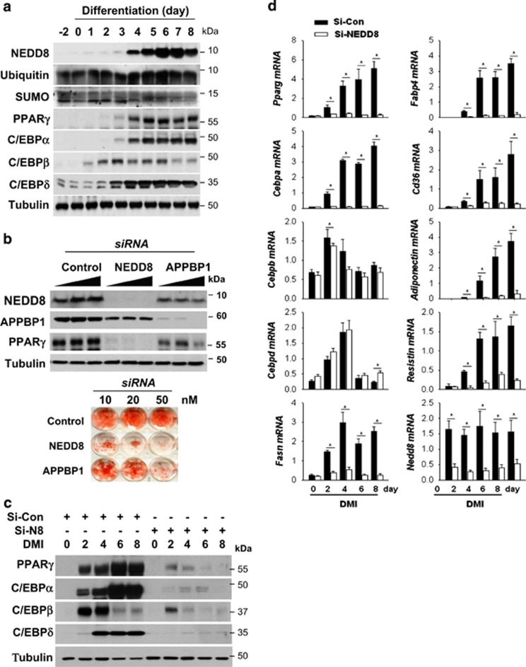
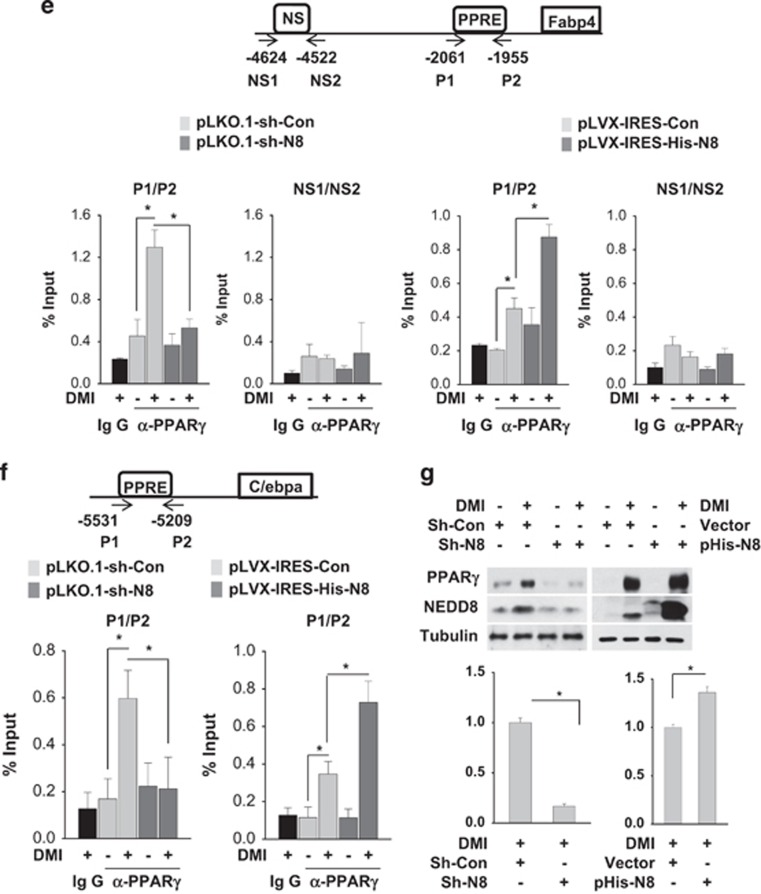
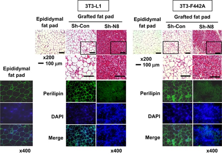
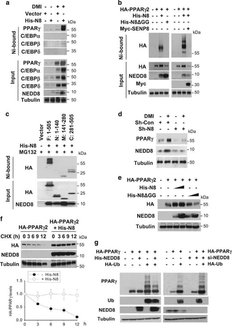
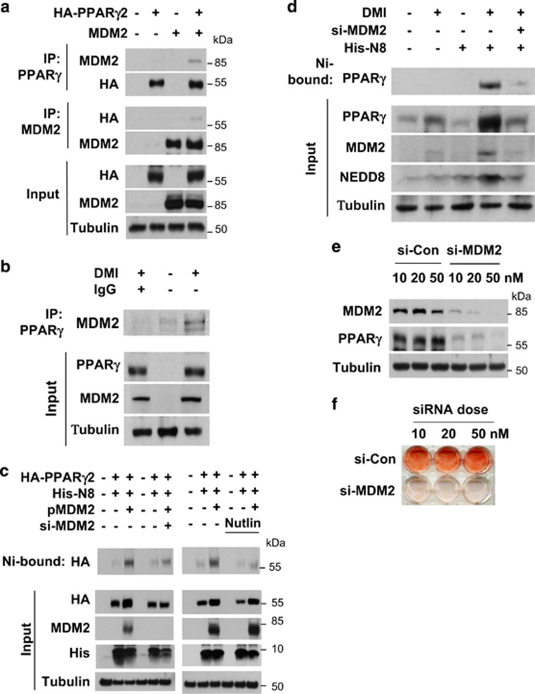
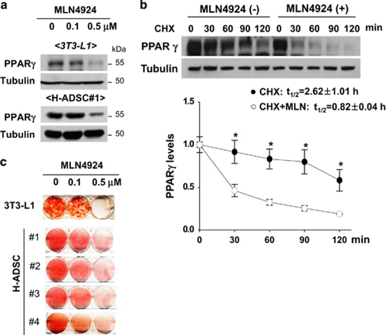
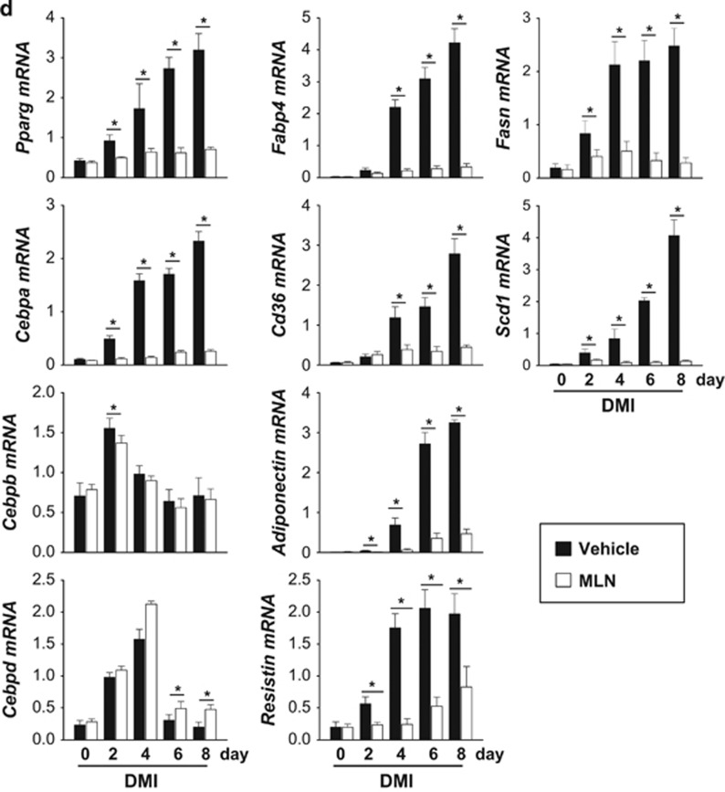
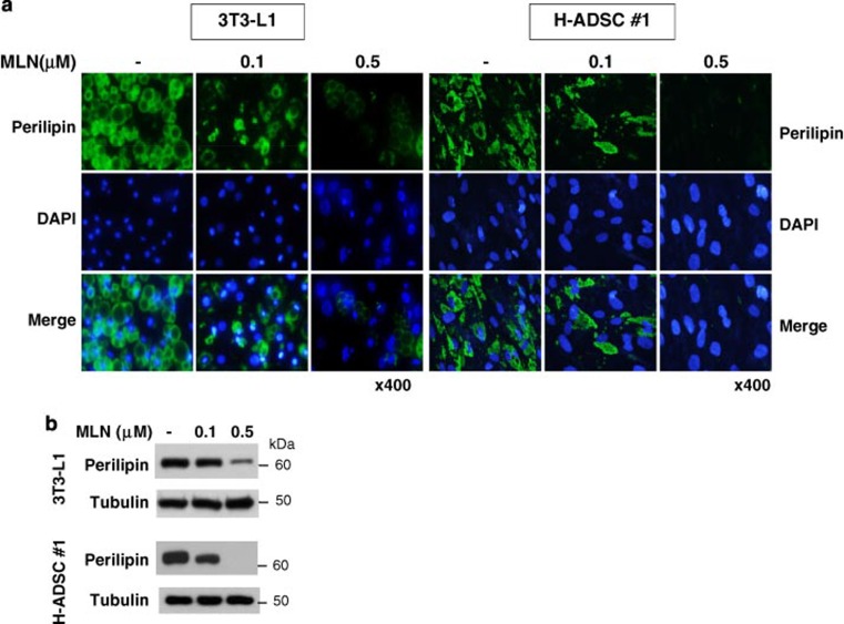
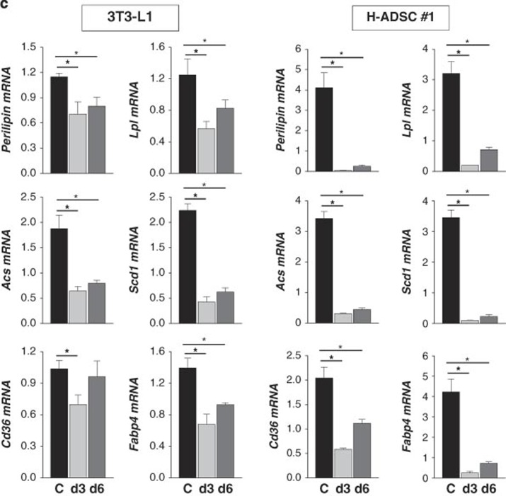
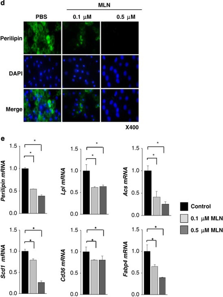
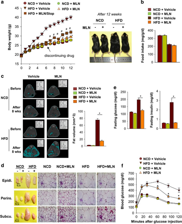
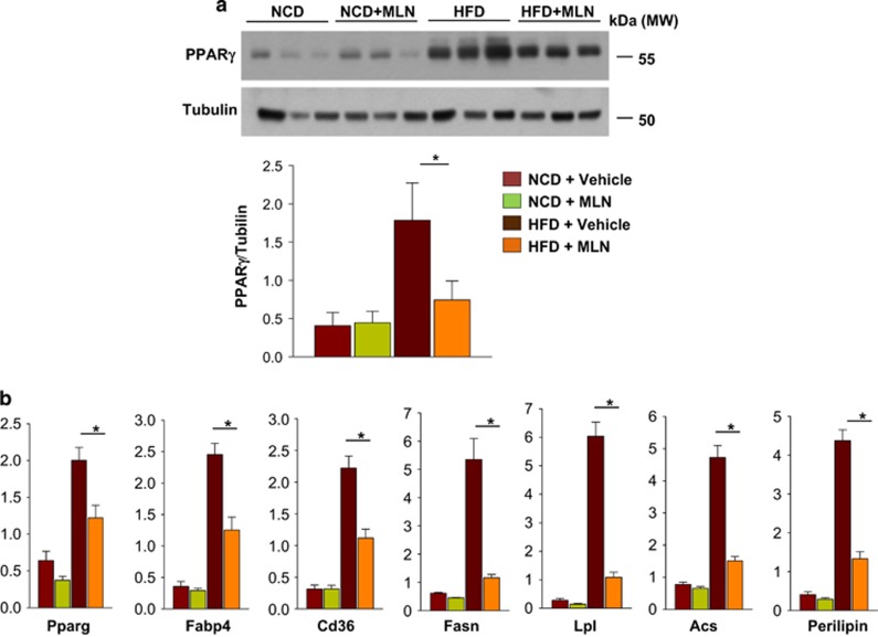
The preadipocyte-to-adipocyte differentiation (adipogenesis) is a key process in fat mass increase and thus it is regarded as a compelling target for preventing or treating obesity. Of adipogenic hormone receptors, peroxisome proliferator-activated receptor gamma (PPARγ) has crucial roles in adipogenesis and lipid accumulation within adipocytes. Here we demonstrate that the NEDD8 (neuronal precursor cell expressed, developmentally downregulated 8)-based post-translation modification (neddylation) of PPARγ is essential for adipogenesis. During adipogenesis, NEDD8 is robustly induced in preadipocytes and conjugates with PPARγ, leading to PPARγ stabilization. When the neddylation process was blocked by NEDD8-targeting siRNAs (or viral vectors) or an inhibitor MLN4924, adipocyte differentiation and fat tissue development were substantially impaired. We also demonstrate that MLN4924 effectively prevents the high-fat diet-induced obesity and glucose intolerance in mice. This study provides a better understanding of how the PPARγ signaling pathway starts and lasts during adipogenesis and a potential anti-obesity strategy that targets the neddylation of PPARγ.


Figures

Figure 1
Figure 1
NEDD8 induction is essential for adipocyte differentiation. (a) NEDD8 and adipogenic transcription factors are induced in 3T3-L1 cells under differentiation. 3T3-L1 cells were stimulated with DMI for the indicated time, and protein expression was evaluated by western blotting. (b) Adipocyte differentiation is retarded by blocking neddylation. 3T3-L1 cells were transfected transiently with si-Control, si-NEDD8 or si-APPBP1 (10, 20 or 50 nM) and then stimulated with DMI for 8 days. Western blotting (upper panel) and Oil Red O staining (lower panel) were performed. (c) PPARγ and C/EBPs are downregulated by NEDD8 knockdown. 3T3-L1 cells were transfected with 50 nM of si-control or si-NEDD8, stabilized for 2 days, and then stimulated by DMI. C/EBPs and PPARγ protein levels were determined by immunoblot analysis in cell lysates and β-tubulin was used as a loading control. (d) NEDD8 is required for the expressions of adipogenic genes. 3T3-L1 cells were transfected with si-control (Si-Con) or si-NEDD8 (Si-N8) and then stimulated with DMI for the indicated lengths of time. Total RNA was purified from the cells, and RT-qPCR was performed to measure the expression levels of Pparg, Cebpa, Cebpb, Cebpd, Fasn, Fabp4, Cd36, adiponectin, resistin, and Nedd8. Expression levels are shown relative to those of 18S RNA (mean+S.D.). Data are presented as the means+S.D. (n=3); *P<0.05. (e and f) NEDD8 promotes PPARγ binding to the promoters of its target genes. Stable 3T3-L1 cell lines expressing non-targeting shRNA (pLKO.1-sh-Con), NEDD8-targeting shRNA (pLKO.1-sh-N8), pLVX-IRES, and pLVX-IRES-His-N8 vectors were stimulated with DMI, and chromatin was cross-linked and immunoprecipitated using an anti-PPARγ antibody. The precipitated Cebpa (f) and Fabp4 promoters (e) were amplified and quantified by qPCR. The results (mean+S.D., n=3) are expressed as percentages of the input level. PPRE, PPARγ response element; NS, non-specific element. (g) PPARγ protein is expressed depending on NEDD8 levels. Cell lysates stimulated with DMI in the stable 3T3-L1 cell lines expressing non-targeting shRNA (pLKO.1-sh-Con), NEDD8-targeting shRNA (pLKO.1-sh-N8), pLVX-IRES, and pLVX-IRES-His-N8 vectors were analyzed by immunoblot analysis. β-Tubulin was used as a loading control. The intensities of PPARγ protein bands were quantified using Image J. Data are presented as the means+S.D. (n=3); *P<0.05
Figure 1
Figure 1
NEDD8 induction is essential for adipocyte differentiation. (a) NEDD8 and adipogenic transcription factors are induced in 3T3-L1 cells under differentiation. 3T3-L1 cells were stimulated with DMI for the indicated time, and protein expression was evaluated by western blotting. (b) Adipocyte differentiation is retarded by blocking neddylation. 3T3-L1 cells were transfected transiently with si-Control, si-NEDD8 or si-APPBP1 (10, 20 or 50 nM) and then stimulated with DMI for 8 days. Western blotting (upper panel) and Oil Red O staining (lower panel) were performed. (c) PPARγ and C/EBPs are downregulated by NEDD8 knockdown. 3T3-L1 cells were transfected with 50 nM of si-control or si-NEDD8, stabilized for 2 days, and then stimulated by DMI. C/EBPs and PPARγ protein levels were determined by immunoblot analysis in cell lysates and β-tubulin was used as a loading control. (d) NEDD8 is required for the expressions of adipogenic genes. 3T3-L1 cells were transfected with si-control (Si-Con) or si-NEDD8 (Si-N8) and then stimulated with DMI for the indicated lengths of time. Total RNA was purified from the cells, and RT-qPCR was performed to measure the expression levels of Pparg, Cebpa, Cebpb, Cebpd, Fasn, Fabp4, Cd36, adiponectin, resistin, and Nedd8. Expression levels are shown relative to those of 18S RNA (mean+S.D.). Data are presented as the means+S.D. (n=3); *P<0.05. (e and f) NEDD8 promotes PPARγ binding to the promoters of its target genes. Stable 3T3-L1 cell lines expressing non-targeting shRNA (pLKO.1-sh-Con), NEDD8-targeting shRNA (pLKO.1-sh-N8), pLVX-IRES, and pLVX-IRES-His-N8 vectors were stimulated with DMI, and chromatin was cross-linked and immunoprecipitated using an anti-PPARγ antibody. The precipitated Cebpa (f) and Fabp4 promoters (e) were amplified and quantified by qPCR. The results (mean+S.D., n=3) are expressed as percentages of the input level. PPRE, PPARγ response element; NS, non-specific element. (g) PPARγ protein is expressed depending on NEDD8 levels. Cell lysates stimulated with DMI in the stable 3T3-L1 cell lines expressing non-targeting shRNA (pLKO.1-sh-Con), NEDD8-targeting shRNA (pLKO.1-sh-N8), pLVX-IRES, and pLVX-IRES-His-N8 vectors were analyzed by immunoblot analysis. β-Tubulin was used as a loading control. The intensities of PPARγ protein bands were quantified using Image J. Data are presented as the means+S.D. (n=3); *P<0.05
Figure 2
Figure 2
NEDD8 is essential for adipose tissue differentiation of grafted preadipocytes. 3T3-L1 and 3T3-F442A preadipocytes, which stably expressed pLKO.1-sh-Control (Sh-Con) or pLKO.1-sh-NEDD8 (Sh-N8), were grafted into the subcutis of the abdomens of 9-week-old male BALB/c nude mice. Grafted fat pads were excised 5 weeks after grafting and then stained with hematoxylin and eosin (upper panel) or stained immunofluorescently with an anti-perilipin antibody (lower panel). Nuclei were stained with DAPI. Epididymal fat tissues were excised from the same mice and used as a positive control for well-differentiated fat tissues
Figure 3
Figure 3
Neddylated PPARγ is stabilized by inhibition of its ubiquitination. (a) PPARγ is conjugated with NEDD8. 3T3-L1 cells stably expressing pLVX-IRES or pLVX-IRES-His-NEDD8 were differentiated with DMI and subjected to western blotting after pull-down purification using a Ni2+ column under denaturing conditions. (b) Ectopically expressed PPARγ2 is neddylated. HEK293 cells were cotransfected with pHA-PPARγ2, pHis-NEDE8, pHis-NEDD8ΔGG, and pMyc-SENP8 plasmids in the indicated combinations. Proteins isolated using Ni2+ were analyzed by western blotting. (c) Identification of neddylated domains of PPARγ. HEK293 cells were cotransfected with pHis-NEDE8 and one of four plasmids for HA-PPARγ2 fragments. PPARγ2 fragments were isolated using Ni2+ and then analyzed by western blotting. The amino acids of expressed peptides are indicated at the top panel. (d) NEDD8 is required for PPARγ expression. 3T3-L1 cells stably expressing sh-control (Sh-Con) or shNEDD8 (Sh-N8) were stimulated with DMI, and then cell lysates were subjected to western blotting. (e) NEDD8 promotes the expression of ectopic PPARγ. HEK293 cells were cotransfected with pHA- PPARγ2 and pHis-NEDD8 or pHis-NEDD8ΔGG, and then cell lysates were analyzed by western blotting using an anti-HA antibody. (f) PPARγ is stabilized by NEDD8. HEK293 cells were transfected with pHA-PPARγ2 and/or pHis-NEDE8, and then incubated with cycloheximide for the indicated time. Cell lysates were subjected to western blotting using anti-HA or anti-NEDD8 antibody (upper panel). Band intensities (mean±S.D., n=3) on blots were analyzed using ImageJ 1.36b and plotted (lower panel). (g) Neddylation competes with ubiquitination in PPARγ. HEK293 cells were cotransfected with the indicated plasmids or siRNAs. Then they were stabilized for 48 h. After incubated with 10 μM MG132 for 8 h, cells were subjected to immunoblot analyses. β-Tubulin was determined as a loading control
Figure 4
Figure 4
PPARγ neddylation is mediated by MDM2. (a) Ectopically expressed MDM2 interacts with PPARγ. HEK293 cells were cotransfected with pHA-PPARγ2 and pcMDM2. Proteins in cell lysates were immunoprecipitated by anti-PPARγ or anti-MDM2 antibody, and immunoprecipitates were analyzed by western blotting. (b) Endogenous MDM2 interacts with PPARγ. 3T3-L1 cells were differentiated by DMI for 2 days, and then cell lysates were subjected to immunoprecipitation with PPARγ antibody. Immunoprecipitates were analyzed by western blotting. (c) Ectopic MDM2 induces the neddylation of PPARγ. HEK293 cells were cotransfected with pHA-PPARγ2, pHis-NEDE8, pcMDM2, and/or si-MDM2. His-NEDD8-conjugated PPARγ was purified using a Ni2+ affinity column and analyzed by western blotting. Protein expressions (Input) were examined to verify transfection efficiency. (d) MDM2 neddylates PPARγ endogenously. 3T3-L1 cells were cotransfected with the NEDD8 plasmid and si-Control or si-MDM2. Transfected cells were treated with DMI to stimulate adipogenesis, and then lysed in a denaturing condition. Neddylated PPARγ was pulled-down with Ni2+ affinity resin, and then analyzed by immunoblotting. β-Tubulin was used as an input control. (e) MDM2 is required for PPARγ expression. 3T3-L1 cells were transfected with si-Control or si-MDM2, and then cell lysates were subjected to western blotting for checking MDM2 or PPARγ expression. (f) MDM2 is required for lipid storage in adipocytes. 3T3-L1 cells, which had been transfected with si-control or si-MDM2 at the indicated concentrations, were differentiated with DMI for 8 days and subjected to Oil Red O staining. (g) Knockdown of MDM2 partially blocks clonal expansion in 3T3-L1 preadipocytes. 3T3-L1 cells, which had been transfected with non-targeting or MDM2-targeting siRNAs, were cultured to reach confluence, and the cells were further cultured for two days. Cells were treated with DMI and incubated for the indicated times. Cell numbers were counted using a hemocytometer and presented relatively to that on day 0. Each bar represents the mean+S.D. from three independent experiments. *denotes P<0.05. (h) Two-day post-confluent 3T3-L1 preadipocytes, which had been transfected with siRNAs, were incubated with DMI. On the indicated times, cells were stained with propidium iodide and subjected to flow cytometry to analyze cell cycle. The data are presented as the mean values from three independent experiments
Figure 4
Figure 4
PPARγ neddylation is mediated by MDM2. (a) Ectopically expressed MDM2 interacts with PPARγ. HEK293 cells were cotransfected with pHA-PPARγ2 and pcMDM2. Proteins in cell lysates were immunoprecipitated by anti-PPARγ or anti-MDM2 antibody, and immunoprecipitates were analyzed by western blotting. (b) Endogenous MDM2 interacts with PPARγ. 3T3-L1 cells were differentiated by DMI for 2 days, and then cell lysates were subjected to immunoprecipitation with PPARγ antibody. Immunoprecipitates were analyzed by western blotting. (c) Ectopic MDM2 induces the neddylation of PPARγ. HEK293 cells were cotransfected with pHA-PPARγ2, pHis-NEDE8, pcMDM2, and/or si-MDM2. His-NEDD8-conjugated PPARγ was purified using a Ni2+ affinity column and analyzed by western blotting. Protein expressions (Input) were examined to verify transfection efficiency. (d) MDM2 neddylates PPARγ endogenously. 3T3-L1 cells were cotransfected with the NEDD8 plasmid and si-Control or si-MDM2. Transfected cells were treated with DMI to stimulate adipogenesis, and then lysed in a denaturing condition. Neddylated PPARγ was pulled-down with Ni2+ affinity resin, and then analyzed by immunoblotting. β-Tubulin was used as an input control. (e) MDM2 is required for PPARγ expression. 3T3-L1 cells were transfected with si-Control or si-MDM2, and then cell lysates were subjected to western blotting for checking MDM2 or PPARγ expression. (f) MDM2 is required for lipid storage in adipocytes. 3T3-L1 cells, which had been transfected with si-control or si-MDM2 at the indicated concentrations, were differentiated with DMI for 8 days and subjected to Oil Red O staining. (g) Knockdown of MDM2 partially blocks clonal expansion in 3T3-L1 preadipocytes. 3T3-L1 cells, which had been transfected with non-targeting or MDM2-targeting siRNAs, were cultured to reach confluence, and the cells were further cultured for two days. Cells were treated with DMI and incubated for the indicated times. Cell numbers were counted using a hemocytometer and presented relatively to that on day 0. Each bar represents the mean+S.D. from three independent experiments. *denotes P<0.05. (h) Two-day post-confluent 3T3-L1 preadipocytes, which had been transfected with siRNAs, were incubated with DMI. On the indicated times, cells were stained with propidium iodide and subjected to flow cytometry to analyze cell cycle. The data are presented as the mean values from three independent experiments
Figure 5
Figure 5
MLN4924 blocks adipocyte differentiation by destabilizing PPARγ. (a) MLN4924 downregulates PPARγ. 3T3-L1 cells or human adipose tissue-derived stem cells were treated with 0.1 or 0.5 μM MLN4924 for 2 days, and then stimulated with DMI for 8 days. Cell lysates were subjected to western blotting. (b) MLN4924 destabilizes PPARγ protein. 3T3-L1 cells were treated by DMI to stimulate differentiation and treated with MLN4924 for 24 h, and then cells were further treated with cycloheximide for the indicated times. PPARγ protein levels were determined by immunoblot analysis (upper). Band intensities were quantified using ImageJ. Half-lives (t1/2) were calculated from the slopes of first-order decay curves (lower). (c) MLN4924 blocks adipogenesis. 3T3-L1 cells and four human adipose tissue-derived stem cells were treated with 0.1 or 0.5 μM MLN4924 for 2 days, and then stimulated with DMI for 8 days. Cells were subjected to Oil Red O staining. (d) MLN4924 inhibits the expression of adipogenic genes. 3T3-L1 cells were differentiated by DMI with vehicle or 0.5 μM MLN4924 for the indicated time, and then RT-qPCRs were performed to evaluate the expression of Pparg, Cebpa, Cebpb, Cebpd, Fabp4, Cd36, adiponectin, resistin, Fasn, and Scd1 mRNAs. The mRNA levels (mean+S.D., n=3) are shown as relative values to 18S RNA levels. *P<0.05
Figure 5
Figure 5
MLN4924 blocks adipocyte differentiation by destabilizing PPARγ. (a) MLN4924 downregulates PPARγ. 3T3-L1 cells or human adipose tissue-derived stem cells were treated with 0.1 or 0.5 μM MLN4924 for 2 days, and then stimulated with DMI for 8 days. Cell lysates were subjected to western blotting. (b) MLN4924 destabilizes PPARγ protein. 3T3-L1 cells were treated by DMI to stimulate differentiation and treated with MLN4924 for 24 h, and then cells were further treated with cycloheximide for the indicated times. PPARγ protein levels were determined by immunoblot analysis (upper). Band intensities were quantified using ImageJ. Half-lives (t1/2) were calculated from the slopes of first-order decay curves (lower). (c) MLN4924 blocks adipogenesis. 3T3-L1 cells and four human adipose tissue-derived stem cells were treated with 0.1 or 0.5 μM MLN4924 for 2 days, and then stimulated with DMI for 8 days. Cells were subjected to Oil Red O staining. (d) MLN4924 inhibits the expression of adipogenic genes. 3T3-L1 cells were differentiated by DMI with vehicle or 0.5 μM MLN4924 for the indicated time, and then RT-qPCRs were performed to evaluate the expression of Pparg, Cebpa, Cebpb, Cebpd, Fabp4, Cd36, adiponectin, resistin, Fasn, and Scd1 mRNAs. The mRNA levels (mean+S.D., n=3) are shown as relative values to 18S RNA levels. *P<0.05
Figure 6
Figure 6
MLN4924 blocks lipid storage in adipocytes. (a) MLN4924 reduces lipid accumulation in mid-differentiated adipocytes. After 3T3-L1 and H-ADSC cells were stimulated with DMI for 5 days, mid-differentiated cells were treated with MLN4924 and further differentiated until day 21 (3T3-L1 cells) or day 30 (H-ADSCs). On the last day, cell morphology was analyzed by phase contrast microscopy, and cells were then subjected to immunofluorescence staining with anti-perilipin antibody. Nuclei were stained with DAPI. (b) Cell lysates from 3T3-L1 and H-ADSCs were subjected to western blotting using anti-perilipin antibody. (c) MLN4924 reduces the expressions of lipid-storing genes. 3T3-L1 and H-ADSC cells were stimulated with DMI, and then MLN4924 (0.5 μM) were administered into culture media from differentiation day 3 (d3) or day 6 (d6). After differentiation of 20-days, RNAs were prepared from the adipocytes, and the expressions of Perilipin, Acs, Cd36, Lpl, Scd1 and Fabp4 were quantified by RT-qPCR. The mRNA levels (mean+S.D., n=3) are presented as relative values to 18S RNA levels. *P<0.05. (d) MLN4924 reduces lipid accumulation in differentiated adipocytes. After 3T3-L1 cells were differentiated by DMI treatment for 8 days, the adipocytes were treated with 0.1 or 0.5 μM MLN4924 for 8 more days. On the last day, cells were subjected to immunofluorescence analysis using anti-perilipin antibody. (e) RNAs were prepared from the adipocytes, and the cellular levels of Perilipin, Lpl, Acs, Scd1, Cd36 and Fabp4 mRNAs were quantified by RT-qPCR. The mRNA levels (mean+S.D., n=3) are presented as relative values to 18S RNA levels. *P<0.05
Figure 6
Figure 6
MLN4924 blocks lipid storage in adipocytes. (a) MLN4924 reduces lipid accumulation in mid-differentiated adipocytes. After 3T3-L1 and H-ADSC cells were stimulated with DMI for 5 days, mid-differentiated cells were treated with MLN4924 and further differentiated until day 21 (3T3-L1 cells) or day 30 (H-ADSCs). On the last day, cell morphology was analyzed by phase contrast microscopy, and cells were then subjected to immunofluorescence staining with anti-perilipin antibody. Nuclei were stained with DAPI. (b) Cell lysates from 3T3-L1 and H-ADSCs were subjected to western blotting using anti-perilipin antibody. (c) MLN4924 reduces the expressions of lipid-storing genes. 3T3-L1 and H-ADSC cells were stimulated with DMI, and then MLN4924 (0.5 μM) were administered into culture media from differentiation day 3 (d3) or day 6 (d6). After differentiation of 20-days, RNAs were prepared from the adipocytes, and the expressions of Perilipin, Acs, Cd36, Lpl, Scd1 and Fabp4 were quantified by RT-qPCR. The mRNA levels (mean+S.D., n=3) are presented as relative values to 18S RNA levels. *P<0.05. (d) MLN4924 reduces lipid accumulation in differentiated adipocytes. After 3T3-L1 cells were differentiated by DMI treatment for 8 days, the adipocytes were treated with 0.1 or 0.5 μM MLN4924 for 8 more days. On the last day, cells were subjected to immunofluorescence analysis using anti-perilipin antibody. (e) RNAs were prepared from the adipocytes, and the cellular levels of Perilipin, Lpl, Acs, Scd1, Cd36 and Fabp4 mRNAs were quantified by RT-qPCR. The mRNA levels (mean+S.D., n=3) are presented as relative values to 18S RNA levels. *P<0.05
Figure 7
Figure 7
MLN4924 prevents obesity and glucose intolerance induced by high-fat diet. (a) MLN4924 prevents excess gaining of weight in mice fed with HFD. Mice were divided into five groups (n=8 per group): vehicle treatment in NCD-fed mice and HFD-fed mice, MLN4924 treatment (30 mg/kg) in NCD-fed mice and HFD-fed mice, and MLN4924 withdrawal after 6 weeks treatment in HFD-fed mice. All mice were weighed every week and the body weights (mean±S.E.M.) are plotted as a function of time (left panel). Mice were photographed on the 12th week (right panel). (b) Weekly food intakes of the mice were monitored and the amounts (mean+S.E.M., n=8) are plotted. (c) MLN4924 reduces the volume of abdominal fat tissues. Representative CT images from the optimal single slice with both renal hila are presented (left panel). Mice underwent a CT scan before and after HFD feeding with vehicle or MLN4924 treatment. The volumes of abdominal fat tissues (mean+S.E.M., n=8) were calculated based on three-dimensional CT images (right panel). *P<0.05. (d) MLN4924 prevents HFD-induced overgrowth of fat tissues in mice. Gross appearance (left panel) and hematoxylin and eosin-stained microscopic pictures (right panel) of mouse epididymal, subcutaneous, and perirectal fat tissues and staining of these fat tissues are presented. (e) MLN4924 reduces the burden of insulin in HFD-fed mice. After 12 h of fasting, glucose and insulin levels were measured in serum. All data are presented as the means+S.E.M. (n=8). *P<0.05. (f) MLN4924 ameliorates HFD-induced glucose intolerance. Glucose was administered intraperitoneally after 12 h fasting, and then serum glucose levels (mean±S.E.M., n=8) were measured at the indicated time
Figure 8
Figure 8
MLN4924 inhibits the expressions of PPARγ and lipid-storing genes in fat tissues. (a) MLN4924 downregulates PPARγ in fat tissues. Protein expression of PPARγ in epididymal fat tissues was analyzed by Western blotting (top panel). The band intensities (mean+S.D., n=3) were calculated using ImageJ and plotted in the bottom panel. (b) MLN4924 represses lipid-storing genes in fat tissues. RNAs prepared from epididymal fat tissues were subjected to RT-qPCR to check the expressions of Pparg, Fabp4, Cd36, Perilipin, Fasn, Acs, and Lpl mRNAs. The mRNA levels (mean+S.D., n=3) are shown as relative values to 18S RNA levels. *P<0.05

Similar articles

Cited by

References

    1. Kopelman PG. Obesity as a medical problem. Nature 2000; 404: 635–643. - PubMed
    1. Spalding KL, Arner E, Westermark PO, Bernard S, Buchholz BA, Bergmann O et al. Dynamics of fat cell turnover in humans. Nature 2008; 453: 783–787. - PubMed
    1. Camp HS, Ren D, Leff T. Adipogenesis and fat-cell function in obesity and diabetes. Trends Mol Med 2002; 9: 442–447. - PubMed
    1. Cao Z, Umek RM, McKnight SL. Regulated expression of three C/EBP isoforms during adipose conversion of 3T3-L1 cells. Genes Dev 1991; 5: 1538–1552. - PubMed
    1. Steger DJ, Grant GR, Schupp M, Tomaru T, Lefterova MI, Schug J et al. Propagation of adipogenic signals though an epigenomic transition state. Genes Dev 2010; 24: 1035–1044. - PMC - PubMed

Publication types

MeSH terms