Metagenomic Survey of Viral Diversity Obtained from Feces of Subantarctic and South American Fur Seals
- PMID: 26986573
- PMCID: PMC4795697
- DOI: 10.1371/journal.pone.0151921
Metagenomic Survey of Viral Diversity Obtained from Feces of Subantarctic and South American Fur Seals
Abstract
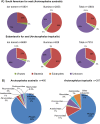
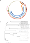
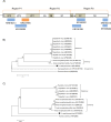
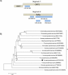
The Brazilian South coast seasonally hosts numerous marine species, observed particularly during winter months. Some animals, including fur seals, are found dead or debilitated along the shore and may harbor potential pathogens within their microbiota. In the present study, a metagenomic approach was performed to evaluate the viral diversity in feces of fur seals found deceased along the coast of the state of Rio Grande do Sul. The fecal virome of two fur seal species was characterized: the South American fur seal (Arctocephalus australis) and the Subantarctic fur seal (Arctocephalus tropicalis). Fecal samples from 10 specimens (A. australis, n = 5; A. tropicalis, n = 5) were collected and viral particles were purified, extracted and amplified with a random PCR. The products were sequenced through Ion Torrent and Illumina platforms and assembled reads were submitted to BLASTx searches. Both viromes were dominated by bacteriophages and included a number of potentially novel virus genomes. Sequences of picobirnaviruses, picornaviruses and a hepevirus-like were identified in A. australis. A rotavirus related to group C, a novel member of the Sakobuvirus and a sapovirus very similar to California sea lion sapovirus 1 were found in A. tropicalis. Additionally, sequences of members of the Anelloviridae and Parvoviridae families were detected in both fur seal species. This is the first metagenomic study to screen the fecal virome of fur seals, contributing to a better understanding of the complexity of the viral community present in the intestinal microbiota of these animals.
Conflict of interest statement
Figures





Similar articles
-
Spleen and lung virome analysis of South American fur seals (Arctocephalus australis) collected on the southern Brazilian coast.Infect Genet Evol. 2021 Aug;92:104862. doi: 10.1016/j.meegid.2021.104862. Epub 2021 Apr 10. Infect Genet Evol. 2021. PMID: 33848685
-
Characterization of the faecal bacterial community of wild young South American (Arctocephalus australis) and Subantarctic fur seals (Arctocephalus tropicalis).FEMS Microbiol Ecol. 2016 Mar;92(3):fiw029. doi: 10.1093/femsec/fiw029. Epub 2016 Feb 15. FEMS Microbiol Ecol. 2016. PMID: 26880785
-
Molecular Detection of Circovirus and Adenovirus in Feces of Fur Seals (Arctocephalus spp.).Ecohealth. 2017 Mar;14(1):69-77. doi: 10.1007/s10393-016-1195-8. Epub 2016 Nov 1. Ecohealth. 2017. PMID: 27803979 Free PMC article.
-
The enigma of picobirnaviruses: viruses of animals, fungi, or bacteria?Curr Opin Virol. 2022 Jun;54:101232. doi: 10.1016/j.coviro.2022.101232. Epub 2022 May 26. Curr Opin Virol. 2022. PMID: 35644066 Review.
-
Anomalous colour in Neotropical mammals: a review with new records for Didelphis sp. (Didelphidae, Didelphimorphia) and Arctocephalus australis (Otariidae, Carnivora).Braz J Biol. 2013 Feb;73(1):185-94. doi: 10.1590/s1519-69842013000100020. Braz J Biol. 2013. PMID: 23644801 Review.
Cited by
-
Metagenomics detection and characterisation of viruses in faecal samples from Australian wild birds.Sci Rep. 2018 Jun 6;8(1):8686. doi: 10.1038/s41598-018-26851-1. Sci Rep. 2018. PMID: 29875375 Free PMC article.
-
Understanding the Genetic Diversity of Picobirnavirus: A Classification Update Based on Phylogenetic and Pairwise Sequence Comparison Approaches.Viruses. 2021 Jul 28;13(8):1476. doi: 10.3390/v13081476. Viruses. 2021. PMID: 34452341 Free PMC article.
-
Detection of adenovirus, papillomavirus and parvovirus in Brazilian bats of the species Artibeus lituratus and Sturnira lilium.Arch Virol. 2019 Apr;164(4):1015-1025. doi: 10.1007/s00705-018-04129-1. Epub 2019 Feb 10. Arch Virol. 2019. PMID: 30740637 Free PMC article.
-
Candidate new rotavirus species in Schreiber's bats, Serbia.Infect Genet Evol. 2017 Mar;48:19-26. doi: 10.1016/j.meegid.2016.12.002. Epub 2016 Dec 6. Infect Genet Evol. 2017. PMID: 27932285 Free PMC article.
-
Current Advances on Virus Discovery and Diagnostic Role of Viral Metagenomics in Aquatic Organisms.Front Microbiol. 2017 Mar 22;8:406. doi: 10.3389/fmicb.2017.00406. eCollection 2017. Front Microbiol. 2017. PMID: 28382024 Free PMC article. Review.
References
-
- Ferreira JM, De Oliveira LR, Wynen L, Bester MN, Guinet C, Moraes-Barros N, et al. Multiple origins of vagrant Subantarctic fur seals: A long journey to the Brazilian coast detected by molecular markers. Polar Biol. 2008;31: 303–308. 10.1007/s00300-007-0358-z - DOI
-
- Moura JF, Siciliano S. Straggler subantarctic fur seal (Arctocephalus tropicalis) on the coast of Rio de Janeiro State, Brazil. Lat Am J Aquat Mamm. 2007;6: 103–107. 10.5597/lajam00114 - DOI
-
- Pinedo MC. Ocorrência de pinípedes na costa brasileira. Gracia Orla Série Zool. 1990;15: 37–48.
-
- Oliveira A, Kolesnikovas CKM, Serafini PP, Moreira LMP, Pontalti M, Simões-Lopes PC, et al. Occurrence of pinnipeds in Santa Catarina between 2000 and 2010. Lat Am J Aquat Mamm. 2011;9: 145–149.
-
- Simões-Lopes PC, Drebmer CJ, Ott PH. Nota sobre os otariidae e phocidae (mammalia: carnivora) da costa norte do Rio Grande do Sul e Santa catarina, Brasil. Biociências. 1995. pp. 173–181.
Publication types
MeSH terms
Grants and funding
LinkOut - more resources
Full Text Sources
Other Literature Sources

