. 2016 Mar 15;11(3):e0151602.
doi: 10.1371/journal.pone.0151602. eCollection 2016.

Mechanosensory and ATP Release Deficits following Keratin14-Cre-Mediated TRPA1 Deletion Despite Absence of TRPA1 in Murine Keratinocytes

Affiliations

Mechanosensory and ATP Release Deficits following Keratin14-Cre-Mediated TRPA1 Deletion Despite Absence of TRPA1 in Murine Keratinocytes

Katherine J Zappia et al. PLoS One. .

Abstract

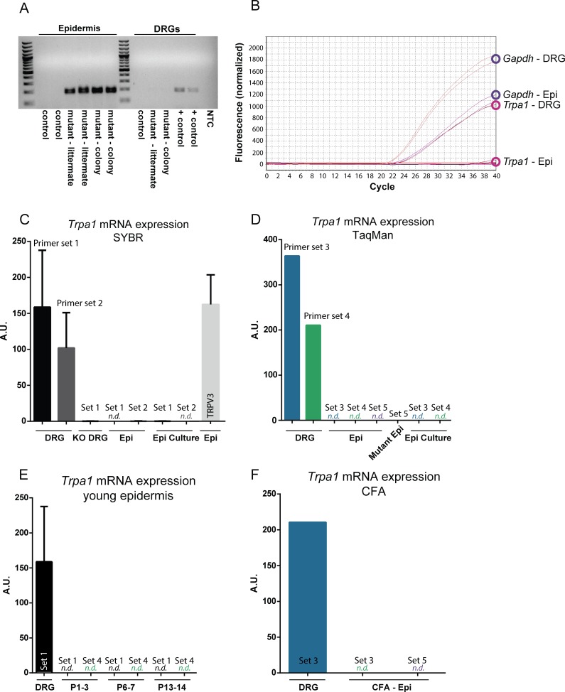
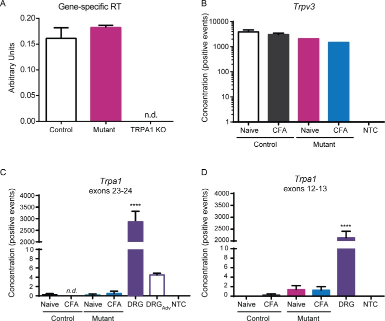
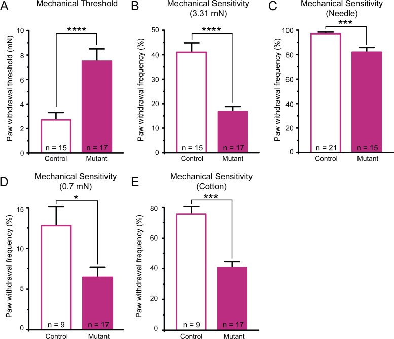
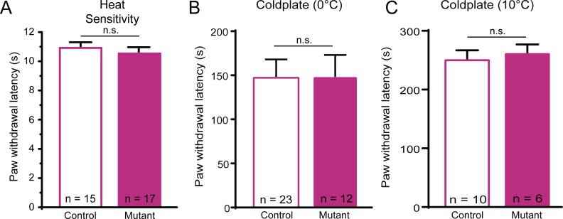
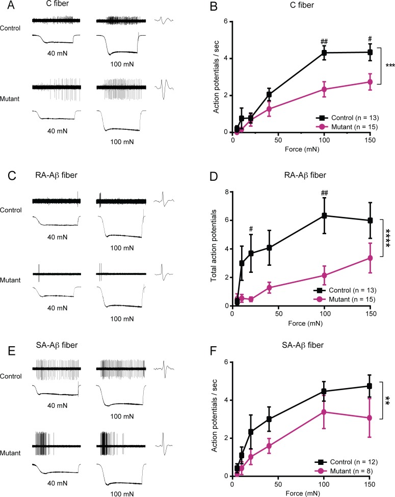
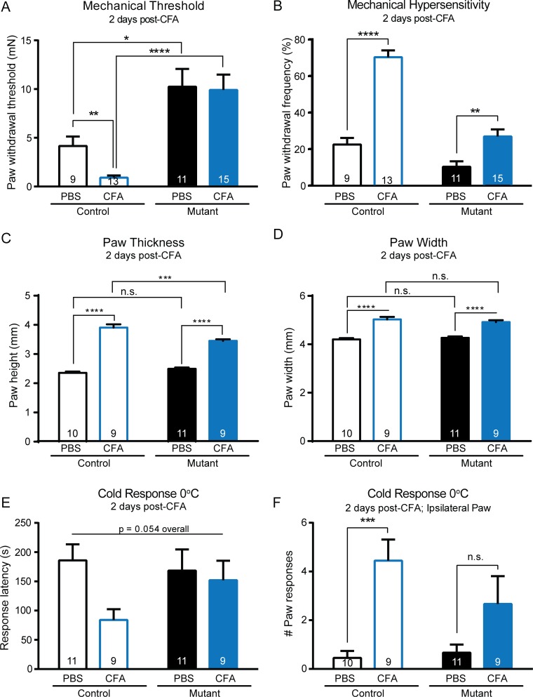
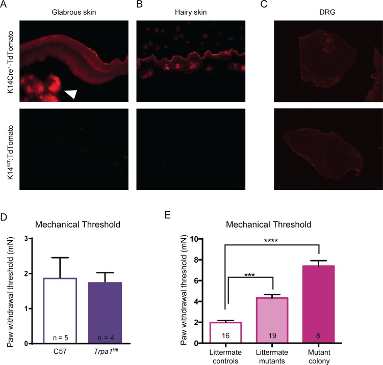
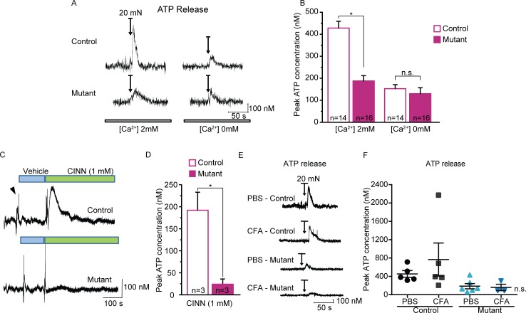
Keratinocytes are the first cells that come into direct contact with external tactile stimuli; however, their role in touch transduction in vivo is not clear. The ion channel Transient Receptor Potential Ankyrin 1 (TRPA1) is essential for some mechanically-gated currents in sensory neurons, amplifies mechanical responses after inflammation, and has been reported to be expressed in human and mouse skin. Other reports have not detected Trpa1 mRNA transcripts in human or mouse epidermis. Therefore, we set out to determine whether selective deletion of Trpa1 from keratinocytes would impact mechanosensation. We generated K14Cre-Trpa1fl/fl mice lacking TRPA1 in K14-expressing cells, including keratinocytes. Surprisingly, Trpa1 transcripts were very poorly detected in epidermis of these mice or in controls, and detection was minimal enough to preclude observation of Trpa1 mRNA knockdown in the K14Cre-Trpa1fl/fl mice. Unexpectedly, these K14Cre-Trpa1fl/fl mice nonetheless exhibited a pronounced deficit in mechanosensitivity at the behavioral and primary afferent levels, and decreased mechanically-evoked ATP release from skin. Overall, while these data suggest that the intended targeted deletion of Trpa1 from keratin 14-expressing cells of the epidermis induces functional deficits in mechanotransduction and ATP release, these deficits are in fact likely due to factors other than reduction of Trpa1 expression in adult mouse keratinocytes because they express very little, if any, Trpa1.


Conflict of interest statement

Competing Interests: The authors have declared that no competing interests exist.

Figures

Fig 1
Fig 1. Trpa1 mRNA is poorly detected in mouse epidermal keratinocytes.
(A) Following Cre-mediated recombination of genomic Trpa1 DNA, an excision product is amplified in the epidermis of K14Cre-Trpa1fl/fl mice (left), but not in the control (K14Cre-Trpa1+/+) mice. Mutant mice (K14Cre-Trpa1fl/fl) mice were either from an independently maintained colony (mutant-colony), or generated as littermates from heterozygous breeders (mutant—littermate). No excision product was observed in the DRGs of either control or mutant mice. Positive control band was obtained from DRG tissue from an AdvillinCre-Trpa1fl/fl mouse (right). For panels B-F, mRNA was isolated from DRG and epidermis from control mice. (B) Amplification plot showing Gapdh and Trpa1 mRNA transcript amplification. Gapdh was consistently detected and quantified in both DRG and epidermal samples (top). Trpa1 mRNA was strongly detected in DRG samples; however, the same PCR protocol did not detect measurable Trpa1 in epidermal samples (bottom). (C) Two different primer sets to detect Trpa1 were effective in measuring Trpa1 from DRG samples using SYBR Green qPCR, but did not amplify Trpa1 from epidermal samples or cultured epidermal keratinocytes. Primer set 1 targets exons 22–23 within the deleted pore region of Trpa1; primer set 2 targets exons 17–19, upstream of the deleted pore region. (D) Three sets of Taqman primer-probes were similarly unable to detect Trpa1 transcripts in control epidermis. Primer set 3 targets exons 13–14, set 4 targets exons 22–23, and set 5 targets exons 23–24 of Trpa1. (E) Neither qPCR for exons 22–23 (set 1) nor exons 22–23 (set 4) were capable of detecting Trpa1 in neonatal mouse epidermis. (F) Two days after hindpaw injection of CFA, Trpa1 remained undetected in epidermis. N.d. denotes transcript not detected.
Fig 2
Fig 2. No Trpa1 knockdown is observed in epidermis of mutant (K14Cre-Trpa1fl/fl) mice compared to controls (K14Cre-Trpa1+/+).
(A) Gene-specific primers to improve reverse transcription of Trpa1 were used prior to targeted amplification of Trpa1. This sensitive method detected a very small amount of Trpa1 transcripts within hindpaw epidermis, and the amplification was equivalent between control and mutant samples. (B-D) Quantitative digital-droplet PCR was performed on samples isolated from epidermis of control and mutant mice. (B) TRPV3 was efficiently detected in all samples tested. No template control (NTC) bars denote ddPCR performed with no cDNA added. (C) ddPCR for exons 23–24 of Trpa1 (within the loxp-flanked region) detected ample transcripts within the DRG samples. On average, less than one positive event per entire reaction volume was detected in both control and mutant samples. A control sample of DRG tissue from an AdvillinCre-Trpa1fl/fl mouse shows that Cre-mediated recombination in a different tissue (sensory neurons) reduces Trpa1 mRNA detection (DRGAdv). (D) ddPCR for exons 12–13 detected on average less than one positive event per epidermal sample. N.d. denotes transcript not detected. **** P<0.0001, compared to every other bar.
Fig 3
Fig 3. Mutant mice (K14Cre-Trpa1fl/fl) exhibit decreased behavioral sensitivity to noxious and gentle mechanical stimuli.
(A) Paw withdrawal responses revealed markedly elevated thresholds in mutant mice. (B) Mutant mice had significantly fewer responses to repeated application of a 3.31 mN von Frey filament. (C) Mutant mice responded less frequently to repeated applications of a spinal needle. (D). Mutant mice responded less frequently to repeated, punctate application of a 0.7 mN von Frey filament to the plantar hindpaw. (E) Mutant mice exhibited decreased responses to gentle stroking puffed-out cotton swab applied to the hindpaw. * P<0.05; *** P<0.001, **** P<0.0001. Data reported as mean ± s.e.m.
Fig 4
Fig 4. Mutant mice have normal behavioral thermal sensitivity.
(A) Mutant and control mice did not differ in latency to respond to a heat lamp directed at the plantar hindpaw. (B-C) Mutant and control mice exhibited similar paw lift response latency to a cold plate at either 0°C (B) or 10°C (C). Data reported as mean ± s.e.m. N.s. denotes not significant.
Fig 5
Fig 5. Mutant mice exhibited decreased mechanically-evoked action potential firing.
Glabrous skin-sural nerve preparations were dissected from mutant and control mice. (A) Examples of responses of single C fiber nociceptors to 40 and 100 mN stimuli, shown for both a control fiber (top) and a mutant fiber (bottom). (B) Average action potential firing rate of mutant fibers compared to control was significantly reduced in C fibers innervating glabrous skin. (C) Sample RA-Aβ fiber responses from control (top) and mutant (bottom) glabrous skin recordings at forces of 40 and 100 mN. (D) Mutant RA-Aβ fibers fired significantly fewer total action potentials compared to control fibers. By 100 mN, there was a 73.8% reduction in the action potential firing rate in mutants compared to the firing rate in controls at 100 mN. (E) Example responses of a control (top) and mutant (bottom) SA-Aβ fiber in response to 40 and 100 mN stimulation. (F) Action potential firing rate in mutant SA-Aβ fibers was significantly decreased compared to control fibers, with a cumulative 31% reduction in action potentials fired across all forces tested. ** P<0.01; *** P<0.001, **** P<0.0001 comparing overall responses between control and mutant fibers. # P<0.05, ## P<0.01 comparing action potential firing at a single force. Data reported as mean ± s.e.m.
Fig 6
Fig 6. Mutant mice exhibit minimal inflammatory allodynia and hyperalgesia two days after CFA injection.
(A) Following injection of CFA into the hindpaw, control mice exhibited a marked increase in mechanical sensitivity following CFA injection into the hindpaw as indicated by the reduction in paw withdrawal threshold. In contrast, no changes in threshold were observed in mutant mice treated with CFA. After inflammation, there remained a striking difference in paw withdrawal threshold between mutant and control animals. Analysis was performed using a 2-way ANOVA with post hoc analysis via Mann-Whitney U tests with Bonferroni adjustment. (B) As indicated by a significant increase in paw withdrawal frequency to repeated application of a 3.31 mN force (left), control mice displayed hypersensitivity after CFA injection. Mutant mice exhibited a smaller, yet significant, increase in withdrawal frequency (right). (C) CFA injection induced an increase in the injected paw thickness of both control and mutant mice. Following CFA, control paws were significantly thicker that mutant paws, though the effect size was limited. (D) Similarly, CFA increased paw width in both control and mutant mice. (E) There were no overall differences in paw withdrawal latency to a 0°C cold plate, though there was a strong trend suggesting CFA may have decreased cold withdrawal latency only in the control mice. (F) CFA greatly increased the number of paw lifts and responses to a 0°C cold plate in control mice, and did not significantly increase paw responses in mutant mice. * P<0.05, ** P<0.01; *** P<0.001, **** P<0.0001.
Fig 7
Fig 7. Cre recombinase activity was visualized in epidermal tissues of K14Cre-tdTomato reporter mice.
(A-B) tdTomato reporter fluorescence was observed in the epidermis of both glabrous and hairy skin sections, as expected. In glabrous skin, reporter fluorescence was also observed in sebaceous glands (arrowhead). Bottom row presents the lack of fluorescence in the tdTomatoLSL mice in the absence of the K14Cre allele. (C) No tdTomato reporter fluorescence was detected in the DRG of either control or mutant animals, suggesting no ectopic Cre recombinase activity was present in these sensory neurons. (D) Importantly, in the absence of the K14Cre allele, the Trpa1fl/fl mice did not show any mechanical sensory deficit compared to wildtype C57BL/6 mice. (E) Mutant animals from the same litters as controls exhibited significant elevated mechanical thresholds (littermate mutants vs. littermate controls). Furthermore, although there was a trend for the mutant animals from the independent colony to have an even greater mechanosensitivity deficit, their mechanical thresholds were not statistically different from those of littermate mutant animals (p = 0.0503). Analysis was performed via Kruskall-Wallis and a Dunn’s post hoc test. *** P< 0.001, **** P< 0.0001.
Fig 8
Fig 8. Decreased mechanically- and chemically-evoked ATP release in mutant glabrous skin.
(A) Example traces of ATP release transient measured from control (top) and mutant (bottom) glabrous skin in response to a 10s, 20 mN mechanical stimulation (applied when indicated by arrows). (B) Peak ATP measurements were markedly decreased from the mutant skin compared to control skin. ATP responses from both control and mutant skin were greatly reduced when performed using a calcium-free extracellular solution. (C) Sample ATP release traces in response to application of 1 mM cinnamaldehyde (CINN; indicated by green bars). (D) Chemically-induced ATP release was greatly reduced in glabrous skin of mutant animals. (E) Examples of ATP recordings from control and mutant glabrous skin excised from mice treated with either PBS or CFA. (F) Maximal ATP release was not significantly different between PBS and CFA-treated animals of either genotype; a single animal treated with CFA did show a substantially elevated peak ATP response. * P<0.05. Data reported as mean ± s.e.m.
Fig 9
Fig 9. Small-diameter sensory neurons from mutant mice have relatively few deficits in chemical activation.
(A) Calcium imaging of small-diameter DRG neurons revealed no difference in the percentage of neurons responding to the 100 μM of the TRPA1 agonist, cinnamaldehyde, when comparing control and mutant neurons. (B) Magnitude response to cinnamaldehyde was similar between naïve control and mutant small-diameter neurons. (C-D) A similar proportion of control and mutant small-diameter neurons responded to another TRPA1 agonist, mustard oil (100 μM; 3 mice per genotype). However, there was a subtle decrease in the magnitude calcium response in mutant compared to control neurons. (E-F) Control and mutant sensory neurons responded similarly to application of acidic solutions of pH 6.0 (E) or pH 5.0 (F). (G) Combining across multiple experiments, the magnitude response to a depolarizing stimulus of 50 mM K+ was similar between control and mutant small-diameter neurons. ** P<0.01.
Fig 10
Fig 10. Microarray analysis of control and mutant epidermal and DRG tissues.
RNA was isolated from epidermis and DRGs of both control and mutant mice; three biological replicates were included per genotype and treatment group. (A) Few genes were dysregulated between mutant and control epidermis in naïve animals. (B) Key genes related to sensory function were similarly expressed in control and mutant epidermis. A.U. denotes arbitrary unit. (C) Only a small number of genes were differentially regulated in mutant and control DRGs. (D) Genes linked to sensory function were expressed at similar levels in control and mutant DRG samples. (E) CFA-induced inflammation revealed a much broader set of expression differences between control and mutant epidermis. (F) A set of genes regulated by IFNα was explored; many of these genes appeared to be downregulated in the mutant epidermis compared to control.

Similar articles

Cited by

References

    1. Azorin N, Raoux M, Rodat-Despoix L, Merrot T, Delmas P, Crest M. ATP signalling is crucial for the response of human keratinocytes to mechanical stimulation by hypo-osmotic shock. Exp Dermatol. 2011;20(5):401–7. 10.1111/j.1600-0625.2010.01219.x - DOI - PubMed
    1. Tsutsumi M, Inoue K, Denda S, Ikeyama K, Goto M, Denda M. Mechanical-stimulation-evoked calcium waves in proliferating and differentiated human keratinocytes. Cell Tissue Res. 2009;338(1):99–106. 10.1007/s00441-009-0848-0 - DOI - PubMed
    1. Pang Z, Sakamoto T, Tiwari V, Kim Y- S, Yang F, Dong X, et al. Selective keratinocyte stimulation is sufficient to evoke nociception in mice. Pain. 2015;156(4):656–65. 10.1097/j.pain.0000000000000092 - DOI - PubMed
    1. Baumbauer KM, DeBerry JJ, Adelman PC, Miller RH, Hachisuka J, Lee KH, et al. Keratinocytes can modulate and directly initiate nociceptive responses. Elife. 2015;4:1–14. - PMC - PubMed
    1. Zylka MJ, Rice FL, Anderson DJ. Topographically distinct epidermal nociceptive circuits revealed by axonal tracers targeted to Mrgprd. Neuron. 2005;45(1):17–25. - PubMed

Publication types

MeSH terms