

Invited review: Small GTPases and their GAPs
- PMID: 26972107
- PMCID: PMC5439442
- DOI: 10.1002/bip.22833
Invited review: Small GTPases and their GAPs
Abstract
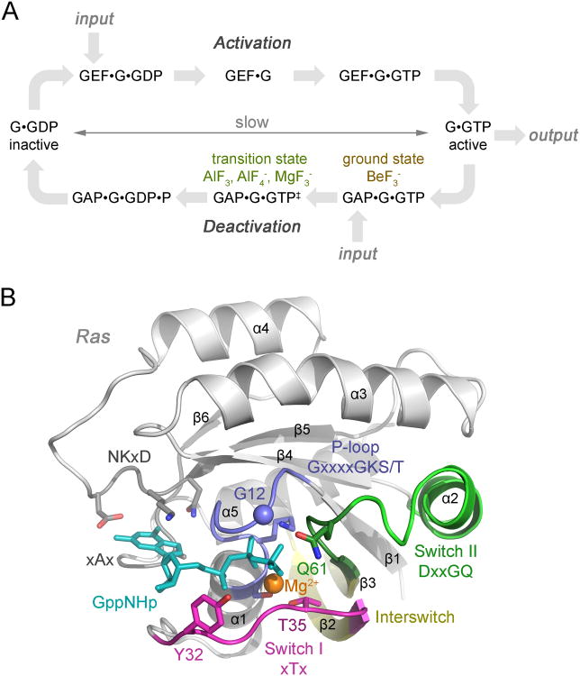
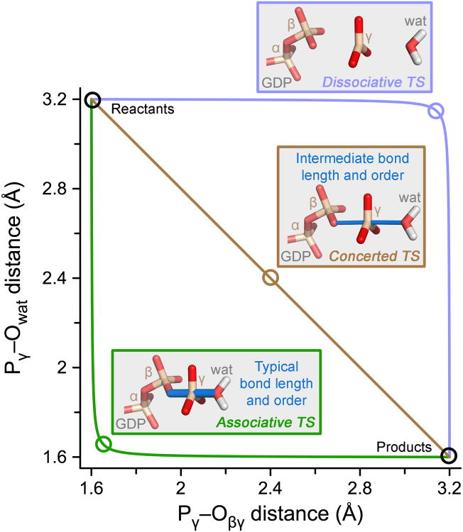
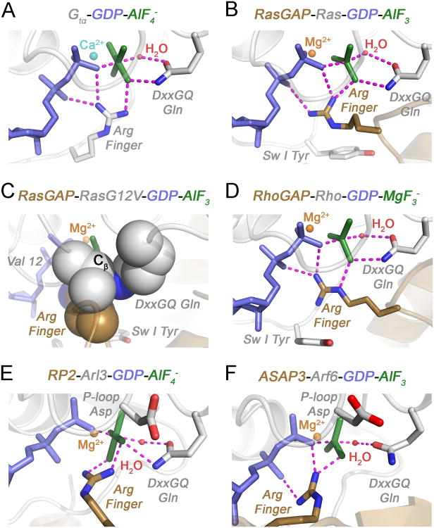
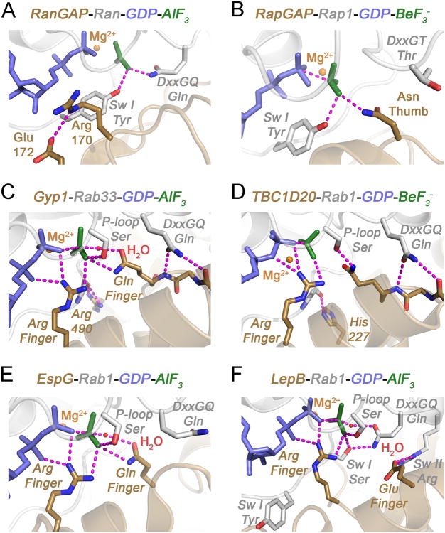
Widespread utilization of small GTPases as major regulatory hubs in many different biological systems derives from a conserved conformational switch mechanism that facilitates cycling between GTP-bound active and GDP-bound inactive states under control of guanine nucleotide exchange factors (GEFs) and GTPase activating proteins (GAPs), which accelerate slow intrinsic rates of activation by nucleotide exchange and deactivation by GTP hydrolysis, respectively. Here we review developments leading to current understanding of intrinsic and GAP catalyzed GTP hydrolytic reactions in small GTPases from structural, molecular and chemical mechanistic perspectives. Despite the apparent simplicity of the GTPase cycle, the structural bases underlying the hallmark hydrolytic reaction and catalytic acceleration by GAPs are considerably more diverse than originally anticipated. Even the most fundamental aspects of the reaction mechanism have been challenging to decipher. Through a combination of experimental and in silico approaches, the outlines of a consensus view have begun to emerge for the best studied paradigms. Nevertheless, recent observations indicate that there is still much to be learned. © 2016 Wiley Periodicals, Inc. Biopolymers 105: 431-448, 2016.
Keywords: GAP; GTPase; hydrolysis; mechanism; structure.
© 2016 Wiley Periodicals, Inc.
Figures
Similar articles
-
In vitro GEF and GAP assays.Curr Protoc Cell Biol. 2009 Jun;Chapter 14:Unit 14.9. doi: 10.1002/0471143030.cb1409s43. Curr Protoc Cell Biol. 2009. PMID: 19499504
-
Probing the GTPase cycle with real-time NMR: GAP and GEF activities in cell extracts.Methods. 2012 Aug;57(4):473-85. doi: 10.1016/j.ymeth.2012.06.014. Epub 2012 Jun 28. Methods. 2012. PMID: 22750304
-
The role of Mg2+ cofactor in the guanine nucleotide exchange and GTP hydrolysis reactions of Rho family GTP-binding proteins.J Biol Chem. 2000 Aug 18;275(33):25299-307. doi: 10.1074/jbc.M001027200. J Biol Chem. 2000. PMID: 10843989
-
The guanine nucleotide-binding switch in three dimensions.Science. 2001 Nov 9;294(5545):1299-304. doi: 10.1126/science.1062023. Science. 2001. PMID: 11701921 Review.
-
Structural Insights into the Regulation Mechanism of Small GTPases by GEFs.Molecules. 2019 Sep 11;24(18):3308. doi: 10.3390/molecules24183308. Molecules. 2019. PMID: 31514408 Free PMC article. Review.
Cited by
-
The IFT81-IFT74 complex acts as an unconventional RabL2 GTPase-activating protein during intraflagellar transport.EMBO J. 2023 Sep 18;42(18):e111807. doi: 10.15252/embj.2022111807. Epub 2023 Aug 22. EMBO J. 2023. PMID: 37606072 Free PMC article.
-
Nutrient Signaling and Lysosome Positioning Crosstalk Through a Multifunctional Protein, Folliculin.Front Cell Dev Biol. 2020 Mar 3;8:108. doi: 10.3389/fcell.2020.00108. eCollection 2020. Front Cell Dev Biol. 2020. PMID: 32195250 Free PMC article. Review.
-
The Role of Calmodulin in Tumor Cell Migration, Invasiveness, and Metastasis.Int J Mol Sci. 2020 Jan 24;21(3):765. doi: 10.3390/ijms21030765. Int J Mol Sci. 2020. PMID: 31991573 Free PMC article. Review.
-
Proteomic Analysis Unveils Expressional Changes in Cytoskeleton- and Synaptic Plasticity-Associated Proteins in Rat Brain Six Months after Withdrawal from Morphine.Life (Basel). 2021 Jul 13;11(7):683. doi: 10.3390/life11070683. Life (Basel). 2021. PMID: 34357055 Free PMC article.
-
The Ras dimer structure.Chem Sci. 2021 May 4;12(23):8178-8189. doi: 10.1039/d1sc00957e. Chem Sci. 2021. PMID: 34194708 Free PMC article.
References
Publication types
MeSH terms
Substances
Grants and funding
LinkOut - more resources
Full Text Sources
Other Literature Sources
Miscellaneous
