Review
. 2016 Mar 10;164(6):1136-1150.
doi: 10.1016/j.cell.2016.02.027.

Communication in Neural Circuits: Tools, Opportunities, and Challenges

Affiliations
Review

Communication in Neural Circuits: Tools, Opportunities, and Challenges

Talia N Lerner et al. Cell. .

Abstract

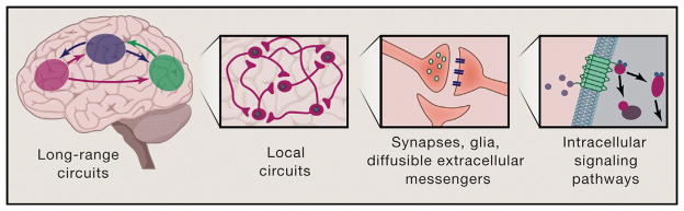
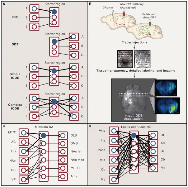
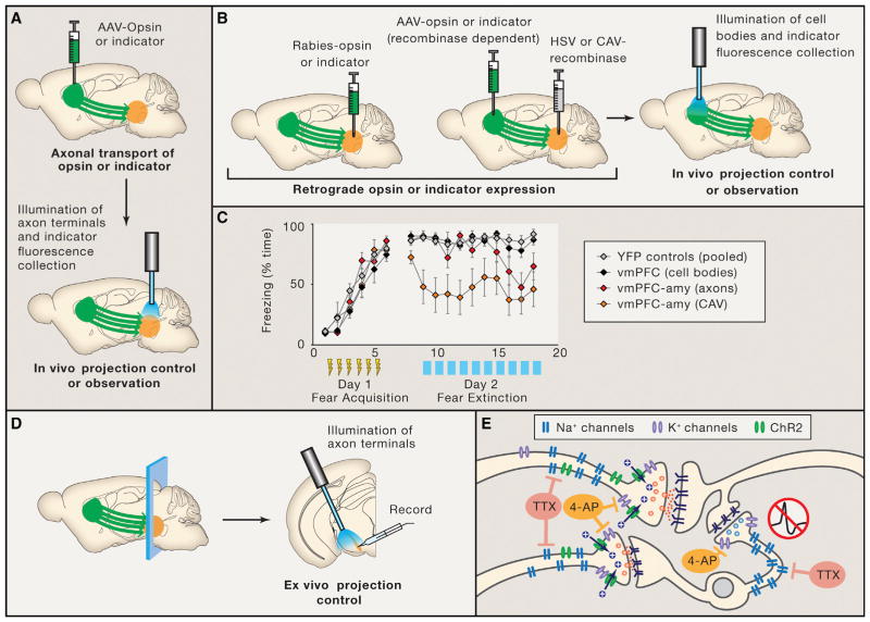
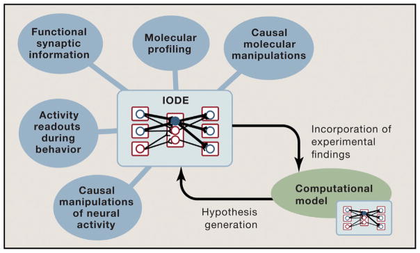
Communication, the effective delivery of information, is fundamental to life across all scales and species. Nervous systems (by necessity) may be most specifically adapted among biological tissues for high rate and complexity of information transmitted, and thus, the properties of neural tissue and principles of its organization into circuits may illuminate capabilities and limitations of biological communication. Here, we consider recent developments in tools for studying neural circuits with particular attention to defining neuronal cell types by input and output information streams--i.e., by how they communicate. Complementing approaches that define cell types by virtue of genetic promoter/enhancer properties, this communication-based approach to defining cell types operationally by input/output (I/O) relationships links structure and function, resolves difficulties associated with single-genetic-feature definitions, leverages technology for observing and testing significance of precisely these I/O relationships in intact brains, and maps onto processes through which behavior may be adapted during development, experience, and evolution.


Figures

Figure 1
Figure 1. Nervous Systems Are Designed for Communication over Many Scales
Nervous systems communicate at the brainwide level, the circuit level, the intercellular (synaptic) level, and the intracellular level (shown left to right). While the latter levels are fundamental to all biological systems, the more complex brainwide and circuit levels of communication distinguish the nervous system and support the unique function of this highly specialized tissue. Opportunities for new discovery in neural communication are abundant across these scales of analysis.
Figure 2
Figure 2. Input/Output-Defined Elements in the Nervous System
(A) Input and output defined elements (highlighted in blue) as nervous system building blocks are schematized. An input-defined element (IDE) is a cell-type defined by location, cell origin, and/or activity of its afferent structures. An output-defined element (ODE) is a cell-type defined by location, cell target, and/or activity of its efferent structures. An I/O-defined element (IODE) is specified by both input and output anatomy and activity as defined above. In a simple case, a cell might serve the purpose of processing and relaying information from input site 1 to output site A. In a more complex case, a cell might integrate and process information from input sites 1 and 2, then relay its output to multiple brain regions (output sites A, B, and C). (B) Intact-system methods for visualizing IODEs. After IODE tracer injections (e.g., those involved in implementing TRIO; Schwarz et al., 2015), whole brains can be clarified (e.g., using CLARITY; adapted with permission from Chung et al., 2013) and intact IODEs can be visualized in the fully intact organ (Lerner et al., 2015, Menegas et al., 2015). Scale bars on optical coronal sections are 1 mm. The IODE visualized here shows inputs from motor cortex and striatum to DLS-projecting midbrain dopamine neurons, as schematized in C (adapted with permission from Lerner et al., 2015). (C and D) IODEs observed in recent studies of the midbrain dopamine (DA) system (B) and the locus coeruleus norepinephrine (NE) system (C). Midbrain dopamine neurons form distinct, though sometimes complex, IODEs (Beier et al., 2015; Lerner et al., 2015), whereas locus coeruleus norepinephrine neurons are not readily distinguishable by either input or output (Schwarz et al., 2015). M1/2, primary and secondary motor cortices; AC, anterior cingulate; DS, dorsal striatum; DLS, dorsolateral striatum; DMS, dorsomedial striatum; NAc, nucleus accumbens; NAc lat, NAc lateral shell; NAc med, NAc medial shell; DR, dorsal raphe; VP, ventral pallidum; mPFC, medial prefrontal cortex; Amy, amygdala; Hy, hypothalamus; Mid, midbrain; Cb, cerebellum; Me, medulla; OB, olfactory bulb; Hi, hippocampus.
Figure 3
Figure 3. Functional Methods for Circuit Element Mapping In Vivo and Ex Vivo
(A) The expression of opsins or activity indicators in axon terminals allows for functional and anatomical circuit element definition in vivo, achieved by specific fiberoptic-based illumination of axon terminals. (B) Specific stimulation or observation of a projection can also be achieved by using a retrograde virus such as rabies, HSV, or CAV to express an opsin or activity indicator only in cells that have a specific efferent target. In this case, cell bodies may be illuminated directly. (C) An example of free mouse behavior during optical control of output-defined elements (ODEs). In this case, the ventromedial prefrontal cortex (vmPFC) to amygdala (amy) projection was manipulated during fear conditioning and extinction using three different strategies. First, non-specific stimulation of vmPFC cell bodies (black symbols); second, stimulation of vmPFC axon terminals in the amygdala (red symbols); third, stimulation of vmPFC cell bodies which send projections to the amygdala (orange symbols; identified by CAV-cre injections in the basomedial amygdala; Adhikari et al., 2015). Increasingly specific output definition of the stimulation circuit element elicits increasingly potent effects on the cued freezing (fear memory) behavior. Yellow bolts indicate six shock-tone pairings given on the training day. Blue bars indicate the time of blue light stimulation of the target circuit element on the extinction day (when tones are played, but no shocks delivered). Gray symbols, YFP (no-opsin) control cohort. Adapted with permission. (D) Expression of opsins in axons’ terminals also allows for optical control of defined circuit elements in the ex vivo slice electrophysiology preparation. (E) Acute slice preparations allow for fine circuit dissection in controlled conditions, for example using TTX/4-AP to definitively isolate monosynaptic connections (Petreanu et al., 2009; Lerner et al., 2015; Adhikari et al., 2015). First, TTX blocks action-potential-dependent release, preventing disynaptic stimulation through non-opsin-expressing neurons. Second, 4-AP increases terminal excitability by blocking K+ channels. Channelrhodopsin (ChR) optical drive then induces action-potential-independent depolarization at ChR-expressing terminals only, while patch clamping of different target cells thus allows definition of afferent fibers as output-defined elements (ODEs).
Figure 4
Figure 4. Organizing Principles for Cross-Modal Investigation of Neural Circuits
IODEs may help provide a behaviorally relevant and experimentally tractable framework for guiding and integrating information about neural circuits across many levels of investigation. Once the basic physical structure of an IODE is understood from the anatomical tools described here (including the most relevant convergence and divergence of information through collaterals when considering a specific behavior), activity information during behavior from physiology, imaging, and molecular datastreams can be collected using targeting tools aligned with the IODE structure and then layered onto the diagram to form a more complete understanding of I/O relationships. Computational analyses may facilitate registration and joint interpretation of information gathered by these disparate techniques, as well as generation of higher-order hypotheses to guide further data collection (e.g., through system-identification strategies; Grosenick et al., 2015). Iterations of this data-collection and hypothesis-generation cycle, and crucially the linking of distinct IODEs into loops and more complex topological structures, may continue until experimental and theoretical concepts converge.
Figure 5
Figure 5. Example: Different Levels of Inspection for Basal Ganglia Circuitry
(A) Simplified diagram of basal ganglia (BG) circuitry depicts the “direct” and “indirect” pathways, which have opposing influences on BG output. While the concepts of the direct and indirect pathways have yielded important insights, the reality of BG circuitry is much more complex. (B–D) Examples of additional circuit complexity in the BG. (B) Dopamine neurons in the substantia nigra pars compacta (SNc) project not only to the striatum but to other BG nuclei. These dopamine neurons also receive direct projections back from these nuclei. (C) Information need not loop all the way through the cortico-BG-thalamic circuitry. Several shortcuts are available, including the one pictured in which the globus pallidus external segment (GPe) sends projections back to the cortex (Saunders et al., 2015). (D) The “direct” and “indirect” pathways are not absolute. For example, some “direct pathway” striatal neurons also send collaterals to the GPe (Cazorla et al., 2014). A more sophisticated understanding of BG circuit dynamics may emerge as we build testable hypotheses based on a more realistic picture of the circuitry as shown in (B–D). Such an approach will be facilitated by defining, controlling, and observing cells based on their input and output properties in the intact functioning system (see Figure 4).

Similar articles

Cited by

References

    1. Adhikari A, Lerner TN, Finkelstein J, Pak S, Jennings JH, Davidson TJ, Ferenczi E, Gunaydin LA, Mirzabekov JJ, Ye L, et al. Basomedial amygdala mediates top-down control of anxiety and fear. Nature. 2015;527:179–185. - PMC - PubMed
    1. Akerboom J, Chen TW, Wardill TJ, Tian L, Marvin JS, Mutlu S, Calderón NC, Esposti F, Borghuis BG, Sun XR, et al. Optimization of a GCaMP calcium indicator for neural activity imaging. J Neurosci. 2012;32:13819–13840. - PMC - PubMed
    1. Alexander GE, DeLong MR, Strick PL. Parallel organization of functionally segregated circuits linking basal ganglia and cortex. Annu Rev Neurosci. 1986;9:357–381. - PubMed
    1. Atasoy D, Betley JN, Li WP, Su HH, Sertel SM, Scheffer LK, Simpson JH, Fetter RD, Sternson SM. A genetically specified connectomics approach applied to long-range feeding regulatory circuits. Nat Neurosci. 2014;17:1830–1839. - PMC - PubMed
    1. Ballesteros-Yáñez I, Benavides-Piccione R, Elston GN, Yuste R, DeFelipe J. Density and morphology of dendritic spines in mouse neocortex. Neuroscience. 2006;138:403–409. - PubMed

Publication types

LinkOut - more resources