

Serum amyloid A1: Structure, function and gene polymorphism
- PMID: 26945629
- PMCID: PMC5683722
- DOI: 10.1016/j.gene.2016.02.044
Serum amyloid A1: Structure, function and gene polymorphism
Abstract
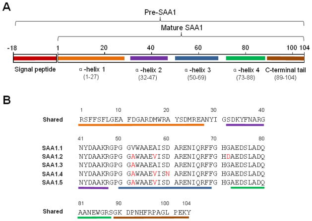
Inducible expression of serum amyloid A (SAA) is a hallmark of the acute-phase response, which is a conserved reaction of vertebrates to environmental challenges such as tissue injury, infection and surgery. Human SAA1 is encoded by one of the four SAA genes and is the best-characterized SAA protein. Initially known as a major precursor of amyloid A (AA), SAA1 has been found to play an important role in lipid metabolism and contributes to bacterial clearance, the regulation of inflammation and tumor pathogenesis. SAA1 has five polymorphic coding alleles (SAA1.1-SAA1.5) that encode distinct proteins with minor amino acid substitutions. Single nucleotide polymorphism (SNP) has been identified in both the coding and non-coding regions of human SAA1. Despite high levels of sequence homology among these variants, SAA1 polymorphisms have been reported as risk factors of cardiovascular diseases and several types of cancer. A recently solved crystal structure of SAA1.1 reveals a hexameric bundle with each of the SAA1 subunits assuming a 4-helix structure stabilized by the C-terminal tail. Analysis of the native SAA1.1 structure has led to the identification of a competing site for high-density lipoprotein (HDL) and heparin, thus providing the structural basis for a role of heparin and heparan sulfate in the conversion of SAA1 to AA. In this brief review, we compares human SAA1 with other forms of human and mouse SAAs, and discuss how structural and genetic studies of SAA1 have advanced our understanding of the physiological functions of the SAA proteins.
Keywords: Acute-phase response; Gene polymorphism; Induced expression; Inflammation; Lipid metabolism; Serum amyloid A.
Copyright © 2016 Elsevier B.V. All rights reserved.
Conflict of interest statement
The authors have declared no conflicts of interest.
Figures
Similar articles
-
Structural mechanism of serum amyloid A-mediated inflammatory amyloidosis.Proc Natl Acad Sci U S A. 2014 Apr 8;111(14):5189-94. doi: 10.1073/pnas.1322357111. Epub 2014 Mar 24. Proc Natl Acad Sci U S A. 2014. PMID: 24706838 Free PMC article.
-
Effect of amino acid variations in the central region of human serum amyloid A on the amyloidogenic properties.Biochem Biophys Res Commun. 2014 Jan 31;444(1):92-7. doi: 10.1016/j.bbrc.2014.01.029. Epub 2014 Jan 16. Biochem Biophys Res Commun. 2014. PMID: 24440699
-
AA amyloidosis-resistant CE/J mice have Saa1 and Saa2 genes that encode an identical SAA isoform.Amyloid. 2014 Mar;21(1):1-8. doi: 10.3109/13506129.2013.852529. Epub 2013 Nov 14. Amyloid. 2014. PMID: 24228751
-
Intrinsic Stability, Oligomerization, and Amyloidogenicity of HDL-Free Serum Amyloid A.Adv Exp Med Biol. 2015;855:117-34. doi: 10.1007/978-3-319-17344-3_5. Adv Exp Med Biol. 2015. PMID: 26149928 Review.
-
[Serum amyloid A (SAA)--pathogenicity and implication of appearance in plasma].Rinsho Byori. 2006 May;54(5):509-12. Rinsho Byori. 2006. PMID: 16789422 Review. Japanese.
Cited by
-
SAA1 regulates pro-labour mediators in term labour by activating YAP pathway.Mol Cell Biochem. 2021 Jul;476(7):2791-2801. doi: 10.1007/s11010-021-04125-1. Epub 2021 Mar 15. Mol Cell Biochem. 2021. PMID: 33719002
-
Advances in development of biomarkers for brain damage and ischemia.Mol Biol Rep. 2024 Jul 13;51(1):803. doi: 10.1007/s11033-024-09708-x. Mol Biol Rep. 2024. PMID: 39001884 Free PMC article. Review.
-
Sample Pooling and Inflammation Linked to the False Selection of Biomarkers for Neurodegenerative Diseases in Top-Down Proteomics: A Pilot Study.Front Mol Neurosci. 2018 Dec 18;11:477. doi: 10.3389/fnmol.2018.00477. eCollection 2018. Front Mol Neurosci. 2018. PMID: 30618622 Free PMC article.
-
Structural Basis for Lipid Binding and Function by an Evolutionarily Conserved Protein, Serum Amyloid A.J Mol Biol. 2020 Mar 27;432(7):1978-1995. doi: 10.1016/j.jmb.2020.01.029. Epub 2020 Feb 6. J Mol Biol. 2020. PMID: 32035904 Free PMC article.
-
Delineating the glioblastoma stemness by genes involved in cytoskeletal rearrangements and metabolic alterations.World J Stem Cells. 2023 May 26;15(5):302-322. doi: 10.4252/wjsc.v15.i5.302. World J Stem Cells. 2023. PMID: 37342224 Free PMC article. Review.
References
-
- Abe-Dohmae S, Kato KH, Kumon Y, Hu W, Ishigami H, Iwamoto N, Okazaki M, Wu CA, Tsujita M, Ueda K, Yokoyama S. Serum amyloid A generates high density lipoprotein with cellular lipid in an ABCA1- or ABCA7-dependent manner. J Lipid Res. 2006;47:1542–50. - PubMed
-
- Atarashi K, Tanoue T, Ando M, Kamada N, Nagano Y, Narushima S, Suda W, Imaoka A, Setoyama H, Nagamori T, Ishikawa E, Shima T, Hara T, Kado S, Jinnohara T, Ohno H, Kondo T, Toyooka K, Watanabe E, Yokoyama S, Tokoro S, Mori H, Noguchi Y, Morita H, Ivanov, Sugiyama T, Nunez G, Camp JG, Hattori M, Umesaki Y, Honda K. Th17 Cell Induction by Adhesion of Microbes to Intestinal Epithelial Cells. Cell. 2015;163:367–80. - PMC - PubMed
Publication types
MeSH terms
Substances
Grants and funding
LinkOut - more resources
Full Text Sources
Other Literature Sources
Miscellaneous
