. 2016 Mar 3;61(5):705-719.
doi: 10.1016/j.molcel.2016.02.009.

Mitochondria-Translocated PGK1 Functions as a Protein Kinase to Coordinate Glycolysis and the TCA Cycle in Tumorigenesis

Affiliations

Mitochondria-Translocated PGK1 Functions as a Protein Kinase to Coordinate Glycolysis and the TCA Cycle in Tumorigenesis

Xinjian Li et al. Mol Cell. .

Abstract

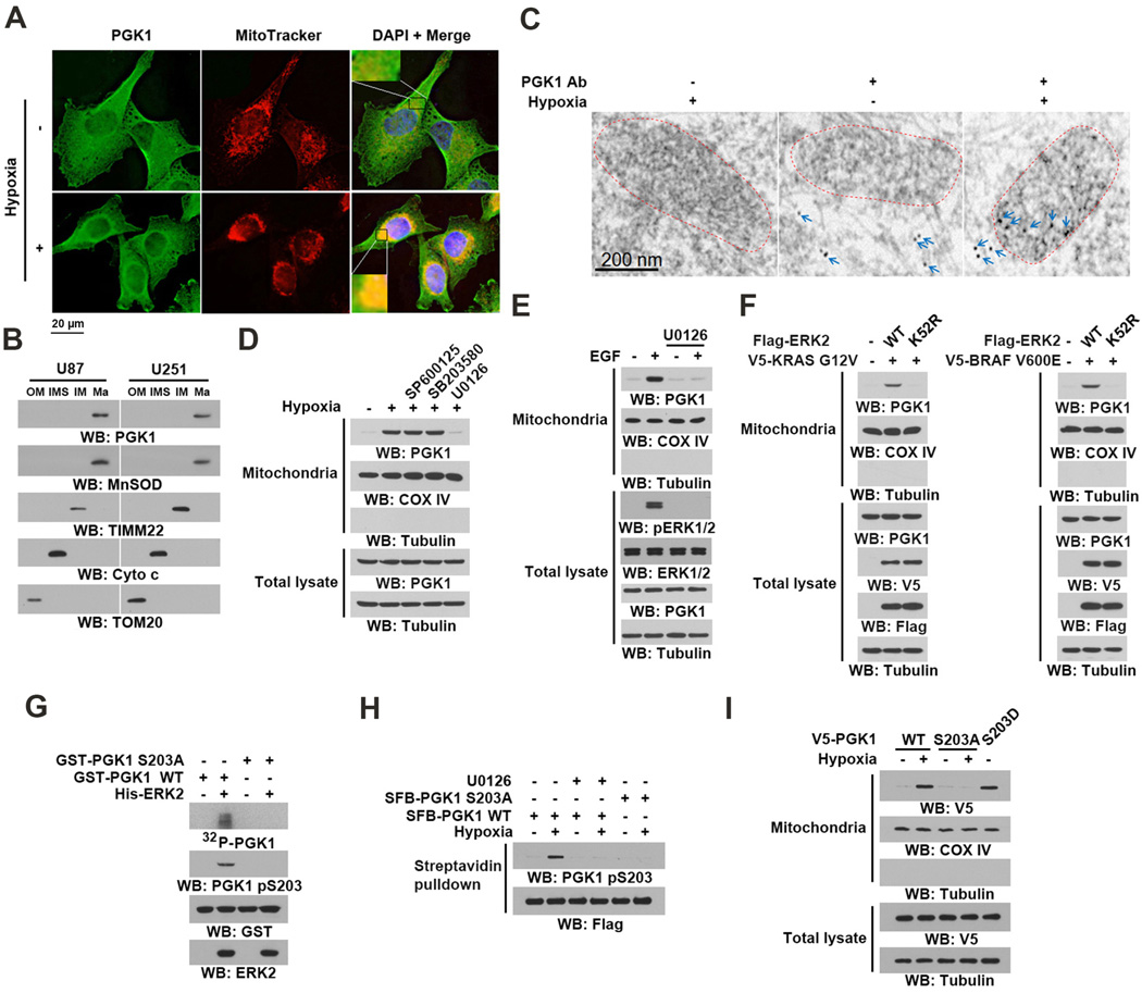
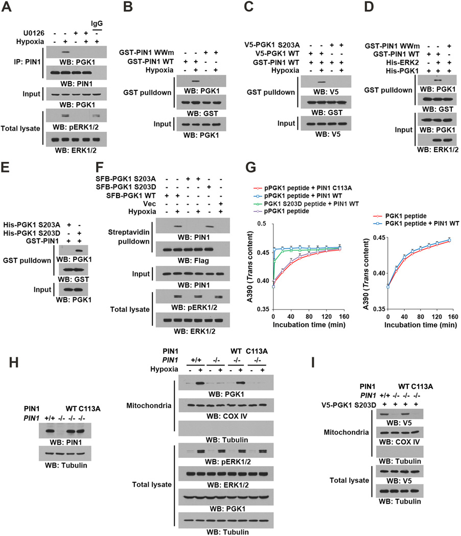
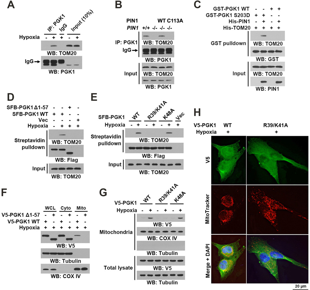
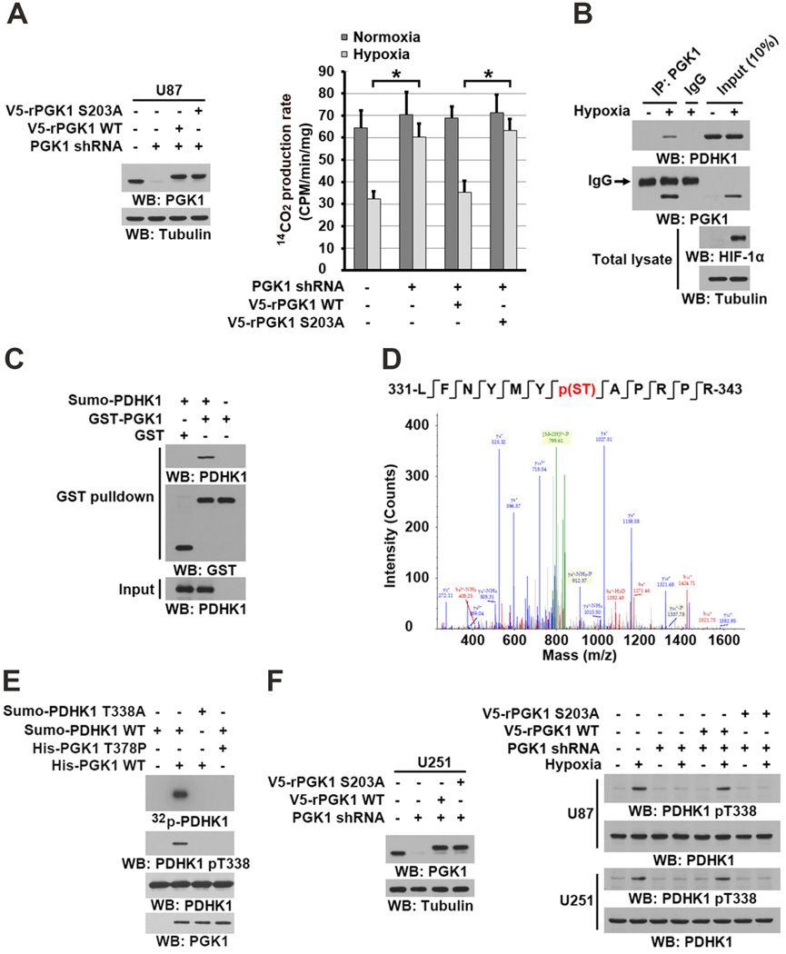
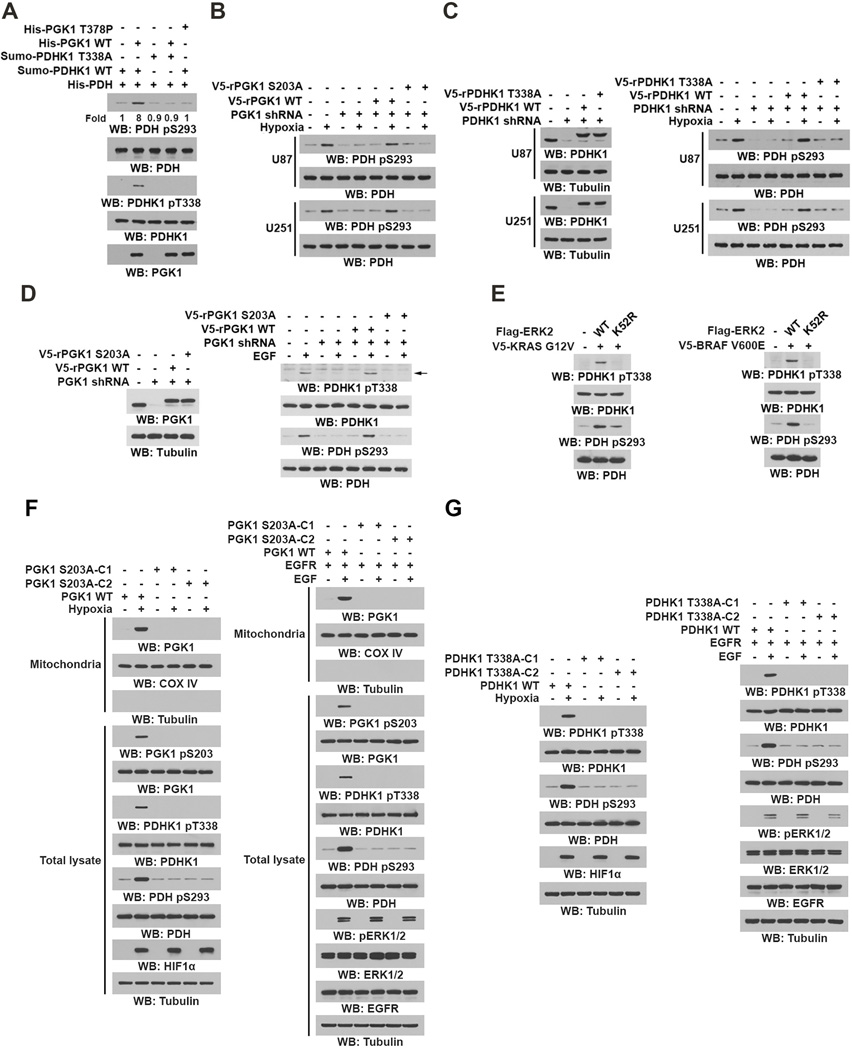
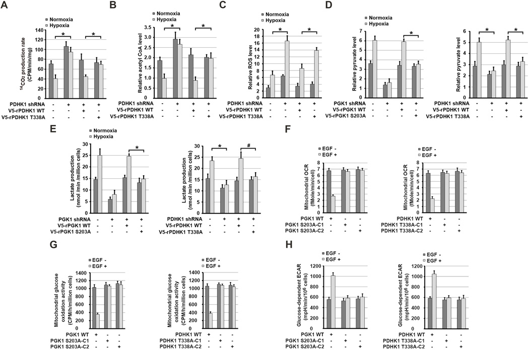
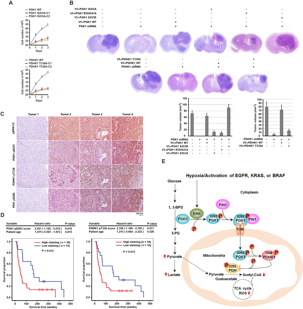
It is unclear how the Warburg effect that exemplifies enhanced glycolysis in the cytosol is coordinated with suppressed mitochondrial pyruvate metabolism. We demonstrate here that hypoxia, EGFR activation, and expression of K-Ras G12V and B-Raf V600E induce mitochondrial translocation of phosphoglycerate kinase 1 (PGK1); this is mediated by ERK-dependent PGK1 S203 phosphorylation and subsequent PIN1-mediated cis-trans isomerization. Mitochondrial PGK1 acts as a protein kinase to phosphorylate pyruvate dehydrogenase kinase 1 (PDHK1) at T338, which activates PDHK1 to phosphorylate and inhibit the pyruvate dehydrogenase (PDH) complex. This reduces mitochondrial pyruvate utilization, suppresses reactive oxygen species production, increases lactate production, and promotes brain tumorigenesis. Furthermore, PGK1 S203 and PDHK1 T338 phosphorylation levels correlate with PDH S293 inactivating phosphorylation levels and poor prognosis in glioblastoma patients. This work highlights that PGK1 acts as a protein kinase in coordinating glycolysis and the tricarboxylic acid (TCA) cycle, which is instrumental in cancer metabolism and tumorigenesis.

Keywords: B-Raf; EGFR; K-Ras; PDH; PDHK1; PGK1; glycolysis; hypoxia; mitochondria; phosphorylation; tumorigenesis.


Figures

Figure 1
Figure 1. Mitochondrial Translocation of PGK1 Is Mediated by ERK1/2-Dependent Phosphorylation
(B, D–I), Immunoblotting and IP analyses were carried out using antibodies against the indicated proteins. (A) U87 cells were stimulated with or without hypoxia for 6 h and stained with an anti-PGK1 antibody, MitoTracker, and DAPI. (B) U87 and U251 cells were stimulated with hypoxia for 6 h. Proteins from mitochondrial outer membrane (OM), intermembrane space (IMS), inner membrane (IM), and matrix (Ma) were isolated. (C) U87 cells were stimulated with or without hypoxia for 6 h. Electron microscopic immunogold analysis with anti-PGK1 antibody was performed. Arrows indicate representative staining of mitochondrial PGK1. Dashed circles indicate mitochondria. (D) Mitochondria fractions and total cell lysates were prepared from U87 cells pretreated with SP600125 (25 µM), SB203580 (10 µM), or U0126 (20 µM) for 30 min before being treated with hypoxia for 6 h. Cytosolic tubulin was used as a control. (E) EGFR-overxpressed U87 (U87/EGFR) cells pretreated with U0126 (20 µM) for 30 min were stimulated with or without EGF (100 ng/ml) for 6 h. Mitochondria fractions and total cell lysates were prepared. (F) V5-KRAS G12V and the indicated Flag-ERK2 proteins were stably expressed in BxPC-3 cells (left panel). V5-BRAF V600E and the indicated Flag-ERK2 proteins were stably expressed in CHL1 cells (right panel). Mitochondria fractions and total cell lysates were prepared. (G) In vitro kinase assays were carried out by mixing purified active ERK2 with purified WT GST-PGK1 or GST-PGK1 S203A in the presence of [γ-32P]ATP. The reaction mixture was separated for autoradiography and immunoblotting analyses. (H) U87 cells expressing the indicated SFB-PGK1 proteins were pretreated with or without U0126 (20 µM) for 30 min before hypoxic stimulation for 6 h. Streptavidin agarose beads were used to pull down SFB-tagged proteins. (I) U87 cells expressing the indicated V5-tagged PGK1 proteins were treated with or without hypoxia for 6 h. Mitochondria fractions and total cell lysates were prepared. See also Figures S1 and S2.
Figure 2
Figure 2. PIN1 Binds to and cis–trans Isomerizes Phosphorylated PGK1 for Mitochondrial Translocation of PGK1
(A–F, H–I) Immunoblotting and IP analyses were carried out using antibodies against the indicated proteins. (A) U87 cells were pretreated with or without U0126 (20 µM) for 30 min before hypoxic stimulation for 6 h. (B) U87 cells were treated with or without hypoxic stimulation for 6 h. A GST pull-down assay with the indicted GST-proteins was performed. GST-PIN1 WWm: GST-PIN1 WW mutant. (C) U87 cells expressing the indicated PGK1 proteins were treated with or without hypoxic stimulation for 6 h. A GST pull-down assay with GST-PIN1 proteins was performed. (D) An in vitro protein kinase assay was performed by mixing purified recombinant PGK1 with or without purified active His-ERK2, which was followed by a GST pull-down assay with the indicated GST-proteins. (E) A GST pull-down assay was performed by mixing GST-PIN1 and the indicated purified recombinant PGK1 proteins. (F) U87 cells expressing the indicated SFB-PGK1 proteins were treated with or without hypoxia for 6 h. A pull-down assay with streptavidin agarose beads was conducted. (G) cis–trans isomerization assays were performed by mixing synthesized phosphorylated or nonphosphorylated oligopeptides of PGK1 containing the S203P204 motif or an oligopeptide of PGK1 containing the D203P204 motif with purified wild-type GST-PIN1 or GST-PIN1 C113A mutant. Data represent the means ± SD of three independent experiments. (H) PIN1−/− cells were reconstituted to express the indicated PIN1 proteins (left panel). The total cell lysates and motochondrial fractions were prepared from the indicated cells treated with or without hypoxia for 6 h (right panel). (I) V5–PGK1 S203D was expressed in PIN1+/+ cells and PIN1−/− cells with or without reconstituted WT PIN1 or PIN1 C113A. Total cell lysates and mitochondrial fractions of the cells were prepared. See also Figure S2.
Figure 3
Figure 3. PIN1 Regulates Binding of PGK1 to the TOM Complex
(A–G) Immunoblotting and IP analyses were carried out using antibodies against the indicated proteins. (A) U87 cells were treated with or without hypoxia for 6 h. Total cell lysates were prepared. (B) PIN1+/+ and the indicated PIN1−/− cells were treated with hypoxia for 6 h. (C) A GST pull-down assay was performed by mixing purified recombinant WT GST-PGK1 or GST-PGK1 S203D with His-TOM20 in the presence or absence of purified His-PIN1. (D, E) U87 cells expressing the indicated SFB-PGK1 proteins were treated with or without hypoxia for 6 h. Streptavidin agarose beads were used to pull down SFB-tagged proteins. (F, G) U87 cells expressing the indicated V5-tagged PGK1 proteins were treated with or without hypoxia for 6 h. Total cell lysate, cytosolic, and mitochondrial fractions were prepared. (H) U87 cells expressing the indicated V5-PGK1 proteins were stimulated with or without hypoxia for 6 h and stained with an anti-V5 antibody, MitoTracker, and DAPI. See also Figure S2.
Figure 4
Figure 4. Mitochondrial PGK1 Phosphorylates PDHK1
(A–C, E–F) Immunoblotting analyses were performed with the indicated antibodies. (A) U87 cells with or without PGK1 shRNA and with or without reconstituted expression of WT rPGK1 or rPGK1 S203A were stimulated with or without hypoxia for 6 h. Isolated mitochondrial fractions were mixed with 14C-labeled pyruvate, ADP, and malate under normoxic condtions. 14C-CO2 production rate was measured. Data represent the means ± SD of three independent experiments. *p < 0.01. (B) U87 cells were stimulated with or without hypoxia for 6 h. Mitochondrial fractions of these cells were prepared. IP analyses with an anti-PGK1 antibody were performed. (C) GST pull-down analyses were performed by mixing bacterially purified SUMO-PDHK1 proteins with purified GST or GST-PGK1. (D) In vitro phosphorylation analyses were performed by mixing bacterially purified His-PGK1 and SUMO-PDHK1 in the presence of ATP. The mass spectrometry results of a fragment spectrum of a peptide at m/z 756.346 (mass error, ±4.2 ppm) matched to the doubly charged peptide 331-LFNYMYp(ST)APRPR-343, suggesting that S337 or T338 was phosphorylated. The Mascot score was 49, Expectation Value: 4.7E-004; the SEQUEST score for this match was Xcorr = 3.5. (E) In vitro phosphorylation analyses with autoradiography were performed by mixing purified WT PGK1 or PGK1 T378P with purified WT PDHK1 or PDHK1 T338A in the presence of [γ-32P]ATP. (F) U251 and U87 cells with or without PGK1 shRNA and with or without reconstituted expression of WT rPGK1 or rPGK1 S203A were stimulated with or without hypoxia for 6 h. See also Figure S3.
Figure 5
Figure 5. PDHK1 Phosphorylation by PGK1 Activates PDHK1
Immunoblotting analyses were performed with the indicated antibodies. (A) Bacterially purified His-PDH (E1α) with purified WT PDHK1 or PDHK1 T338A was mixed with purified WT PGK1 or PGK1 T378P. In vitro phosphorylation analyses were performed. (B, C) U87 and U251 cells expressing PGK1 shRNA (B) or PDHK1 shRNA (C) with or without reconstituted expression of the indicated proteins were stimulated with or without hypoxia for 6 h. (D) U87/EGFR cells expressing PGK1 shRNA and with or without reconstituted expression of WT rPGK1 or rPGK1 S203A were stimulated with or without EGF (100 ng/ml) for 6 h. (E) BxPC-3 cells were stably transfected with or without vectors expressing V5-KRAS G12V and the indicated Flag-ERK2 proteins (left panel). CHL1 cells were stably transfected with or without vectors expressing V5-BRAF V600E and the indicated Flag-ERK2 proteins (right panel). (F, G) Parental U87 cells and the indicated clones of U87 cells with PGK1 S203A (F) or PDHK1 T338A (G) knock-in were stimulated with or without hypoxia (left panel) or EGF (100 ng/ml) (right panel) for 6 h. Mitochondrial fractions and total cell lysates were prepared. See also Figure S4.
Figure 6
Figure 6. PGK1-Mediated PDHK1 Phosphorylation Inhibits Mitochondrial Pyruvate Metabolism, Induces Hypoxia-Induced ROS Production, and Promotes Glycolysis
(A–H) Data represent the means ± SD of three independent experiments. *p < 0.01, #p <0.05. (A, B) U87 cells expressing PDHK1 shRNA with or without reconstituted expression of WT rPDHK1 or rPDHK1 T338A were stimulated with or without hypoxia for 6 h. Mitochondrial fractions of the cells were prepared and activity of PDH complex–mediated conversion of 14C-pyruvate into 14C-CO2 was measured (A). Levels of mitochondrial acetyl-CoA were measured (B). (C) U87 cells expressing PDHK1 shRNA with or without reconstituted expression of the indicated proteins were stimulated with or without hypoxia for 24 h. Levels of mitochondrial ROS were measured. (D, E) U87 cells expressing PGK1 shRNA (left panel) or PDHK1 shRNA (right panel) with or without reconstituted expression of the indicated proteins were cultured in no-serum DMEM during hypoxia for 6 h. Levels of cytosolic pyruvate level were measured (D). The media were collected for analysis of lactate production (E). (F and H) Serum-starved parental U87 cells and the indicated clones of U87 cells with PGK1 S203A (left panel) or PDHK1 T338A (right panel) knock-in were treated with or without EGF (100 ng/ml) for 6 h. The glucose-dependent mitochondrial OCR (F) and ECAR (H) in these cells were measured. (G) Serum-starved parental U87 cells and the indicated clones of U87 cells with PGK1 S203A (left panel) or PDHK1 T338A (right panel) knock-in were treated with or without EGF (100 ng/ml) for 6 h and then incubated with 5 mM glucose spiked with 0.001 mM U-14C glucose, 1-14C glucose, or 6-14C glucose for 2 h. The radioactivity levels of the cells were normalized according to cell number. See also Figure S5.
Figure 7
Figure 7. Mitrochondrial PGK1-Dependent PDHK1 Phosphorylation Promotes Cell Proliferation and Brain Tumorigenesis and Indicates a Poor Prognosis in GBM Patients
(A) Parental U87 cells (2 × 10⁵) and the indicated clones of U87 cells with PGK1 S203A (upper panel) or PDHK1 T338A (lower panel) knock-in were plated for 3 days under hypoxic conditions. The cells were then collected and counted. The data are presented as the means ± SD from three independent experiments. (B) U87 cells with or without PGK1 shRNA or PDHK1 shRNA expression and with or without reconstituted expression of the indicated proteins were intracranially injected into athymic nude mice. H&E-stained coronal brain sections show representative tumor xenografts. Tumor volume was calculated. (C) IHC staining with phospho-ERK1/2, PGK1 pS203, PDHK1 pT338, and PDH pS293 antibodies was performed on 50 human primary GBM specimens. Representative photos of four tumors are shown. (D) The survival time for 50 patients with low (1–4 staining scores, blue curve) versus high (4.1–8 staining scores, red curve) phosphorylation levels of PGK1 S203 (low, 14 patients; high, 36 patients) and PDHK1 T338 (low, 16 patients; high, 34 patients) were compared. The table shows the multivariate analysis results after adjustment for patient age, indicating the significance level of the association of PGK1 S203 (p = 0.016) and PDHK1 T338 (p = 0.017) phosphorylation with patient survival. Empty circles represent censored data from patients alive at last clinical follow-up. (E) A Mechanism of Mitochondrial PGK1-Coordinated Glycolysis and TCA Cycle in Tumorigenesis. Broken arrows: inhibited directions or reactions. See also Figures S6, S7.

Similar articles

Cited by

References

    1. Ahmad SS, Glatzle J, Bajaeifer K, Buhler S, Lehmann T, Konigsrainer I, Vollmer JP, Sipos B, Ahmad SS, Northoff H, et al. Phosphoglycerate kinase 1 as a promoter of metastasis in colon cancer. International journal of oncology. 2013 - PubMed
    1. Ai J, Huang H, Lv X, Tang Z, Chen M, Chen T, Duan W, Sun H, Li Q, Tan R, et al. FLNA and PGK1 are two potential markers for progression in hepatocellular carcinoma. Cellular physiology and biochemistry : international journal of experimental cellular physiology, biochemistry, and pharmacology. 2011;27:207–216. - PubMed
    1. Bernstein BE, Hol WG. Crystal structures of substrates and products bound to the phosphoglycerate kinase active site reveal the catalytic mechanism. Biochemistry. 1998;37:4429–4436. - PubMed
    1. Chacinska A, Koehler CM, Milenkovic D, Lithgow T, Pfanner N. Importing mitochondrial proteins: machineries and mechanisms. Cell. 2009;138:628–644. - PMC - PubMed
    1. Chiarelli LR, Morera SM, Bianchi P, Fermo E, Zanella A, Galizzi A, Valentini G. Molecular insights on pathogenic effects of mutations causing phosphoglycerate kinase deficiency. PloS one. 2012;7:e32065. - PMC - PubMed

Publication types

MeSH terms

Substances