. 2016 Feb 29;212(5):499-513.
doi: 10.1083/jcb.201507009.

ALIX and ESCRT-I/II function as parallel ESCRT-III recruiters in cytokinetic abscission

Affiliations

ALIX and ESCRT-I/II function as parallel ESCRT-III recruiters in cytokinetic abscission

Liliane Christ et al. J Cell Biol. .

Abstract

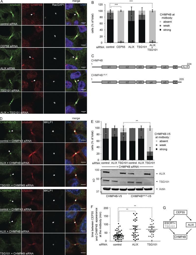
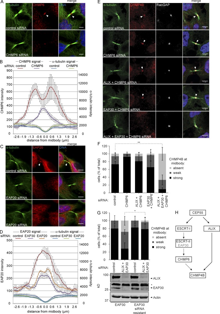
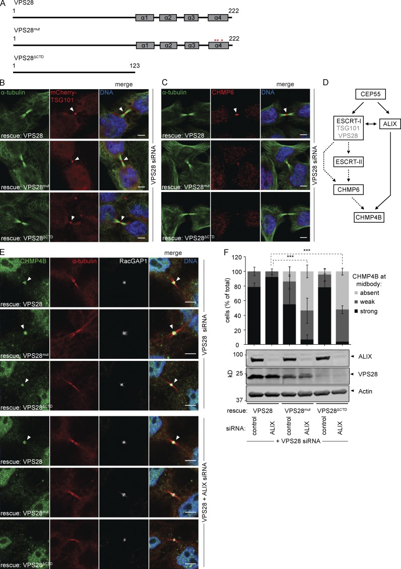
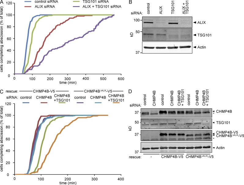
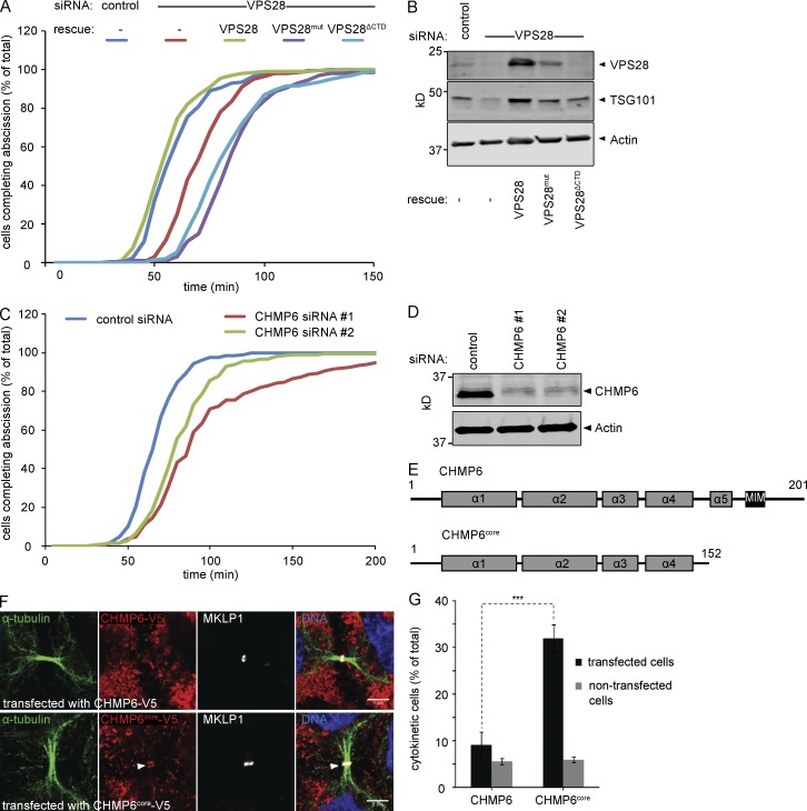
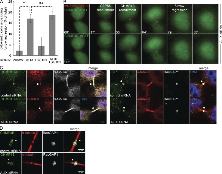
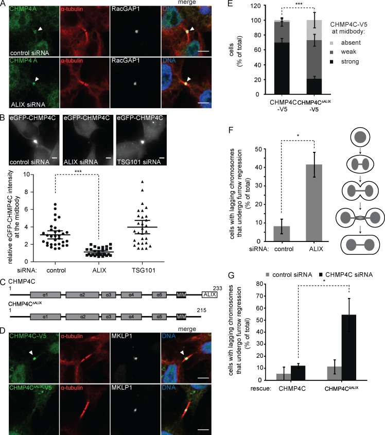
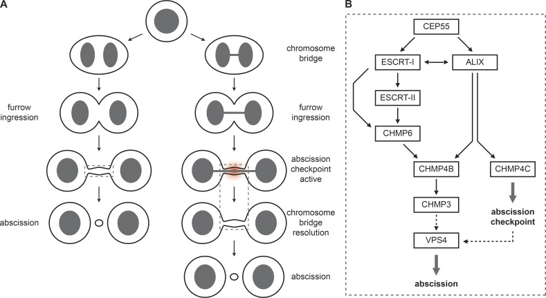
Cytokinetic abscission, the final stage of cell division where the two daughter cells are separated, is mediated by the endosomal sorting complex required for transport (ESCRT) machinery. The ESCRT-III subunit CHMP4B is a key effector in abscission, whereas its paralogue, CHMP4C, is a component in the abscission checkpoint that delays abscission until chromatin is cleared from the intercellular bridge. How recruitment of these components is mediated during cytokinesis remains poorly understood, although the ESCRT-binding protein ALIX has been implicated. Here, we show that ESCRT-II and the ESCRT-II-binding ESCRT-III subunit CHMP6 cooperate with ESCRT-I to recruit CHMP4B, with ALIX providing a parallel recruitment arm. In contrast to CHMP4B, we find that recruitment of CHMP4C relies predominantly on ALIX. Accordingly, ALIX depletion leads to furrow regression in cells with chromosome bridges, a phenotype associated with abscission checkpoint signaling failure. Collectively, our work reveals a two-pronged recruitment of ESCRT-III to the cytokinetic bridge and implicates ALIX in abscission checkpoint signaling.


Figures

Figure 1.
Figure 1.
ALIX and ESCRT-I constitute parallel CHMP4B recruitment arms. (A) Confocal images showing CHMP4B at the cytokinetic bridge of HeLa cells stained for DNA (Hoechst), α-tubulin, CHMP4B, and the midbody marker RacGAP1 upon siRNA treatment as indicated. Bars, 5 µm. (B) Quantification of CHMP4B localization at the midbody (error bars indicate SEM; n > 30 cells from three independent experiments; unpaired t test; ***, P < 0.001). (C) Schematic view of the CHMP4B constructs used in D and E. (D) Confocal images of HeLa cells stably expressing CHMP4B-V5 or CHMP4BΔALIX-V5 stained for DNA (Hoechst), α-tubulin, V5, and MKLP1 upon siRNA treatment as indicated. Bars, 5 µm. (E) Quantification of the CHMP4B or CHMP4BΔALIX signal at the midbody in cells stably expressing CHMP4B-V5 or CHMP4BΔALIX-V5 (error bars indicate SEM; n = 15 cells from three independent experiments; unpaired t test; **, P < 0.01). Knockdown of TSG101 and ALIX is shown by Western blot. (F) Live imaging of CHMP4B enrichment at the midbody relative to CEP55 in HeLa cells stably expressing mCherry-CEP55 and CHMP4B-eGFP (error bars indicate mean with 95% confidence interval; n ≥ 27 cells from five independent experiments; control siRNA 35 ± 23.2 min, ALIX siRNA 72 ± 51.4 min, TSG101 siRNA 71 ± 34.7 min (± SD); mixed factor model; **, P ≤ 0.01). (G) Schematic view of cytokinetic CHMP4B recruitment.
Figure 2.
Figure 2.
CHMP6 and ESCRT-II localize to the cytokinetic bridge and contribute to CHMP4B recruitment. (A and C) Confocal images of HeLa cells stained for DNA (Hoechst), α-tubulin, CHMP6, and EAP20 upon siRNA treatment as indicated. Bars, 5 µm. (B and D) Intensity of CHMP6, EAP20, and α-tubulin along the intercellular bridge (error bars indicate SEM from three independent experiments; n = 30 cells; mean CHMP6 intensity in the bridge upon CHMP6 siRNA relative to control siRNA [set to 1, ± SEM] 0.47 ± 0.07 [P = 0.02], mean EAP20 intensity upon EAP30 siRNA relative to control siRNA 0.59 ± 0.05 [P = 0.01], mean EAP20 intensity upon EAP20 siRNA relative to control siRNA 0.69 ± 0.03 [P = 0.01]; P-values obtained using one-sample t test). (E) Confocal images showing CHMP4B at the cytokinetic bridge of HeLa cells stained for DNA (Hoechst), α-tubulin, CHMP4B, and the midbody marker RacGAP1 upon siRNA treatment as indicated. Bars, 5 µm. (F) Quantification of cells with or without CHMP4B at the midbody (error bars indicate SEM; n > 30 cells from four independent experiments; unpaired t test; **, P < 0.01) upon siRNA treatment as indicated. (G) Quantification of CHMP4B recruitment to the midbody in HeLa cells stably expressing siRNA sensitive or resistant EAP30 (error bars indicate SEM; n ≥ 24 cells from three independent experiments; unpaired t test; *, P < 0.05). Knockdown of ALIX and EAP30 is shown by Western blot (*, nonspecific immunoreactivity). (H) Schematic view of cytokinetic CHMP4B recruitment.
Figure 3.
Figure 3.
VPS28 binds ESCRT-II and CHMP6 to recruit CHMP4B. (A) Schematic view of the VPS28 constructs used. (B) Confocal images of HeLa cells stably expressing siRNA-resistant VPS28 alleles and mCherry-TSG101 upon siRNA treatment as indicated, stained for DNA (Hoechst), α-tubulin, and mCherry. Bars, 5 µm. (C) Confocal images of CHMP6 recruitment in HeLa cells stably expressing VPS28 alleles. Bars, 5 µm. (D) Schematic view of cytokinetic CHMP4B recruitment pathways. (E) Confocal images of CHMP4B recruitment in HeLa cells stably expressing VPS28 alleles. Bars, 5 µm. (F) Quantification of CHMP4B at the midbody (error bars indicate SEM; n ≥ 23 cells from four independent experiments; unpaired t test; ***, P < 0.001). Knockdown of ALIX and expression of siRNA-resistant VPS28 is shown by Western blot.
Figure 4.
Figure 4.
ALIX and TSG101 cooperatively control abscission timing. (A) Live imaging of HeLa cells stably expressing Histone2B-mCherry and eGFP–α-tubulin upon siRNAs treatment as indicated. Cumulative frequency plot showing the time interval between furrow ingression and abscission (n > 100 cells from three independent experiments; control, 74 ± 17.3 min; ALIX siRNA, 177 ± 78.7 min; TSG101 siRNA, 98 ± 28.4 min; ALIX + TSG101 siRNA, 338.2 ± 228.9 min [± SD]; ALIX relative to control, P < 0.001; TSG101 relative to control, P = 0.04; ALIX+TSG101 relative to ALIX, P = 0.002; ALIX+TSG101 relative to all others, P < 0.001; P-values obtained using mixed factor model). (B) Knockdown efficiency of ALIX and TSG101. (C) Live imaging of HeLa cells stably expressing Histone2B-mCherry, eGFP–α-tubulin, and CHMP4B-V5 or CHMP4BΔALIX-V5 upon siRNA treatment as indicated. Cumulative frequency plot showing the time interval between furrow ingression and abscission (n > 100 cells from three independent experiments; CHMP4B: control, 82 ± 19.9 min; CHMP4B siRNA, 74 ± 14.5 min; CHMP4B+TSG101 siRNA, 110 ± 25.6 min; CHMP4BΔALIX control, 80 ± 24.0 min; CHMP4B siRNA, 84 ± 24.7 min; CHMP4B+TSG101 siRNA, 153 ± 50.4 min [± SD]; CHMP4B with control siRNA or CHMP4B siRNA relative to CHMP4B with CHMP4B+TSG101 siRNA, P < 0.05; CHMP4BΔALIX with CHMP4B+TSG101 siRNA relative to all others, P < 0.001; P-values obtained using mixed factor model). (D) Knockdown efficiency of CHMP4B and TSG101 as well as expression of CHMP4B-V5 or CHMP4BΔALIX-V5 (*, nonspecific immunoreactivity).
Figure 5.
Figure 5.
Depletion of CHMP6 and overexpression of dominant-negative CHMP6 results in abscission delay. (A) Cumulative frequency plot showing the time interval between furrow ingression and abscission in HeLa cells stably expressing Histone2B-mCherry, eGFP–α-tubulin, and VPS28 alleles upon siRNA treatment as indicated (n ≥ 100 cells per treatment from four independent experiments; control, 61 ± 15.2 min; VPS28 siRNA, 73 ± 15.0 min; VPS28 with VPS28 siRNA, 58 ± 14.0 min; VPS28mut with VPS28 siRNA, 88 ± 18.1 min; VPS28ΔCTD with VPS28 siRNA, 86 ± 23.3 min [± SD]; control and VPS28 rescue relative to VPS28 siRNA, P < 0.01; control and VPS28 rescue relative to VPS28mut and VPS28ΔCTD, P < 0.01; VPS28 siRNA relative to VPS28mut and VPS28ΔCTD, P < 0.01; P-values obtained using mixed factor model). (B) Knockdown efficiency of VPS28 and codepletion of TSG101. (C) Cumulative frequency plot showing the time interval between furrow ingression and abscission in HeLa cells stably expressing Histone2B-mCherry and eGFP–α-tubulin upon siRNA treatment as indicated (n ≥ 200 cells per treatment from seven independent experiments; control, 69 ± 14.9 min; CHMP6 siRNA#1, 104 ± 53.7 min; CHMP6 siRNA#2, 85 ± 28.2 min [± SD]; CHMP6 1 relative to control, P < 0.001; CHMP6 2 relative to control, P < 0.001; P-values obtained using mixed factor model). (D) Knockdown efficiency of CHMP6. (E) Schematic view of the CHMP6 constructs used in F and G. (F) Confocal images of HeLa cells transfected with wild-type or truncated CHMP6 and stained for DNA (Hoechst), α-tubulin, and MKLP1. Bars, 5 µm. CHMP6core localizes to the midbody. (G) HeLa cells were transfected with wild-type or truncated CHMP6 and analyzed by high-throughput widefield microscopy. The mean of the total cell population is shown (error bars indicate SEM from six independent experiments; n > 1,000 cells per experiment; unpaired t test; ***, P < 0.001).
Figure 6.
Figure 6.
ALIX, but not TSG101, controls furrow regression. (A) Live imaging of HeLa cells stably expressing Histone2B-mCherry and eGFP–α-tubulin upon siRNA treatment as indicated. Percentage of cells undergoing furrow regression (error bars indicate SEM; n > 90 cells from three independent experiments; unpaired t test; **, P < 0.01; n.s., not significant). (B) Live imaging of HeLa cells stably expressing mCherry-CEP55 and CHMP4B-eGFP upon siRNA treatment as indicated. Selected frames of ALIX-depleted cells undergoing cytokinesis, showing enrichment of CHMP4B at the midbody at time of furrow regression (top, merged channels; bottom, CHMP4B-eGFP channel alone. Bar, 5 µm. (C) Confocal images of HeLa cells transfected with indicated siRNAs and stained for DNA (Hoechst), α-tubulin, RacGAP1, GFP, CHMP3, and IST1, as indicated. Bars, 5 µm. (left) HeLa cells stably expressing CHMP4B-eGFP. (D) Localization of CHMP4B to the secondary ingression of HeLa cells is unaffected by ALIX knockdown. Bars, 3 µm.
Figure 7.
Figure 7.
CHMP4C recruitment to the midbody relies on ALIX. (A) CHMP4A recruitment is maintained in the absence of ALIX. Confocal images of HeLa cells upon siRNA treatment as indicated, stained for DNA (Hoechst), CHMP4A, α-tubulin, and RacGAP1. Bars, 5 µm. (B) CHMP4C recruitment to the midbody is perturbed in the absence of ALIX. Live imaging of HeLa cells stably expressing inducible eGFP-CHMP4C upon siRNA treatment as indicated. Images and quantification of the relative intensity of eGFP-CHMP4C at the midbody are shown (bars indicate mean with 95% confidence interval; n ≥ 30 cells from four independent experiments; mixed factor model; ***, P < 0.001). Bars, 5 µm. (C) Schematic view of the CHMP4C constructs used in D–F. (D) Confocal images of HeLa cells stably expressing CHMP4C-V5 or CHMP4CΔALIX-V5, stained for DNA (Hoechst), V5, α-tubulin, and MKLP1. Bars, 5 µm. (E) Quantification of full-length or truncated CHMP4C-V5 at the midbody (error bars indicate SEM; n > 50 cells from six independent experiments; unpaired t test; ***, P < 0.001). (F) Depletion of ALIX increases frequency of binucleation in the presence of lagging chromosomes. Live imaging of HeLa cells stably expressing Histone2B-mCherry and eGFP-α-tubulin upon siRNA treatment as indicated. Quantification of furrow regression in cells with lagging chromosomes as depicted in the cartoon (error bars indicate SEM; n = 24 cells from three independent experiments; unpaired t test; *, P < 0.05). (G) Live imaging of HeLa cells stably expressing Histone2B-mCherry, eGFP-α-tubulin, and siRNA-resistant CHMP4C or CHMP4CΔALIX upon CHMP4C siRNA treatment. Quantification of furrow regression in cells with lagging chromosomes (error bars indicate SEM; n ≥ 15 cells from three independent experiments; unpaired t test; *, P < 0.05).
Figure 8.
Figure 8.
ESCRT-III recruitment model. (A) Cartoon illustrating cytokinesis in the absence or presence of chromosome segregation defects. (B) Model for ESCRT-III recruitment during cytokinetic abscission and checkpoint signaling. CHMP4B, the main component of the ESCRT-III filaments, is recruited via two parallel arms, namely CEP55–ALIX or CEP55–ESCRT-I–ESCRT-II–CHMP6. This recruitment is essential for timing of cytokinetic abscission. In contrast, recruitment of the abscission checkpoint regulator CHMP4C relies on ALIX, indicating that ALIX represent a dual functionality as abscission regulator and checkpoint signaling node.

Comment in

Similar articles

Cited by

References

    1. Adell M.A., and Teis D.. 2011. Assembly and disassembly of the ESCRT-III membrane scission complex. FEBS Lett. 585:3191–3196. 10.1016/j.febslet.2011.09.001 - DOI - PMC - PubMed
    1. Agromayor M., and Martin-Serrano J.. 2013. Knowing when to cut and run: mechanisms that control cytokinetic abscission. Trends Cell Biol. 23:433–441. 10.1016/j.tcb.2013.04.006 - DOI - PubMed
    1. Babst M., Katzmann D.J., Estepa-Sabal E.J., Meerloo T., and Emr S.D.. 2002a Escrt-III: an endosome-associated heterooligomeric protein complex required for mvb sorting. Dev. Cell. 3:271–282. 10.1016/S1534-5807(02)00220-4 - DOI - PubMed
    1. Babst M., Katzmann D.J., Snyder W.B., Wendland B., and Emr S.D.. 2002b Endosome-associated complex, ESCRT-II, recruits transport machinery for protein sorting at the multivesicular body. Dev. Cell. 3:283–289. 10.1016/S1534-5807(02)00219-8 - DOI - PubMed
    1. Bache K.G., Stuffers S., Malerød L., Slagsvold T., Raiborg C., Lechardeur D., Wälchli S., Lukacs G.L., Brech A., and Stenmark H.. 2006. The ESCRT-III subunit hVps24 is required for degradation but not silencing of the epidermal growth factor receptor. Mol. Biol. Cell. 17:2513–2523. 10.1091/mbc.E05-10-0915 - DOI - PMC - PubMed

Publication types

MeSH terms

Substances