. 2016 Apr 22;60(5):2834-48.
doi: 10.1128/AAC.02700-15. Print 2016 May.

Cross Talk between Nucleotide Synthesis Pathways with Cellular Immunity in Constraining Hepatitis E Virus Replication

Affiliations

Cross Talk between Nucleotide Synthesis Pathways with Cellular Immunity in Constraining Hepatitis E Virus Replication

Yijin Wang et al. Antimicrob Agents Chemother. .

Abstract

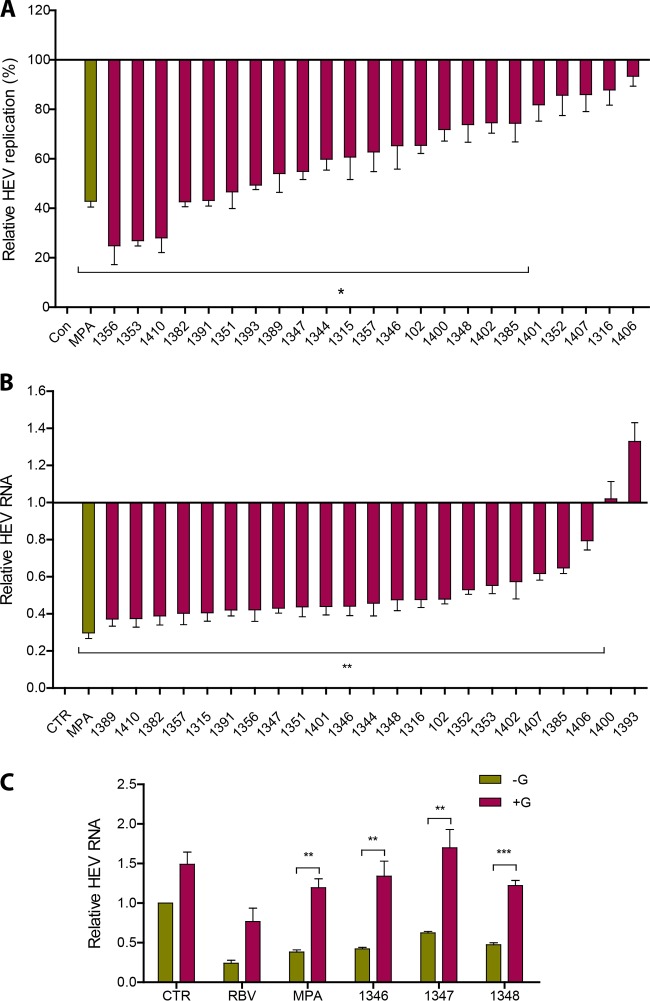
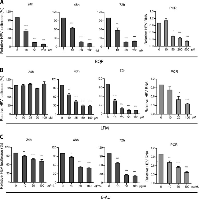
Viruses are solely dependent on host cells to propagate; therefore, understanding virus-host interaction is important for antiviral drug development. Since de novo nucleotide biosynthesis is essentially required for both host cell metabolism and viral replication, specific catalytic enzymes of these pathways have been explored as potential antiviral targets. In this study, we investigated the role of different enzymatic cascades of nucleotide biosynthesis in hepatitis E virus (HEV) replication. By profiling various pharmacological inhibitors of nucleotide biosynthesis, we found that targeting the early steps of the purine biosynthesis pathway led to the enhancement of HEV replication, whereas targeting the later step resulted in potent antiviral activity via the depletion of purine nucleotide. Furthermore, the inhibition of the pyrimidine pathway resulted in potent anti-HEV activity. Interestingly, all of these inhibitors with anti-HEV activity concurrently triggered the induction of antiviral interferon-stimulated genes (ISGs). Although ISGs are commonly induced by interferons via the JAK-STAT pathway, their induction by nucleotide synthesis inhibitors is completely independent of this classical mechanism. In conclusion, this study revealed an unconventional novel mechanism of cross talk between nucleotide biosynthesis pathways and cellular antiviral immunity in constraining HEV infection. Targeting particular enzymes in nucleotide biosynthesis represents a viable option for antiviral drug development against HEV. HEV is the most common cause of acute viral hepatitis worldwide and is also associated with chronic hepatitis, especially in immunocompromised patients. Although often an acute and self-limiting infection in the general population, HEV can cause severe morbidity and mortality in certain patients, a problem compounded by the lack of FDA-approved anti-HEV medication available. In this study, we have investigated the role of the nucleotide synthesis pathway in HEV infection and its potential for antiviral drug development. We show that targeting the later but not the early steps of the purine synthesis pathway exerts strong anti-HEV activity. In particular, IMP dehydrogenase (IMPDH) is the most important anti-HEV target of this cascade. Importantly, the clinically used IMPDH inhibitors, including mycophenolic acid and ribavirin, have potent anti-HEV activity. Furthermore, targeting the pyrimidine synthesis pathway also exerts potent antiviral activity against HEV. Interestingly, antiviral effects of nucleotide synthesis pathway inhibitors appear to depend on the medication-induced transcription of antiviral interferon-stimulated genes. Thus, this study reveals an unconventional novel mechanism as to how nucleotide synthesis pathway inhibitors can counteract HEV replication.


Figures

FIG 1
FIG 1
Exogenous guanosine stimulated HEV replication. (A) Schematic overview of de novo biosynthesis of purine nucleotide. PRPP, 5-phosphoribosyl-1-pyrophosphate; PRA, 5-phosphoribosylamine; GAR, glycinamide ribonucleotide; FGAR, formyl-GAR; AICAR, 5-aminoimidazole-4-carboxamide ribonucleotide. (B) Huh7 cell-based subgenomic HEV replicons containing the luciferase reporter gene were treated for 24 h, 48 h, and 72 h with a dose range of guanosine (n = 4). Data are presented as means ± standard errors of the means (SEM). Meanwhile, Huh7 cells with the infectious HEV containing the full-length p6 genome were treated for 48 h with a dose range of guanosine (n = 5). Data were normalized to two housekeeping genes (GAPDH and RP2) and are presented relative to the control (CTR) (set as 1). Data represent means ± SEM. *, P < 0.05; **, P < 0.01; ***, P < 0.001.
FIG 2
FIG 2
Exogenous uridine does not affect HEV replication. (A) Schematic overview of de novo biosynthesis of pyrimidine nucleotide. (B) Huh7 cell-based subgenomic HEV replicon containing the luciferase reporter gene was treated for 24 h, 48 h, and 72 h with a dose range of uridine (n = 5). Data are presented as means ± SEM.
FIG 3
FIG 3
Inhibitors of IMP synthesis cascade stimulate HEV replication. The Huh7 cells containing subgenomic HEV replicons with luciferase reporter genes were incubated with increasing doses of 6-TG (A), lometrexol (B), and MTX (C). The luciferase activity was determined at 24 h, 48 h, and 72 h. Accordingly, Huh7 cells infected with full-length HEV were treated with increasing doses of 6-TG (A), lometrexol (B), and MTX (C). The HEV RNA level was quantified by qRT-PCR after 48 h. Data were normalized to two housekeeping genes and are presented relative to the control (CTR) (set as 1). Data represent means ± SEM from five to eight experiments. *, P < 0.05; **, P < 0.01; ***, P < 0.001.
FIG 4
FIG 4
Silencing of enzymes involved in IMP synthesis cascade facilitates HEV replication. (A) Huh7 cells were transduced with lentiviral shRNAs to stably silence the corresponding genes for PPAT, GART, and ATIC (a set of independent shRNA clones targeting each gene was used). Huh7 cells transduced with lentiviral shRNA targeting GFP (shCTR) were used as a control. The efficiency of gene knockdown was analyzed by qRT-PCR. (B) Silencing of PPAT, GART, and ATIC resulted in significant elevation of viral RNA upon inoculation of HEV. HEV RNA levels were determined 72 h after inoculation. (C) Silencing of PPAT, GART, and ATIC abrogated the pro-HEV effects of 6-TG, lometrexol, and MTX. Data were normalized to that for cells without treatment with the three compounds (green bar; set as 1). All data were normalized to two housekeeping genes and are presented relative to the control (CTR) (set as 1) (means ± SEM from four to eight experiments). *, P < 0.05; **, P < 0.01; ***, P < 0.001.
FIG 5
FIG 5
IMPDH inhibitors potently inhibit HEV replication by depletion of the purine nucleotide pool. (A) Huh7 HEV replicon luciferase cells were treated with 23 specific IMPDH inhibitors (10 μM) with MPA as a positive control. Luciferase activity was quantified at 24 h after treatment (n = 3). (B) Huh7 cells harboring full-length HEV were treated with 23 specific IMPDH inhibitors with MPA as a positive control. HEV RNA levels were measured by qRT-PCR at 48 h after treatment (n = 5). (C) Supplementation of guanosine abrogated the anti-HEV effects of 3 representative IMPDH inhibitors (1346, 1347, and 1348) (n = 5). Ribavirin (RBV) and MPA served as positive controls. Data were normalized to two housekeeping genes and are presented relative to the control (CTR) (set as 1) (means ± SEM). *, P < 0.05; **, P < 0.01; ***, P < 0.001.
FIG 6
FIG 6
Inhibition of pyrimidine nucleotide synthesis suppresses HEV replication. Huh7 cells containing subgenomic HEV replicons with luciferase report genes were treated with increasing doses of BQR (A), LFM (B), and 6-AU (C). The luciferase activity was determined after 24 h, 48 h, and 72 h. Accordingly, Huh7 cells harboring infectious HEV also were treated with increasing doses of BQR (A), LFM (B), and 6-AU (C). HEV RNA was quantified by qRT-PCR after 48 h of treatment. Data were normalized to two housekeeping genes and are presented relative to the control (CTR) (set as 1). Data represent means ± SEM from four to seven experiments. *, P < 0.05; **, P < 0.01; ***, P < 0.001.
FIG 7
FIG 7
Anti-HEV activity by BQR can be attributed to the inhibition of its target, DHODH. (A) Huh7 cells were transduced with lentiviral shRNA to stably silent DHODH (DHODH shRNA positive). Huh7 cells transduced with lentiviral shRNA targeting GFP were used as a control (DHODH shRNA negative). DHODH knockdown was assessed by qRT-PCR (n = 3). DHODH knockdown resulted in the significant inhibition of HEV replication. HEV RNA levels were determined 72 h after HEV inoculation (n = 6). (B) DHODH knockdown abrogated the anti-HEV effect of BQR (n = 7). Data were normalized to cells without BQR treatment (green bar; set as 1). All data were normalized to two housekeeping genes and are presented relative to the control (shCTR) (set as 1) (means ± SEM). **, P < 0.01; ***, P < 0.001.
FIG 8
FIG 8
Uridine reverses the anti-HEV activity mediated by pyrimidine inhibition. The Huh7 subgenomic HEV replicon was incubated with BQR (A), LFM (B), and 6-AU (C) and supplemented with increasing doses of uridine. After 72 h, luciferase activity was determined. Accordingly, Huh7 cells harboring full-length HEV RNA were treated with BQR (A), LFM (B), and 6-AU (C) and supplemented with 200 μM uridine. HEV viral RNA was assessed by qRT-PCR 48 h after treatment. Data were normalized to two housekeeping genes and are presented relative to the control (CTR) (set as 1). Data represent means ± SEM from four to seven experiments. *, P < 0.05; **, P < 0.01; ***, P < 0.001.
FIG 9
FIG 9
Inhibition of IMPDH stimulates ISG expression through purine nucleotide deprivation. (A) Huh7 cells infected with HEV were treated with MPA or 3 other IMPDH inhibitors (1346, 1347, and 1348). The expression of a panel of ISGs was determined by qRT-PCR after 48 h of treatment. Data were normalized to basal ISG expression without treatment (gray bar; set as 1). (B) Supplementation of guanosine abrogated the induction of ISGs by IMPDH inhibitors. The expression of ISGs was determined by qRT-PCR 48 h after treatment. Data were normalized to basal ISG expression without treatment (purple bar; set as 1). All data were normalized to two housekeeping genes and represent means ± SEM from four experiments.
FIG 10
FIG 10
Inhibition of pyrimidine synthesis stimulates ISG expression through pyrimidine nucleotide depletion. (A) Huh7 cells infected with HEV were treated with BQR or 6-AU. After 48 h, the expression of a panel of ISGs was determined by qRT-PCR. Data were normalized to basal ISG expression without treatment (gray bar; set as 1). (B) Supplementation of uridine completely abrogated the induction of ISGs by BQR (B) or 6-AU (C).The expression of ISGs was determined by qRT-PCR at 48 h after treatment. Data were normalized to basal ISG expression without treatment (purple bar; set as 1). All data were normalized to two housekeeping genes and represent means ± SEM from five experiments. *, P < 0.05; **, P < 0.01.
FIG 11
FIG 11
ISG induction and the anti-HEV activity triggered by nucleotide synthesis inhibitors are independent of the JAK-STAT signaling pathway. The induction of ISGs (A and C) and the anti-HEV effects (B and D) by MPA were quantified in the presence or absence of JAK inhibitor 1 (A and B) or CP-690550 (CP) (C and D). The induction of ISGs was normalized to basal ISG expression without MPA treatment (purple bar; set as 1). The relative HEV RNA levels were normalized to cells without treatment of MPA (set as 1). Similarly, the induction of ISGs (E and G) and the anti-HEV effects (F and H) mediated by BQR were quantified in the presence or absence of JAK inhibitor 1 (E and F) or CP (G and H). The induction of ISGs was normalized to basal ISG expression without BQR treatment (purple bar; set as 1). The relative HEV RNA levels were normalized to cells without BQR treatment (set as 1). Data were normalized to two housekeeping genes and represent means ± SEM from three or four experiments.

Similar articles

Cited by

References

    1. Kamar N, Bendall R, Legrand-Abravanel F, Xia NS, Ijaz S, Izopet J, Dalton HR. 2012. Hepatitis E. Lancet 379:2477–2488. doi:10.1016/S0140-6736(11)61849-7. - DOI - PubMed
    1. Kamar N, Selves J, Mansuy JM, Ouezzani L, Peron JM, Guitard J, Cointault O, Esposito L, Abravanel F, Danjoux M, Durand D, Vinel JP, Izopet J, Rostaing L. 2008. Hepatitis E virus and chronic hepatitis in organ-transplant recipients. N Engl J Med 358:811–817. doi:10.1056/NEJMoa0706992. - DOI - PubMed
    1. Zhou X, de Man RA, de Knegt RJ, Metselaar HJ, Peppelenbosch MP, Pan Q. 2013. Epidemiology and management of chronic hepatitis E infection in solid organ transplantation: a comprehensive literature review. Rev Med Virol 23:295–304. doi:10.1002/rmv.1751. - DOI - PubMed
    1. Kamar N, Garrouste C, Haagsma EB, Garrigue V, Pischke S, Chauvet C, Dumortier J, Cannesson A, Cassuto-Viguier E, Thervet E, Conti F, Lebray P, Dalton HR, Santella R, Kanaan N, Essig M, Mousson C, Radenne S, Roque-Afonso AM, Izopet J, Rostaing L. 2011. Factors associated with chronic hepatitis in patients with hepatitis E virus infection who have received solid organ transplants. Gastroenterology 140:1481–1489. doi:10.1053/j.gastro.2011.02.050. - DOI - PubMed
    1. van der Eijk AA, Pas SD, Cornelissen JJ, de Man RA. 2014. Hepatitis E virus infection in hematopoietic stem cell transplant recipients. Curr Opin Infect Dis 27:309–315. doi:10.1097/QCO.0000000000000076. - DOI - PubMed

Publication types

MeSH terms