Review
. 2016 Jun 8;116(11):6424-62.
doi: 10.1021/acs.chemrev.5b00548. Epub 2016 Feb 29.

Dynamic Protein Interaction Networks and New Structural Paradigms in Signaling

Affiliations
Review

Dynamic Protein Interaction Networks and New Structural Paradigms in Signaling

Veronika Csizmok et al. Chem Rev. .

Abstract

Understanding signaling and other complex biological processes requires elucidating the critical roles of intrinsically disordered proteins (IDPs) and regions (IDRs), which represent ∼30% of the proteome and enable unique regulatory mechanisms. In this review, we describe the structural heterogeneity of disordered proteins that underpins these mechanisms and the latest progress in obtaining structural descriptions of conformational ensembles of disordered proteins that are needed for linking structure and dynamics to function. We describe the diverse interactions of IDPs that can have unusual characteristics such as "ultrasensitivity" and "regulated folding and unfolding". We also summarize the mounting data showing that large-scale assembly and protein phase separation occurs within a variety of signaling complexes and cellular structures. In addition, we discuss efforts to therapeutically target disordered proteins with small molecules. Overall, we interpret the remodeling of disordered state ensembles due to binding and post-translational modifications within an expanded framework for allostery that provides significant insights into how disordered proteins transmit biological information.


Figures

Figure 1
Figure 1. The role of disordered proteins/regions in signalling pathways
a. Signal transduction from receptors to the nucleus: Disorder regions (red line) of the cytoplasmic tails of receptors (green, magenta, blue circle) often serve as regulatory sites, participating in numerous intra- and intermolecular interactions. From the receptor, the signal is often propagated by consecutive phosphorylation and activation of a kinase cascade (green, yellow rectangle and blue ellipsoid), with PTMs usually occurring in disorder regions and the signal often lands on disordered transcription factor (see also Fig 1f). b. Multivalent interactions in signal transduction pathways: Disordered regions contain multiple recognition motifs (magenta rectangle) for binding to multiple modular binding domains of partner molecule (blue ellipsoids), potentially leading to sharp 'liquid-liquid' phase separation and enhancing activity. c. Scaffold proteins in signalling pathway: Signal transduction is often mediated by disordered scaffold protein, which also contains folded domains (green circle, blue ellipsoid). The disordered scaffold protein has a large capacity for binding, enabling the integration of different signals by orchestrating the interactions between different components, such as a transcription factor (orange rectangle) and modification enzymes (yellow square and magenta hexagon). d. Translation initiation pathway: Assembly of the translation initiation complex can be blocked by a partly or fully disordered protein. This process is regulated by phosphorylations (red circles), on the inhibitor inducing a binding-incompatible folded domain, leads to dissociation of the disordered inhibitor from one of the initiation factors (green rectangle), assembly of the complex (green rectangle with blue, yellow ellipsoids and orange rectangle) and translation of the mRNA (grey line). Dissociation of the initiation complex from mRNA and the small subunit of the ribosome yields the final, active ribosome (light blue). e. Nuclear transport: Transport from the cytoplasm to the nucleus proceeds through the nuclear pore complex (blue), which is comprised of many different nucleoporins. Nucleoporins have significant disordered regions, which can form a phase-separated elastic hydrogel that acts as the permeability barrier. f. Transcriptional regulation: Besides a folded DNA-binding domain (green-blue helix), most transcription factors are comprised of long disordered regions that are involved in regulation and binding. g. Cell cycle inhibition: Cell cycle regulation is often mediated by disordered proteins. The process is regulated by PTMs on cell cycle inhibitors, including phosphorylation and subsequent ubiquitination, which leads to degradation of the disordered inhibitor by the proteosome (magenta-green cylinder) and the activation of the CDK-cyclin complex (yellow-blue ellipsoids).
Figure 2
Figure 2. Regulatory (R) region of CFTR acts as a dynamic integrator
The regulatory (R) region (red line) of the cystic fibrosis conductance transmembrane regulator (CFTR) forms highly dynamic complexes with different intra- and inter-molecular partners targeting the same or largely overlapping segments and these interactions are largely dependent on the phosphorylation state of R region. Non-phosphorylated R region binds to the nucleotide-binding domains (NBDs) of CFTR (magenta rectangles) and inhibits their dimerization and consequently channel opening (grey box). The segments bound to NBDs show higher α-helical propensity than the rest of R region, and interestingly, phosphorylation on several different sites in R region destabilizes helical structure in an order-to-disorder transition in these segments. Phosphorylated R region shows much lower affinity to NBDs, which can bind to other partners such as the STAS domain of SLC26A3 and enables the dimerization of NBDs and channel opening (pink box). The interaction of R region with STAS is critical to ensure the close physical proximity and reciprocal activation of CFTR and the chloride/bicarbonate exchanger, SLC26A3. Phosphorylated R region also binds to 14-3-3, and this interaction is crucial for the normal CFTR trafficking from the endoplasmatic reticulum (blue box). Binding of different partners that likely exchanging on and off of the same binding segments of R region facilitates the integration of stimuli from different pathways and supports the role of the R region as a dynamic integrator. Adapted from ref .
Figure 3
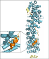
Figure 3. Molecular mimicry
Unrelated disordered proteins/regions can adopt the same conformations when bound to the same partner, which gives rise to the phenomenon called molecular mimicry. The disordered regions of E-cadherin (PDB code: 1I7W, shown in yellow) and TCF3 (PDB code:1G3J, shown in orange) make identical hydrophobic interactions with β-catenin (shown in blue), despite the difference in local secondary structure (shown in the enlargement). This suggests that binding to β-catenin in a highly disordered, extended structural state is biologically advantageous and highly conserved.
Figure 4
Figure 4. Order-to-disorder transitions and functional implications of structural flexibility in the cell cycle inhibitor p27
a. Structure of p27 kinase inhibitory domain (p27-KID) bound to the cyclin-dependent kinase 2 (Cdk2)/cyclin A complex. Key sub-domains within p27-KID are indicated: D1, LH and D2. b. Schematic illustrating sequential folding of p27 upon binding to Cdk2/cyclin A. The binding of sub-domain D1 to cyclin A is fast, followed by slow binding and folding of sub-domain D2 to Cdk2. The number of residues shown to fold (R) by isothermal titration calorimetry during each of the different binding reactions is indicated. c. Schematic illustration of the phosphorylation/ubiquitination cascade that regulates p27 activity and stability. Phosphorylation on tyrosine 88 (Y88) by non-receptor tyrosine kinases (NRTKs) locally displaces a localized region of p27 from the ATP binding pocket of Cdk2, partially re-activating its catalytic activity (Step 1). Partially active Cdk2 phosphorylates threonine 187 (T187) within the flexible p27 C-terminus (p27-C; Step 2), creating a phospho-degron binding site for the E3 ubiquitin ligase, SCFSkp2, that promotes p27 poly-ubiquitination on several lysine residues (indicated by diamonds; Step 3). Once poly-ubiquitinated, p27 is selectively degraded by the 26S proteasome (Step 4), freeing fully active Cdk2/cyclin complexes. Adapted from refs , , .
Figure 5
Figure 5. Schematic illustration of the allosteric mechanism of PUMA binding-dependent displacement of p53 from BCL-xL
Through a π-stacking interaction between Trp71 at its N-terminus and His113 at the C-terminus of α-helix 3 in BCL-xL, PUMA unfolds α3 in BCL-xL and disrupts its interface with p53. Upon release from the inhibitory interaction with BCL-xL, cytosolic p53 can exert a pro-apoptotic function. Adapted from ref .
Figure 6
Figure 6. Order-to-disorder transitions in activation of the apoptotic effector BAK by BH3-only proteins
a. Structure of BAK in its free, inactive state (left) and in complex with a helix-stabilized BID BH3 peptide (BID-SAHB). The binding of BID-SAHB displaced residues within the BH1 and BH3 domains of BAK. b. Schematic illustration of the sequence of interactions and conformational rearrangements that lead to BAK activation and oligomerization, MOMP and apoptosis. Adapted from ref .
Figure 7
Figure 7. Order-to-disorder transitions and conformational switches in activation of the apoptotic effector BAX by BH3-only proteins or cytosolic p53
a. Structure of BAX. The two surface representations on the right highlight the ‘trigger’ interaction sites with BH3 activators or p53. b. Schematic illustration of the sequence of interactions and conformational rearrangements that lead to BAX activation by BH3-only activators (top) or cytosolic p53 (bottom) resulting in apoptosis. Adapted from ref .
Figure 8
Figure 8. Molten globule state regulates NFĸB signalling
The transcriptional activity of NFĸBs, which form either homo- or heterodimers (green-blue), is tightly regulated by IĸBs (magenta). In the cytoplasm they form a tight complex in which the disordered nuclear localization sequence of NFĸB (dark green) binds the first two ankyrin repeats (AR) of IĸB, and the disordered C-terminal PEST region of IĸB (purple) interacts with the Rel homology domain of NFĸB. Distinct extracellular stimuli lead to the phosphorylation and subsequent ubiquitination and degradation of IĸB by the proteosome. Once the NFĸB is released from IĸB, it translocates into the nucleus and regulates the transcription of different gene targets. Free IĸB, which has characteristics of a molten-globule state, can also enter into the nucleus and promote dissociation of NFĸB from the DNA (grey box). The disordered PEST domain and partially folded C-terminal AR (ARs 5 and 6, shown in light violet) of IĸB have important role in this “stripping” process. The fifth and sixth ARs undergo coupled folding and binding in the ternary complex, and the disordered PEST region competes for the binding to the DNA-binding site of the NFĸB, ultimately leading to dissociation from DNA.
Figure 9
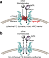
Figure 9. Schematic illustration of the function of disordered FG Nups in determining the selectivity of transport through the nuclear pore complex (NPC)
a. Wild-type cohesive FG domains that form hydrogels in vitro establish a selective barrier that is permeable to nuclear transport receptor (NTR)-bound cargo but impermeable to other, non-NTR-bound biomolecules. b. Mutated Nups lacking FG repeats that are non-cohesive do not form a selective barrier and are permeable to both NTR-bound and –unbound biomolecules. Adapted from ref .
Figure 10
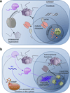
Figure 10. Schematic illustration of the cellular functions that are altered upon expression of the Arf tumor suppressor
a. In a normal cell, Mdm2 mediates rapid ubiquitination and degradation of p53, and nucleophosmin (Npm) mediates ribosome biogenesis in the nucleolus and shuttling of pre-ribosomal particles from the nucleus to cytoplasm. b. Under oncogenic stress, Arf is activated and mediates tumor suppression through several mechanisms. In one, Arf binds and sequesters Mdm2 in nucleoli through formation of fibril-like aggregates. This activates p53. In another, Arf binds Npm in the nucleolus, inhibiting its role in ribosome biogenesis. This interaction occurs within the liquid-like environment of the nucleolus.
Figure 11
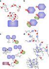
Figure 11. Allostery in disordered protein interactions
a. Allosteric communication often occurs in proteins with multiple phosphorylation sites, which may result in ultrasensitive binding. In this case, every site, even it is not bound (red circles) has an effect on binding of the other sites (pink circle), through long-range electrostatic or structural effects, and each phosphosite increases the probability of the binding of all the others by lowering the energy of bound states. b. Post-translational modifications such as phosphorylation or interactions with a partner (orange asterisk) may shift the conformational ensemble of a region (red) to a more ordered or to a more disordered state. The redistribution of the conformational ensemble for that region may facilitate the binding of another partner (green oval) through allostery. c. Allosteric communication can occur between more distant regions, bound to two or more different partners. Binding to a partner (blue octagons) can be negatively affected by post-translational modifications on another sites (red circles), which promote the binding to a different partner (green circle). In more complicated complexes, such as of hub proteins, binding of additional partner(s) (magenta rectangle) can be facilitated (as in figure) or inhibited by the interaction with the second partner (green circle). d. Allosteric coupling can also manifest in higher-order associated protein states. Post-translational modifications such as methylation (red diamonds) or binding to different partners (not shown) can redistribute the conformational ensemble or energy landscape to inhibit formation of higher-order assemblies.

Similar articles

Cited by

References

    1. Frauenfelder H, Sligar SG, Wolynes PG. The Energy Landscapes and Motions of Proteins. Science. 1991;254:1598–1603. - PubMed
    1. Biehl R, Richter D. Slow Internal Protein Dynamics in Solution. J. Phys. Condens. Matter. 2014;26:503103. - PubMed
    1. Dunker AK, Oldfield CJ, Meng J, Romero P, Yang JY, Chen JW, Vacic V, Obradovic Z, Uversky VN. The Unfoldomics Decade: An Update on Intrinsically Disordered Proteins. BMC Genomics. 2008;9(Suppl 2):S1. - PMC - PubMed
    1. Tompa P. Intrinsically Disordered Proteins: A 10-Year Recap. Trends Biochem. Sci. 2012;37:509–516. - PubMed
    1. Cohen P. The Regulation of Protein Function by Multisite Phosphorylation--a 25 Year Update. Trends Biochem. Sci. 2000;25:596–601. - PubMed

Publication types

MeSH terms

Substances

LinkOut - more resources