

RNA disruption is associated with response to multiple classes of chemotherapy drugs in tumor cell lines
- PMID: 26911141
- PMCID: PMC4765116
- DOI: 10.1186/s12885-016-2197-1
RNA disruption is associated with response to multiple classes of chemotherapy drugs in tumor cell lines
Abstract
Background: Cellular stressors and apoptosis-inducing agents have been shown to induce ribosomal RNA (rRNA) degradation in eukaryotic cells. Recently, RNA degradation in vivo was observed in patients with locally advanced breast cancer, where mid-treatment tumor RNA degradation was associated with complete tumor destruction and enhanced patient survival. However, it is not clear how widespread chemotherapy induced "RNA disruption" is, the extent to which it is associated with drug response or what the underlying mechanisms are.
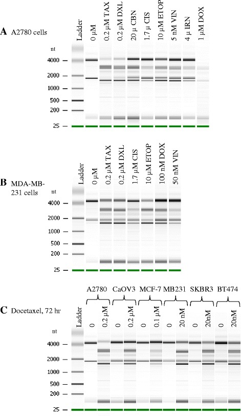
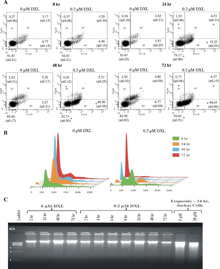
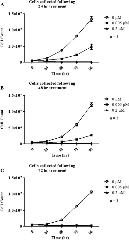
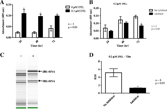
Methods: Ovarian (A2780, CaOV3) and breast (MDA-MB-231, MCF-7, BT474, SKBR3) cancer cell lines were treated with several cytotoxic chemotherapy drugs and total RNA was isolated. RNA was also prepared from docetaxel resistant A2780DXL and carboplatin resistant A2780CBN cells following drug exposure. Disruption of RNA was analyzed by capillary electrophoresis. Northern blotting was performed using probes complementary to the 28S and 18S rRNA to determine the origins of degradation bands. Apoptosis activation was assessed by flow cytometric monitoring of annexin-V and propidium iodide (PI) binding to cells and by measuring caspase-3 activation. The link between apoptosis and RNA degradation (disruption) was investigated using a caspase-3 inhibitor.
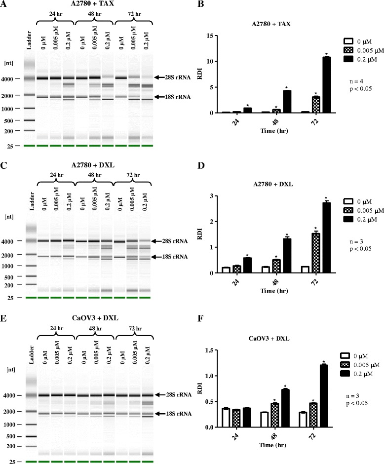
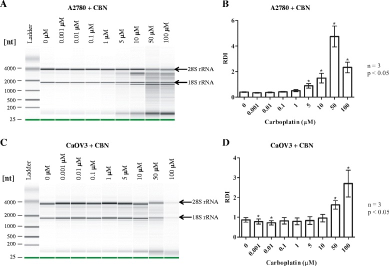
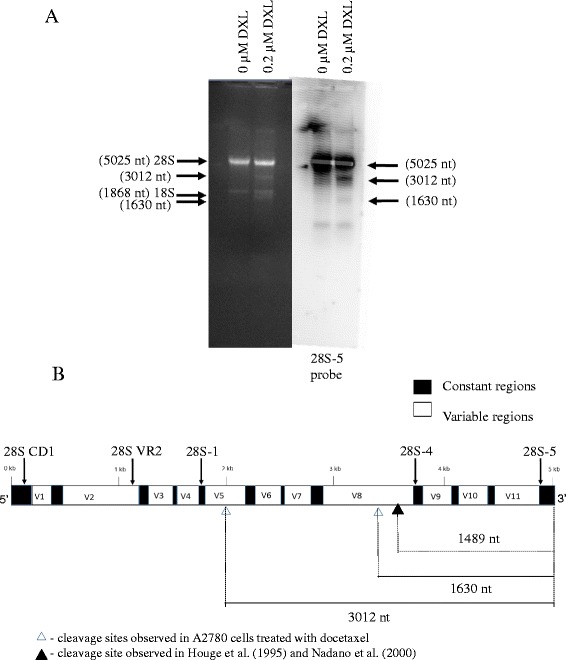
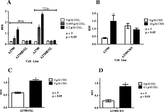
Results: All chemotherapy drugs tested were capable of inducing similar RNA disruption patterns. Docetaxel treatment of the resistant A2780DXL cells and carboplatin treatment of the A2780CBN cells did not result in RNA disruption. Northern blotting indicated that two RNA disruption bands were derived from the 3'-end of the 28S rRNA. Annexin-V and PI staining of docetaxel treated cells, along with assessment of caspase-3 activation, showed concurrent initiation of apoptosis and RNA disruption, while inhibition of caspase-3 activity significantly reduced RNA disruption.
Conclusions: Supporting the in vivo evidence, our results demonstrate that RNA disruption is induced by multiple chemotherapy agents in cell lines from different tissues and is associated with drug response. Although present, the link between apoptosis and RNA disruption is not completely understood. Evaluation of RNA disruption is thus proposed as a novel and effective biomarker to assess response to chemotherapy drugs in vitro and in vivo.
Figures
Similar articles
-
Inhibitory effects of mild hyperthermia plus docetaxel therapy on ER(+/-) breast cancer cells and action mechanisms.J Huazhong Univ Sci Technolog Med Sci. 2013 Dec;33(6):870-876. doi: 10.1007/s11596-013-1214-8. Epub 2013 Dec 13. J Huazhong Univ Sci Technolog Med Sci. 2013. PMID: 24337851
-
Altered DNA methylation is associated with docetaxel resistance in human breast cancer cells.Int J Oncol. 2010 May;36(5):1235-41. doi: 10.3892/ijo_00000607. Int J Oncol. 2010. PMID: 20372798
-
Purine Nucleoside Phosphorylase mediated molecular chemotherapy and conventional chemotherapy: a tangible union against chemoresistant cancer.BMC Cancer. 2011 Aug 24;11:368. doi: 10.1186/1471-2407-11-368. BMC Cancer. 2011. PMID: 21861932 Free PMC article.
-
Docetaxel-induced prostate cancer cell death involves concomitant activation of caspase and lysosomal pathways and is attenuated by LEDGF/p75.Mol Cancer. 2009 Aug 28;8:68. doi: 10.1186/1476-4598-8-68. Mol Cancer. 2009. PMID: 19715609 Free PMC article.
-
miRNA-34a is associated with docetaxel resistance in human breast cancer cells.Breast Cancer Res Treat. 2012 Jan;131(2):445-54. doi: 10.1007/s10549-011-1424-3. Epub 2011 Mar 12. Breast Cancer Res Treat. 2012. PMID: 21399894
Cited by
-
RNA disruption is a widespread phenomenon associated with stress-induced cell death in tumour cells.Sci Rep. 2023 Jan 31;13(1):1711. doi: 10.1038/s41598-023-28635-8. Sci Rep. 2023. PMID: 36720913 Free PMC article.
-
Doxorubicin chemotherapy affects the intracellular and interstitial free amino acid pools in skeletal muscle.PLoS One. 2018 Apr 4;13(4):e0195330. doi: 10.1371/journal.pone.0195330. eCollection 2018. PLoS One. 2018. PMID: 29617462 Free PMC article.
-
Iron-dependent cleavage of ribosomal RNA during oxidative stress in the yeast Saccharomyces cerevisiae.J Biol Chem. 2018 Sep 14;293(37):14237-14248. doi: 10.1074/jbc.RA118.004174. Epub 2018 Jul 18. J Biol Chem. 2018. PMID: 30021840 Free PMC article.
-
Digital-PCR for gene expression: impact from inherent tissue RNA degradation.Sci Rep. 2017 Dec 8;7(1):17235. doi: 10.1038/s41598-017-17619-0. Sci Rep. 2017. PMID: 29222437 Free PMC article.
-
Investigating the impact of RNA integrity variation on the transcriptome of human leukemic cells.3 Biotech. 2022 Aug;12(8):160. doi: 10.1007/s13205-022-03223-1. Epub 2022 Jul 7. 3 Biotech. 2022. PMID: 35814037 Free PMC article.
References
Publication types
MeSH terms
Substances
LinkOut - more resources
Full Text Sources
Other Literature Sources
Medical
Research Materials
Miscellaneous
