Prelamin A processing, accumulation and distribution in normal cells and laminopathy disorders
- PMID: 26900797
- PMCID: PMC4916894
- DOI: 10.1080/19491034.2016.1150397
Prelamin A processing, accumulation and distribution in normal cells and laminopathy disorders
Abstract
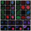
Lamin A is part of a complex structural meshwork located beneath the nuclear envelope and is involved in both structural support and the regulation of gene expression. Lamin A is initially expressed as prelamin A, which contains an extended carboxyl terminus that undergoes a series of post-translational modifications and subsequent cleavage by the endopeptidase ZMPSTE24 to generate lamin A. To facilitate investigations of the role of this cleavage in normal and disease states, we developed a monoclonal antibody (PL-1C7) that specifically recognizes prelamin A at the intact ZMPSTE24 cleavage site, ensuring prelamin A detection exclusively. Importantly, PL-1C7 can be used to determine prelamin A localization and accumulation in cells where lamin A is highly expressed without the use of exogenous fusion proteins. Our results show that unlike mature lamin A, prelamin A accumulates as discrete and localized foci at the nuclear periphery. Furthermore, whereas treatment with farnesylation inhibitors of cells overexpressing a GFP-prelamin A fusion protein results in the formation of large nucleoplasmic clumps, these aggregates are not observed upon similar treatment of cells expressing endogenous prelamin A or in cells lacking ZMPSTE24 expression and/or activity. Finally, we show that specific laminopathy-associated mutations exhibit both positive and negative effects on prelamin A accumulation, indicating that these mutations affect prelamin A processing efficiency in different manners.
Keywords: ZMPSTE24; intracellular flow cytometry (IFC); lamin A; monoclonal antibody; nuclear envelope; post-translational processing; prelamin A; progeriod syndromes.
Figures


Similar articles
-
The farnesyl transferase inhibitor (FTI) lonafarnib improves nuclear morphology in ZMPSTE24-deficient fibroblasts from patients with the progeroid disorder MAD-B.Nucleus. 2023 Dec;14(1):2288476. doi: 10.1080/19491034.2023.2288476. Epub 2023 Dec 5. Nucleus. 2023. PMID: 38050983 Free PMC article.
-
Abolishing the prelamin A ZMPSTE24 cleavage site leads to progeroid phenotypes with near-normal longevity in mice.Proc Natl Acad Sci U S A. 2022 Mar 1;119(9):e2118695119. doi: 10.1073/pnas.2118695119. Proc Natl Acad Sci U S A. 2022. PMID: 35197292 Free PMC article.
-
ZMPSTE24 missense mutations that cause progeroid diseases decrease prelamin A cleavage activity and/or protein stability.Dis Model Mech. 2018 Jul 13;11(7):dmm033670. doi: 10.1242/dmm.033670. Dis Model Mech. 2018. PMID: 29794150 Free PMC article.
-
Prelamin A and ZMPSTE24 in premature and physiological aging.Nucleus. 2023 Dec;14(1):2270345. doi: 10.1080/19491034.2023.2270345. Epub 2023 Oct 26. Nucleus. 2023. PMID: 37885131 Free PMC article. Review.
-
A humanized yeast system to analyze cleavage of prelamin A by ZMPSTE24.Methods. 2019 Mar 15;157:47-55. doi: 10.1016/j.ymeth.2019.01.001. Epub 2019 Jan 6. Methods. 2019. PMID: 30625386 Free PMC article. Review.
Cited by
-
Effect of β-Estradiol on Adipogenesis in a 3T3-L1 Cell Model of Prelamin A Accumulation.Int J Mol Sci. 2024 Jan 20;25(2):1282. doi: 10.3390/ijms25021282. Int J Mol Sci. 2024. PMID: 38279282 Free PMC article.
-
OGT (O-GlcNAc Transferase) Selectively Modifies Multiple Residues Unique to Lamin A.Cells. 2018 May 17;7(5):44. doi: 10.3390/cells7050044. Cells. 2018. PMID: 29772801 Free PMC article.
-
The nuclear envelope: LINCing tissue mechanics to genome regulation in cardiac and skeletal muscle.Biol Lett. 2020 Jul;16(7):20200302. doi: 10.1098/rsbl.2020.0302. Epub 2020 Jul 8. Biol Lett. 2020. PMID: 32634376 Free PMC article.
-
Knockdown of LMNA inhibits Akt/β-catenin-mediated cell invasion and migration in clear cell renal cell carcinoma cells.Cell Adh Migr. 2023 Dec;17(1):1-14. doi: 10.1080/19336918.2023.2260644. Epub 2023 Sep 25. Cell Adh Migr. 2023. PMID: 37749865 Free PMC article.
-
Biology, pathology, and therapeutic targeting of RAS.Adv Cancer Res. 2020;148:69-146. doi: 10.1016/bs.acr.2020.05.002. Epub 2020 Jul 9. Adv Cancer Res. 2020. PMID: 32723567 Free PMC article. Review.
References
-
- Dwyer N, Blobel G. A modified procedure for the isolation of a pore complex-lamina fraction from rat liver nuclei. J Cell Biol 1976; 70:581-91; PMID:986398; http://dx.doi.org/10.1083/jcb.70.3.581 - DOI - PMC - PubMed
-
- Schreiber KH, Kennedy BK. When lamins go bad: nuclear structure and disease. Cell 2013; 152:1365-75; PMID:23498943; http://dx.doi.org/10.1016/j.cell.2013.02.015 - DOI - PMC - PubMed
-
- Gruenbaum Y, Foisner R. Lamins: nuclear intermediate filament proteins with fundamental functions in nuclear mechanics and genome regulation. Annu Rev Biochem 2015; 84:131-64; PMID:25747401; http://dx.doi.org/10.1146/annurev-biochem-060614-034115 - DOI - PubMed
-
- Burke B, Stewart CL. Functional architecture of the cell's nucleus in development, aging, and disease. Curr Top Dev Biol 2014; 109:1-52; PMID:24947235; http://dx.doi.org/10.1016/B978-0-12-397920-9.00006-8 - DOI - PubMed
-
- Davidson PM, Lammerding J. Broken nuclei – lamins, nuclear mechanics, and disease. Trends Cell Biol 2014; 24:247-56; PMID:24309562; http://dx.doi.org/10.1016/j.tcb.2013.11.004 - DOI - PMC - PubMed
Publication types
MeSH terms
Substances
Grants and funding
LinkOut - more resources
Full Text Sources
Other Literature Sources
