. 2016 Apr 14;90(9):4427-4440.
doi: 10.1128/JVI.00219-16. Print 2016 May.

Phosphorylation of Bovine Herpesvirus 1 VP8 Plays a Role in Viral DNA Encapsidation and Is Essential for Its Cytoplasmic Localization and Optimal Virion Incorporation

Affiliations

Phosphorylation of Bovine Herpesvirus 1 VP8 Plays a Role in Viral DNA Encapsidation and Is Essential for Its Cytoplasmic Localization and Optimal Virion Incorporation

Kuan Zhang et al. J Virol. .

Abstract

VP8 is a major tegument protein of bovine herpesvirus 1 (BoHV-1) and is essential for viral replication in cattle. The protein undergoes phosphorylation after transcription through cellular casein kinase 2 (CK2) and a viral kinase, US3. In this study, a virus containing a mutated VP8 protein that is not phosphorylated by CK2 and US3 (BoHV-1-YmVP8) was constructed by homologous recombination in mammalian cells. When BoHV-1-YmVP8-infected cells were observed by transmission electron microscopy, blocking phosphorylation of VP8 was found to impair viral DNA encapsidation, resulting in release of incomplete viral particles to the extracellular environment. Consequently, less infectious virus was produced by the mutant virus than by wild-type (WT) virus. A comparison of mutant and WT VP8 by confocal microscopy revealed that mutant VP8 is nuclear throughout infection while WT VP8 is nuclear early during infection and is associated with the Golgi apparatus at later stages. This, together with the observation that mutant VP8 is present in virions, albeit in smaller amounts, suggests that the incorporation of VP8 may occur at two stages. The first takes place without the need for phosphorylation and before or during nuclear egress of capsids, whereas the second occurs in the Golgi apparatus and requires phosphorylation of VP8. The results indicate that phosphorylated VP8 plays a role in viral DNA encapsidation and in the secondary virion incorporation of VP8. To perform these functions, the cellular localization of VP8 is adjusted based on the phosphorylation status.

Importance: In this study, phosphorylation of VP8 was shown to have a function in BoHV-1 replication. A virus containing a mutated VP8 protein that is not phosphorylated by CK2 and US3 (BoHV-1-YmVP8) produced smaller numbers of infectious virions than wild-type (WT) virus. The maturation and egress of WT and mutant BoHV-1 were studied, showing a process similar to that reported for other alphaherpesviruses. Interestingly, lack of phosphorylation of VP8 by CK2 and US3 resulted in reduced incorporation of viral DNA into capsids during mutant BoHV-1 infection, as well as lower numbers of extracellular virions. Furthermore, mutant VP8 remained nuclear throughout infection, in contrast to WT VP8, which is nuclear at early stages and Golgi apparatus associated late during infection. This correlates with smaller amounts of mutant VP8 in virions and suggests for the first time that VP8 may be assembled into the virions at two stages, with the latter dependent on phosphorylation.


Figures

FIG 1
FIG 1
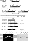
Schematic representations of DNA constructs and the genomes of recombinant viruses. (A) Development of DNA fragments for homologous recombination. The locations of restriction sites are indicated on each construct. (B) Genomes of recombinant viruses. The BoHV-1 genome consists of a unique long (UL) region and a unique short (US) region bracketed by inverted-repeat sequences (IR and TR). The UL48 and UL47 ORFs are highlighted. (C) PCR analysis of the viral genomes. Purified genomic DNA of WT BoHV-1, BoHV-1-YVP8, BoHV-1-YmVP8, and BoHV-1-RVP8 were used as PCR templates. PCR products covering from about 1 kbp of upstream flanking sequence of UL47 to the MluI restriction site of the UL47 gene were amplified using primers 5′-CGTGTTCGTTTCGCTGTACTATGC-3′ and 5′-CAGTAAATCAGGGAGCCCATTGAG-3′. The PCR products were separated in a 1% agarose gel with a DNA marker. (D) Western blot of VP8 proteins. MDBK cells were infected with WT BoHV-1, BoHV-1-YVP8, BoHV-1-YmVP8, or BoHV-1-RVP8 at an MOI of 1 for 20 h. Whole-protein extracts from cell lysates were analyzed by Western blotting using polyclonal anti-VP8 antibody and IRDye 600RD-conjugated secondary antibody. Molecular mass markers are shown on the left.
FIG 2
FIG 2
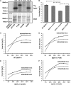
Phosphorylation status of VP8 and growth characteristics of viruses in MDBK cells. (A) Analysis of VP8 proteins by immunoprecipitation. MDBK cells were infected with WT BoHV-1, BoHV-1-YVP8, BoHV-1-YmVP8, and BoHV-1-RVP8 at an MOI of 1 and labeled with [32P]orthophosphate. Cell lysates were collected at 20 hpi and used for VP8 purification by incubation with anti-VP8 polyclonal antibody and protein G Sepharose. (Top) The samples were separated by SDS-PAGE and exposed to Imaging Screen K. (Bottom) The gels were stained with ProtoBlue Safe to indicate the amount of protein loading. The relative difference of each sample from WT VP8 is shown as a percentage at the top. (B) Titration of viruses. MDBK cells were infected with viruses at an MOI of 1. The supernatant and cells were collected at 24 hpi. Viruses from infected cells and supernatants were quantified by plaque titration on MDBK cell monolayers. The data were analyzed by two-tailed t test. The statistical significance of the difference between the values is shown; **, P ≤ 0.01. (C to F) Single-step growth curve of viruses. MDBK cells were infected with viruses at an MOI of 1, and the cell culture medium and cells were harvested separately at the indicated time points. The titer of infectious virus progeny in each sample was determined by plaque assay on MDBK cells. The error bars indicate standard deviations.
FIG 3
FIG 3
Influence of blocking VP8 phosphorylation on virion composition. (A) Solubilized proteins of purified extracellular virions of BoHV-1, BoHV-1-YVP8, and BoHV-1-YmVP8 were analyzed by SDS-PAGE, and the proteins were stained with ProtoBlue Safe. The densities of VP5 and VP8 were analyzed by densitometry. The percentages of YVP8 and YmVP8 in comparison to WT VP8 are indicated below the samples. (B) Western blotting of viral proteins. Solubilized proteins of purified extracellular WT BoHV-1, BoHV-1-YVP8, and BoHV-1-YmVP8 virions were analyzed by Western blotting. The proteins were detected with polyclonal anti-VP5, -VP22, -VP8, and -US3 antibodies and monoclonal anti-gB, -gC, and -gD antibodies, followed by IRDye 680RD goat anti-rabbit IgG and IRDye 800CW goat anti-mouse IgG, respectively. Each protein was probed simultaneously with VP5. The blots were analyzed by densitometry; the top and bottom of each gel show the images taken of VP5 and the tested proteins, respectively.
FIG 4
FIG 4
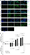
Localization of VP8 proteins in infected MDBK cells. MDBK cell monolayers were infected with WT BoHV-1, BoHV-1-YVP8, or BoHV-1-YmVP8 at an MOI of 5 or mock infected and processed for immunofluorescence staining every 2 h until 8 hpi. (A) VP8 was labeled with polyclonal anti-VP8 antibody and Alexa 488-conjugated antibody. DNA was labeled with DAPI. The arrowheads indicate nuclear VP8, and the arrows indicate cytoplasmic VP8. (B) The cell images for each time point were analyzed with a biological image-processing program, Fiji (34). The average green value within a selected area in an image was calculated by the program. At each time point, the cytoplasm and the nuclei of 10 cells were selected and analyzed. The mean values are shown. The data were analyzed by two-tailed t test. The statistical significance of the difference between the values is shown; *, 0.01 < P ≤ 0.05; **, P ≤ 0.01. The error bars indicate standard deviations.
FIG 5
FIG 5
Colocalization of cytoplasmic VP8 with the Golgi apparatus. MDBK cell monolayers were infected with WT BoHV-1, BoHV-1-YVP8, or BoHV-1-YmVP8 at an MOI of 0.001 until plaques were well developed or mock infected. WT VP8 was labeled with monoclonal anti-VP8 antibody and Alexa 488-conjugated antibody. The Golgi apparatus was identified with polyclonal anti-58K protein antibody and Alexa 633-conjugated antibody. The DNA was labeled with DAPI. The cells were observed under a Leica SP5 confocal microscope. The boxed areas are enlarged in the bottom row.
FIG 6
FIG 6
Coimmunostaining of VP8 and VP5. MDBK cell monolayers were infected with WT BoHV-1, BoHV-1-YVP8, or BoHV-1-YmVP8 at an MOI of 0.001 until plaques were well developed or mock infected. WT VP8 was labeled with monoclonal anti-VP8 antibody and Alexa 488-conjugated antibody. VP5 was identified with polyclonal anti-VP5 antibody and Alexa 633-conjugated antibody. DNA was labeled with DAPI. The cells were observed under a Leica SP5 confocal microscope. The boxed areas are enlarged in the bottom row.
FIG 7
FIG 7
Transmission electron microscopy of cells infected with WT BoHV-1. MDBK cells infected with WT BoHV-1 at an MOI of 1 were collected and processed at 15 hpi and observed with a Philips CM10 transmission electron microscope. The boxed areas in the central image are enlarged on the right and left.
FIG 8
FIG 8
Analysis of the impact of blocking phosphorylation of VP8 on viral DNA content. (A to C) MDBK cells infected with WT BoHV-1, BoHV-1-YVP8, or BoHV-1-YmVP8 at an MOI of 1 were collected and processed at 15 hpi and observed with a Philips CM10 transmission electron microscope. The locations of nuclear capsids are marked with asterisks. Cyt, cytoplasmic area; Nuc, nuclear area. The boxed areas are enlarged in the bottom left corners. (D) The capsids from five views of the nuclear areas infected with WT BoHV-1, BoHV-1-YVP8, or BoHV-1-YmVP8 were counted. The data were analyzed by two-tailed t test. The statistical significance of the difference between the values is shown; **, P ≤ 0.01. The error bars indicate standard deviations.
FIG 9
FIG 9
Transmission electron microscopy of cells infected with BoHV-1-YmVP8. MDBK cells infected with BoHV-1-YmVP8 at an MOI of 1 were collected and processed at 15 hpi and observed with a Philips CM10 transmission electron microscope. The boxed areas from the large images are enlarged on the right and left. Cyt, cytoplasmic area; Nuc, nucleus area; Ext, extracellular area.
FIG 10
FIG 10
Analysis of extracellular viruses. (A) MDBK cells infected with WT BoHV-1, BoHV-1-YVP8, or BoHV-1-YmVP8 at an MOI of 1 were collected and processed at 15 hpi and observed with a Philips CM10 transmission electron microscope. The extracellular viral particles are displayed. (B) The viral particles from five views of extracellular areas of each treatment were counted. The data were analyzed by two-tailed t test. The statistical significance of the difference between the values is shown; *, 0.01 < P ≤ 0.05. The error bars indicate standard deviations.
FIG 11
FIG 11
Gradient sedimentation analysis and TEM observation of extracellular viruses. Extracellular viral particles were loaded onto 10 to 60% potassium sodium tartrate gradients in TNE buffer and sedimented by centrifugation at 25,000 rpm for 2 h at 4°C in a SW41 rotor. The virus bands were visualized by passing a light beam from the top through the tartrate gradient in a dark environment. The double asterisks indicate bands containing complete viruses with higher density, and the single asterisk indicates a band containing incomplete viruses with lower density. (A to D) The virus bands were aspired and observed by TEM. The single arrowheads indicate virions without a DNA core, the double arrowheads indicate virions with an incomplete DNA core, and the triple arrowheads indicate virions with a complete DNA core (bars, 200 nm).

Similar articles

Cited by

References

    1. Levings RL, Roth JA. 2013. Immunity to bovine herpesvirus 1. I. Viral lifecycle and innate immunity. Anim Health Res Rev 14:88–102. doi:10.1017/S1466252313000042. - DOI - PubMed
    1. Bosse JB, Virding S, Thiberge SY, Scherer J, Wodrich H, Ruzsics Z, Koszinowski UH, Enquist LW. 2014. Nuclear herpesvirus capsid motility is not dependent on F-actin. MBio 5:e01909-01914. doi:10.1128/mBio.01909-14. - DOI - PMC - PubMed
    1. Shen S, Jia X, Guo H, Deng H. 2015. Tegument protein ORF33 of a gammaherpesvirus is associated with intranuclear capsids at an early stage of the tegumentation process. J Virol 89:5288–5297. doi:10.1128/JVI.00079-15. - DOI - PMC - PubMed
    1. Yu X, Li W, Liu L, Che Y, Cun W, Wu W, He C, Shao C, Li Q. 2008. Functional analysis of transcriptional regulation of herpes simplex virus type 1 tegument protein VP22. Sci China C Life Sci 51:966–972. doi:10.1007/s11427-008-0127-4. - DOI - PubMed
    1. Kato A, Liu Z, Minowa A, Imai T, Tanaka M, Sugimoto K, Nishiyama Y, Arii J, Kawaguchi Y. 2011. Herpes simplex virus 1 protein kinase Us3 and major tegument protein UL47 reciprocally regulate their subcellular localization in infected cells. J Virol 85:9599–9613. doi:10.1128/JVI.00845-11. - DOI - PMC - PubMed

Publication types

LinkOut - more resources