. 2016 Apr 14;90(9):4441-4453.
doi: 10.1128/JVI.00222-16. Print 2016 May.

A Novel Toll-Like Receptor 9 Agonist, MGN1703, Enhances HIV-1 Transcription and NK Cell-Mediated Inhibition of HIV-1-Infected Autologous CD4+ T Cells

Affiliations

A Novel Toll-Like Receptor 9 Agonist, MGN1703, Enhances HIV-1 Transcription and NK Cell-Mediated Inhibition of HIV-1-Infected Autologous CD4+ T Cells

Rasmus Offersen et al. J Virol. .

Abstract

Toll-like receptor (TLR) agonists are potent enhancers of innate antiviral immunity and may also reverse HIV-1 latency. Therefore, TLR agonists have a potential role in the context of a "shock-and-kill" approach to eradicate HIV-1. Our extensive preclinical evaluation suggests that a novel TLR9 agonist, MGN1703, may indeed perform both functions in an HIV-1 eradication trial. Peripheral blood mononuclear cells (PBMCs) from aviremic HIV-1-infected donors on antiretroviral therapy (ART) that were incubated with MGN1703 ex vivo exhibited increased secretion of interferon alpha (IFN-α) (P= 0.005) and CXCL10 (P= 0.0005) in culture supernatants. Within the incubated PBMC pool, there were higher proportions of CD69-positive CD56(dim)CD16(+)NK cells (P= 0.001) as well as higher proportions of CD107a-positive (P= 0.002) and IFN-γ-producing (P= 0.038) NK cells. Incubation with MGN1703 also increased the proportions of CD69-expressing CD4(+)and CD8(+)T cells. Furthermore, CD4(+)T cells within the pool of MGN1703-incubated PBMCs showed enhanced levels of unspliced HIV-1 RNA (P= 0.036). Importantly, MGN1703 increased the capacity of NK cells to inhibit virus spread within a culture of autologous CD4(+)T cells assessed by using an HIV-1 p24 enzyme-linked immunosorbent assay (ELISA) (P= 0.03). In conclusion, we show that MGN1703 induced strong antiviral innate immune responses, enhanced HIV-1 transcription, and boosted NK cell-mediated suppression of HIV-1 infection in autologous CD4(+)T cells. These findings support clinical testing of MGN1703 in HIV-1 eradication trials.

Importance: We demonstrate that MGN1703 (a TLR9 agonist currently undergoing phase 3 clinical testing for the treatment of metastatic colorectal cancer) induces potent antiviral responses in immune effector cells from HIV-1-infected individuals on suppressive antiretroviral therapy. The significantly improved safety and tolerability profiles of MGN1703 versus TLR9 agonists of the CpG-oligodeoxynucleotide (CpG-ODN) family are due to its novel "dumbbell-shape" structure made of covalently closed, natural DNA. In our study, we found that incubation of peripheral blood mononuclear cells with MGN1703 results in natural killer cell activation and increased natural killer cell function, which significantly inhibited the spread of HIV in a culture of autologous CD4(+)T cells. Furthermore, we discovered that MGN1703-mediated activation can enhance HIV-1 transcription in CD4(+)T cells, suggesting that this molecule may serve a dual purpose in HIV-1 eradication therapy: enhanced immune function and latency reversal. These findings provide a strong preclinical basis for the inclusion of MGN1703 in an HIV eradication clinical trial.


Figures

FIG 1
FIG 1
MGN1703 induces specific release of IFN-α and CXCL10. PBMCs from ART-suppressed HIV-1-infected donors were incubated for 48 h with MGN1703 at the indicated concentrations. Controls included noCG-MGN1703 as a TLR9-specific negative control or cRPMI as an untreated (UT) control. Cytokine and chemokine levels were measured in culture supernatants. (A and B) At 3 μM, MGN1703 induced a 200-fold median increase for IFN-α (A) and a 32-fold increase for CXCL10 (B). (C to E) IL-2 (C) and IL-6 (D) levels were unaffected, while TNF-α production changed by only 1.6-fold (E). Each donor in Fig. 1 to 4 is represented by the same distinct symbol. Lines represent the median.
FIG 2
FIG 2
MGN1703 activates NK cells. PBMCs from ART-suppressed HIV-1-infected donors were incubated for 48 h with MGN1703 at the indicated concentrations. Controls included noCG-MGN1703 as a TLR9-specific negative control or cRPMI as an untreated control. Cells were stained and then assessed by flow cytometry. (A to C) The median percentage of CD69-positive CD56dim (CD56dim CD16+) NK cells increased up to 7.5-fold in response to MGN1703. Also, CD56bright (CD56bright CD16+/−) and CD56neg (CD56neg CD16+) NK cells increased the proportion of CD69-positive cells by 2.2- and 5-fold, respectively (for comparative results for CpG-ODN2006, see Fig. S2 in the supplemental material). (D and E) The proportions of activated CD8+ and CD4+ T cells were significantly increased. (F and G) Marginally more CD8+ T cells were CD25 positive, while the proportion of CD25-positive CD4+ T cells did not increase. (H) Representative flow diagram for one donor (represented by green triangles), showing the gating strategy for CD69 expression on CD56dim NK cells. Each donor in Fig. 1 to 4 is represented by the same distinct symbol. Lines represent the median. SSC, side scatter; FSC, forward scatter.
FIG 3
FIG 3
MGN1703 upregulates NK cell receptors NKp46 and NKG2A. PBMCs from ART-suppressed HIV-1-infected donors were incubated for 48 h with MGN1703 (3 μM). cRPMI was used as an untreated control. Cells were stained and then assessed by flow cytometry. (A) Representative flow diagram for one donor (represented by open black diamonds) showing expression of NK cell receptors on CD56dim NK cells. Initial gating was done as described in the legend of Fig. 2. Insets at the top show FMO controls. (B) On CD56dim NK cells, MGN1703 incubation led to a 1.2-fold median increase in the proportion of cells expressing the activating receptor NKp46. (C and D) For NKR-P1A, there was no significant change (C), while 1.08-fold more CD56dim NK cells increased the expression of the inhibitory receptor NKG2A (D). NKp46 and NKG2A were exclusively upregulated on CD56dim NK cells by MGN1703 but not on CD56bright and CD56neg subsets (see Fig. S3 in the supplemental material). Each donor in Fig. 1 to 4 is represented by the same distinct symbol. Lines represent the median.
FIG 4
FIG 4
Panobinostat does not alter MGN1703 activity. PBMCs from ART-suppressed HIV-1-infected donors were stimulated for 16 h with either MGN1703 (3 μM), CPG-ODN2006 (0.75 μM) (see Fig. S5 in the supplemental material), panobinostat (7.5 nM), or cRPMI as an untreated control. Cells were stained and then assessed by flow cytometry. Cytokine levels were measured in culture supernatants. (A to D) Overall, there was no significant difference between a TLR9 agonist alone and coadministration with panobinostat. Specifically, panobinostat did not inhibit the MGN1703-induced production of IFN-α (A) or CXCL-10 (B). Furthermore, panobinostat did not inhibit the percentage of NK cells expressing NKG2A (C) or CD69 (D). NS, not significant. (E and F) Representative flow diagram for one donor (represented by blue dots) showing expression of NKG2A (E) and CD69 (F) on CD56dim NK cells. Initial gating was done as described in the legend of Fig. 2. Insets show FMO controls. Each donor in Fig. 1 to 4 is represented by the same distinct symbol. Lines represent the median.
FIG 5
FIG 5
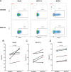
MGN1703 enhances HIV-1 transcription ex vivo. (A and B) ACH2 and U1 cells were stimulated with MGN1703 in 2-fold serial dilutions from 6 nM to 6 μM. HIV-1 p24 antigen (ELISA) was used as a measure of latency reversal. Direct MGN1703 exposure did not increase HIV-1 production in these latently infected cell models. (C) PBMCs from HIV-1-negative donors were stimulated for 48 h with MGN1703. Culture supernatants were then transferred to U1 cells and incubated for 36 h before the level of HIV-1 p24 antigen in the supernatant was quantified. In response to supernatants from MGN1703-stimulated PBMCs, U1 cells were shown to significantly increase median HIV-1 production up to 2.2-fold. (D) PBMCs from ART-suppressed HIV-1-infected donors were incubated for 16 h with MGN1703 (3 μM) or cRPMI as an untreated control, CD4+ T cells were then isolated by negative selection, and RNA was extracted. HIV-1 usRNA levels were measured by using seminested RT-qPCR. MGN1703 significantly enhanced the transcription of HIV-1 usRNA ex vivo by a median of 1.4-fold. Lines represent the median.
FIG 6
FIG 6
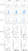
MGN1703 primes NK cells to degranulate and produce IFN-γ upon exposure to target cells. CD107a LAMP-1 expression was used to measure NK cell degranulation. PBMCs from ART-suppressed HIV-1-infected donors were incubated for 48 h with MGN1703 (3 μM), LPS (100 ng/ml) as a positive control, or cRPMI as an untreated control. Cells were washed before being cultured alone or with K562 or ACH2 cells as target cells (ratio of 1:1) for 6 h in the presence of brefeldin A, Golgi Stop, and anti-CD107a. Cells were washed and then stained before being assessed by flow cytometry. (A) Representative flow plot. (B to D) NK cells stimulated with MGN1703 increased intracellular IFN-γ production by 1.89-fold and 1.47-fold for CD107a (B and C) but not significantly for CD107a IFN-γ double-positive cells (D). (E to G) Also in response to ACH2 cells, significantly more MGN1703-treated NK cells than untreated NK cells degranulated (median of 1.86-fold). Lines represent the median.
FIG 7
FIG 7
MGN1703 enhances NK cell-mediated inhibition of HIV-1-producing autologous CD4+ T cells. PBMCs from ART-suppressed HIV-1-infected donors were stimulated for 48 h with MGN1703 (3 μM) or cRPMI as an untreated control. NK cells were subsequently purified by negative magnetic selection and cocultured with autologous HIV-1-infected CD4+ T cells as target cells at a ratio of 1:1. HIV-1-infected CD4+ T cells cocultured without NK cells served as a relative parameter for maximal HIV-1 p24 antigen release, measured on days 1, 3, and 5 by an ELISA. (A and B) Cross-sectional analysis on day 5 revealed significantly less p24 antigen in the culture supernatant for MGN1703-treated NK cells than in untreated NK cells (n = 5; lines represent the median) (A) and a trend toward reduced numbers of p24+ CD4+ T cells in wells with MGN1703-treated NK cells (P = 0.18) (B). To determine this, cells from day 5 (n = 4) were collected, stained for intracellular HIV-1 p24 antigen, and analyzed by flow cytometry (B). CD4+ T cells were gated as live, single, CD3+ CD8neg T cells. Data from all donors were normalized to data for CD4+ T cells cultured without NK cells. (C) By analyzing these data longitudinally, instead of cross-sectionally at day 5, the AUCs encompass p24 antigen release from day 1 through day 5, showing significantly reduced levels of p24 antigen in the supernatants. This confirms the impact of MGN1703 on the ability of NK cells to inhibit viral spread over time. Depicted are the mean values (± standard errors of the means) (n = 5) at each time point, with connecting lines generating a curve for each condition. For each individual, an AUC was calculated for each condition and used to make AUC comparisons. Each donor in panels A and B is represented by the same distinct symbol.

Similar articles

Cited by

References

    1. Alter G, Heckerman D, Schneidewind A, Fadda L, Kadie CM, Carlson JM, Oniangue-Ndza C, Martin M, Li B, Khakoo SI, Carrington M, Allen TM, Altfeld M. 2011. HIV-1 adaptation to NK-cell-mediated immune pressure. Nature 476:96–100. doi:10.1038/nature10237. - DOI - PMC - PubMed
    1. Wren LH, Chung AW, Isitman G, Kelleher AD, Parsons MS, Amin J, Cooper DA, Stratov I, Navis M, Kent SJ. 2013. Specific antibody-dependent cellular cytotoxicity responses associated with slow progression of HIV infection. Immunology 138:116–123. doi:10.1111/imm.12016. - DOI - PMC - PubMed
    1. Sips M, Sciaranghella G, Diefenbach T, Dugast A-S, Berger CT, Liu Q, Kwon D, Ghebremichael M, Estes JD, Carrington M, Martin JN, Deeks SG, Hunt PW, Alter G. 2012. Altered distribution of mucosal NK cells during HIV infection. Mucosal Immunol 5:30–40. doi:10.1038/mi.2011.40. - DOI - PMC - PubMed
    1. Seay K, Church C, Zheng JH, Deneroff K, Ochsenbauer C, Kappes JC, Liu B, Jeng EK, Wong HC, Goldstein H. 2015. In vivo activation of human NK cells by treatment with an interleukin-15 superagonist potently inhibits acute in vivo HIV-1 infection in humanized mice. J Virol 89:6264–6274. doi:10.1128/JVI.00563-15. - DOI - PMC - PubMed
    1. Krieg AM. 2002. CpG motifs in bacterial DNA and their immune effects. Annu Rev Immunol 20:709–760. doi:10.1146/annurev.immunol.20.100301.064842. - DOI - PubMed

Publication types

MeSH terms