. 2016 Feb 15:6:20818.
doi: 10.1038/srep20818.

Critical POU domain residues confer Oct4 uniqueness in somatic cell reprogramming

Affiliations

Critical POU domain residues confer Oct4 uniqueness in somatic cell reprogramming

Wensong Jin et al. Sci Rep. .

Abstract

The POU domain transcription factor Oct4 plays critical roles in self-renewal and pluripotency of embryonic stem cells (ESCs). Together with Sox2, Klf4 and c-Myc, Oct4 can reprogram any other cell types to pluripotency, in which Oct4 is the only factor that cannot be functionally replaced by other POU family members. To investigate the determinant elements of Oct4 uniqueness, we performed Ala scan on all Ser, Thr, Tyr, Lys and Arg of murine Oct4 by testing their capability in somatic cell reprogramming. We uncovered a series of residues that are important for Oct4 functionality, in which almost all of these key residues are within the POU domains making direct interaction with DNA. The Oct4 N- and C-terminal transactivation domains (TADs) are not unique and could be replaced by the Yes-associated protein (YAP) TAD domain to support reprogramming. More importantly, we uncovered two important residues that confer Oct4 uniqueness in somatic cell reprogramming. Our systematic structure-function analyses bring novel mechanistic insight into the molecular basis of how critical residues function together to confer Oct4 uniqueness among POU family for somatic cell reprogramming.


Figures

Figure 1
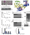
Figure 1. Identification of critical Oct4 residues for somatic cell reprogramming by alanine scanning.
(a) Percentage of GFP+ iPSC formation by Oct4 alanine mutants, shown here are defective ones. For complete data see Supplementary Fig. S1c and Supplementary Table S1). (b) Modeling of Oct4POU:PORE complex. The POUS domain is shown in blue, the POUHD domain in green, the linker in magenta, and the DNA in yellow. K and R residues, except for K147 (4.4 Å), that have atoms within 3.6 Å of DNA (protein-DNA interface) are highlighted and those in contact with the DNA base are labeled with a star (*). The residue numbering in the model is in accord with positions in murine Oct4 (mOct4). This labeling and coloring is maintained throughout this manuscript. (c) Protein stability of reprogramming defective mOct4 K-to-A and R-to-A mutants in MEFs.
Figure 2
Figure 2. Characterization of reprogramming defective mOct4 S-to-A, T-to-A and Y-to-A mutants.
(a) AP staining of reprogramming defective S-to-A, T-to-A and Y-to-A mutants. (b) Model of Oct4POU:PORE complex. The POUS domain is shown in blue, the POUHD domain in green, the linker in magenta, and the DNA in yellow. Ser/Thr/Tyr residues that have atoms within 3.6 Å of DNA (protein-DNA interface) are highlighted and those in contact with the DNA base or phosphates are labeled with two stars or one star, respectively. R150, Y155 and Q211 are highlighted to show their intra-molecular interactions, and hydrogen bonds are represented by red dot lines. Right upper panel shows zoomed-in view of the intra-molecular interactions. (c) Protein stability of reprogramming defective S-to-A, T-to-A and Y-to-A mutants in MEFs. (d) CHX-based assay was used to determine the protein turnover of S186A, as compared to WT, in HEK293 cells. The corresponding immunoblotting is at the right. (e) MG132 and chloroquine-based assays were used to determine the main means by which S186A was degraded. The corresponding immunoblotting is at the right. (f) Enhanced polyubiquitination of S186A mutant compared to WT. (g) Comparison of protein stability of S186A, S186D and S186E mutants in MEFs. (h) The relative transcriptional activity of S186A, S186D and S186E mutants was measured using 6xCR4-Luc and 5xW-Luc reporters, respectively. Data are representative of at least three independent experiments. Statistics analysis was performed using a t-test (mean and s.d. of duplicate assays). (i) Generation of GFP + iPSC colonies by the S186A, S186D and S186E mutants. Data are representative of three independent experiments. Statistics analysis was performed using a t-test (mean and s.d. of duplicate assays).
Figure 3
Figure 3. Characterization of mOct4 critical DNA binding residues.
(a) Model of Oct4POU:PORE complex. The POUS domain is shown in blue, the POUHD domain in green, the linker in magenta, and the DNA in yellow. Residues tested in our collection that have atoms within 3.6 Å of DNA (protein-DNA interface) are highlighted. Those making hydrogen bonds to phosphate or DNA bases are labeled with one star or two stars, respectively. V269 is labeled with three stars for its non-polar contacts with DNA base. R150, T156, D159 and E181 are highlighted to show their intra-molecular interactions. Right upper panel shows zoomed-in view of the interaction between D159 and T156. Right lower panel shows zoomed-in view of the interaction among E181, R150 and Q157. Hydrogen bonds are shown by red dot lines. (b) AP staining of reprogramming defective alanine mutants. (c) Protein stability of reprogramming defective alanine mutants in MEFs. (d) The relative transcriptional activity of reprogramming defective alanine mutants was measured using 6xCR4-Luc and 5xW-Luc reporters, respectively. Data are representative of at least three independent experiments. Statistics analysis was performed using a t-test (mean and s.d. of duplicate assays). (e) Interaction of mOct4 Q174A with Sox2 was compared to that of Oct4 WT with Sox2.
Figure 4
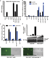
Figure 4. POU domain of Oct4 is the minimal requirement for generating iPS cells.
(a) The number of GFP+ iPSCs colonies formed by mOct4 WT, the indicated deletion mutants, or the YAPTAD fusion proteins was calculated. Data are representative of three independent experiments. Statistics analysis was performed using a t-test (mean and s.d. of duplicate assays). (b) The relative transcriptional activities of mOct4 WT, the indicated deletion mutants, or the YAPTAD fusion proteins were measured using 6xCR4-Luc and 5xW-Luc reporters, respectively. Data are representative of at least three independent experiments. Statistics analysis was performed using a t-test (mean and s.d. of duplicate assays). (c) The relative transcriptional activities of mOct4 WT, POU, POU-YAPTAD and YAPTAD were measured using 6xCR4-Luc and 5xW-Luc reporters, respectively. Data are representative of at least three independent experiments. Statistics analysis was performed using a t-test (mean and s.d. of duplicate assays). (d) The number of GFP+ iPSC colonies formed by increasing doses of mOct4 WT and POU-YAPTAD was calculated. The corresponding protein levels of Oct4 WT and POU-YAPTAD were shown in the lower panel. *:non-specific band (N.S.). (e) iPSC colony formed by POU-YAPTAD under GFP fluorescence and phase contrast. Scale bars, 100 μm. (f) Establishment of iPS cell lines generated by POU-YAPTAD. Scale bars, 250 μm.
Figure 5
Figure 5. Specific residues make Oct4 unique in reprogramming.
(a) Sequence alignment of various murine POU domains showing the unique K170 in Oct4, which is highlighted in yellow. (b) Residue substitution counts surrounding mOct4 K170 among the 16 murine POU domain-containing members. (c) Superimpositions of the POUs helices α2–α3 between Oct4 and Pit, Oct1 or Oct6. The POUs helices α2–α3 of Oct4, Pit1, Oct1 and Oct6 are shown as cartoon in red, blue, green, and cyan, respectively. The unique K170 of Oct4 is highlighted in yellow.

Similar articles

Cited by

  • Phosphorylation of OCT4 Serine 236 Inhibits Germ Cell Tumor Growth by Inducing Differentiation.
    Kim DK, Song B, Han S, Jang H, Bae SH, Kim HY, Lee SH, Lee S, Kim JK, Kim HS, Hong KM, Lee BI, Youn HD, Kim SY, Kang SW, Jang H. Kim DK, et al. Cancers (Basel). 2020 Sep 11;12(9):2601. doi: 10.3390/cancers12092601. Cancers (Basel). 2020. PMID: 32932964 Free PMC article.
  • Permissive epigenomes endow reprogramming competence to transcriptional regulators.
    Kim KP, Choi J, Yoon J, Bruder JM, Shin B, Kim J, Arauzo-Bravo MJ, Han D, Wu G, Han DW, Kim J, Cramer P, Schöler HR. Kim KP, et al. Nat Chem Biol. 2021 Jan;17(1):47-56. doi: 10.1038/s41589-020-0618-6. Epub 2020 Aug 17. Nat Chem Biol. 2021. PMID: 32807969
  • POU3F3-related disorder: Defining the phenotype and expanding the molecular spectrum.
    Rossi A, Blok LS, Neuser S, Klöckner C, Platzer K, Faivre LO, Weigand H, Dentici ML, Tartaglia M, Niceta M, Alfieri P, Srivastava S, Coulter D, Smith L, Vinorum K, Cappuccio G, Brunetti-Pierri N, Torun D, Arslan M, Lauridsen MF, Murch O, Irving R, Lynch SA, Mehta SG, Carmichael J, Zonneveld-Huijssoon E, de Vries B, Kleefstra T, Johannesen KM, Westphall IT, Hughes SS, Smithson S, Evans J, Dudding-Byth T, Simon M, van Binsbergen E, Herkert JC, Beunders G, Oppermann H, Bakal M, Møller RS, Rubboli G, Bayat A. Rossi A, et al. Clin Genet. 2023 Aug;104(2):186-197. doi: 10.1111/cge.14353. Epub 2023 May 10. Clin Genet. 2023. PMID: 37165752 Free PMC article.
  • Reprogramming competence of OCT factors is determined by transactivation domains.
    Kim KP, Wu Y, Yoon J, Adachi K, Wu G, Velychko S, MacCarthy CM, Shin B, Röpke A, Arauzo-Bravo MJ, Stehling M, Han DW, Gao Y, Kim J, Gao S, Schöler HR. Kim KP, et al. Sci Adv. 2020 Sep 2;6(36):eaaz7364. doi: 10.1126/sciadv.aaz7364. Print 2020 Sep. Sci Adv. 2020. PMID: 32917606 Free PMC article.
  • Oct4 redox sensitivity potentiates reprogramming and differentiation.
    Shen Z, Wu Y, Manna A, Yi C, Cairns BR, Evason KJ, Chandrasekharan MB, Tantin D. Shen Z, et al. Genes Dev. 2024 May 21;38(7-8):308-321. doi: 10.1101/gad.351411.123. Genes Dev. 2024. PMID: 38719541 Free PMC article.

References

    1. Downs K. M. Systematic localization of Oct-3/4 to the gastrulating mouse conceptus suggests manifold roles in mammalian development. Dev. Dyn. 237, 464–475 (2008). - PubMed
    1. Takahashi K. & Yamanaka S. Induction of pluripotent stem cells from mouse embryonic and adult fibroblast cultures by defined factors. Cell 126, 663–676 (2006). - PubMed
    1. Radzisheuskaya A. & Silva J. C. Do all roads lead to Oct4? the emerging concepts of induced pluripotency. Trends Cell Biol. 24, 275–284 (2014). - PMC - PubMed
    1. Zeineddine D., Hammoud A. A., Mortada M. & Boeuf H. The Oct4 protein: more than a magic stemness marker. Am. J. Stem Cells 3, 74–82 (2014). - PMC - PubMed
    1. Esch D. et al. A unique Oct4 interface is crucial for reprogramming to pluripotency. Nat. Cell Biol. 15, 295–301 (2013). - PubMed

Publication types

MeSH terms

LinkOut - more resources