Review
. 2016 Jun;17(6):569-92.
doi: 10.1111/tra.12387. Epub 2016 Mar 31.

Principles of Virus Uncoating: Cues and the Snooker Ball

Affiliations
Review

Principles of Virus Uncoating: Cues and the Snooker Ball

Yohei Yamauchi et al. Traffic. 2016 Jun.

Abstract

Viruses are spherical or complex shaped carriers of proteins, nucleic acids and sometimes lipids and sugars. They are metastable and poised for structural changes. These features allow viruses to communicate with host cells during entry, and to release the viral genome, a process known as uncoating. Studies have shown that hundreds of host factors directly or indirectly support this process. The cell provides molecules that promote stepwise virus uncoating, and direct the virus to the site of replication. It acts akin to a snooker player who delivers accurate and timely shots (cues) to the ball (virus) to score. The viruses, on the other hand, trick (snooker) the host, hijack its homeostasis systems, and dampen innate immune responses directed against danger signals. In this review, we discuss how cellular cues, facilitators, and built-in viral mechanisms promote uncoating. Cues come from receptors, enzymes and chemicals that act directly on the virus particle to alter its structure, trafficking and infectivity. Facilitators are defined as host factors that are involved in processes which indirectly enhance entry or uncoating. Unraveling the mechanisms of virus uncoating will continue to enhance understanding of cell functions, and help counteracting infections with chemicals and vaccines.

Keywords: cytoskeleton; endocytosis; low pH; membrane fusion; molecular motor; nuclear import; nuclear pore complex; penetration; signaling; virus structure.


Figures

Figure 1
Figure 1
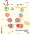
Endocytic pathways involved in virus entry. The majority of viruses use endocytosis for entry 4, 279. The virus‐carrying vesicles and vacuoles often move along microtubules toward the nucleus. Cellular markers for the vesicles are shown within the light‐blue boxes. The pH lowers as the vesicles mature and approach the nucleus. Viruses (not shown) enter the endocytic pathway and respond to cellular cues and facilitators that serve as uncoating signals. Such cues and facilitators are regulated in time and space and control the stepwise uncoating program, as shown in the examples in Figures 2, 3, 4. Abbreviations: EEA1, early endosomal antigen 1; ER, endoplasmic reticulum; ESCRT, endosomal sorting complexes required for transport; LAMP1, lysosome associated membrane protein 1; NPC1, Niemann‐Pick Disease, Type C1; PI3P, Phosphatidylinositol 3‐phosphate; SNX, syntaxin; TGN, trans‐Golgi network.
Figure 2
Figure 2
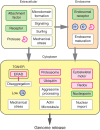
Cellular cues and processes implicated in viral uncoating. The scheme depicts cellular cues (receptors, enzymes and chemicals including ions) and their cellular processes implicated in the stepwise uncoating of incoming virus particles. After binding to the cell surface via receptors and attachment factors, viruses are typically taken up into endocytic vacuoles following activation of signaling. They penetrate endosomes to enter the cytoplasm. Inside endosomes, viruses can be primed via endosome maturation, receptor binding, protease digestion or ions. In some cases, viruses fuse directly at the plasma membrane to enter the cytoplasm. In the cytoplasm, viruses can be exposed to cellular cues and processes which culminate in the completion of uncoating and release of the viral genome from the capsid. The viral genome is transported to the site of replication, which may be in the cytosol, on cytoplasmic membranes or in the nucleus. Receptor cues (green), enzymatic cues (pink), chemical cues (blue) and cellular processes (white) implicated in viral uncoating are indicated. Facilitators are not shown. Abbreviation: ERAD, ER‐associated protein degradation.
Figure 3
Figure 3
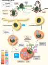
Cues and facilitators of Influenza A virus entry and uncoating. After binding to sialic acids on the cell surface, influenza A virus induces receptor tyrosine kinase (RTK) signaling via EGFR and endocytosis by CME or macropinocytosis 194, 197, 198, 280, 281, 282. CME involves Epsin1 and the virus particle enters Rab5‐positive early endosomes 197. Macropinocytic uptake requires N‐linked glycans on the cell surface, and also involves RTK signaling 194, 195, 196, 198. Endosome maturation and the influx of H+/K+ into the viral core via the M2 channel (shown by H+/K+ with white arrows) primes the virion for uncoating 199, 202, 203. Low pH in Rab7/LAMP1‐positive late endosomes induces HA‐mediated membrane fusion (as shown by H+ with blue arrows) 206. Unanchored ubiquitin chains are exposed to the cytosol, followed by recruitment of HDAC6 and the aggresome processing machinery (including dynein, myosin and the cytoskeleton) to disassemble the capsid shell by mechanical force 25. CD81 and cathepsin W also promote virus fusion 204, 205. Poly‐ubiquitination of matrix protein M1 by E3 ubiquitin ligase Itch is also implicated in uncoating 283. Following capsid disassembly, the vRNPs are released into the cytosol, followed by NLS‐mediated import into the nucleus, viral gene transcription and replication 110. Receptor cues (green), enzymatic cues (pink), chemical cues (blue), facilitators (brown), viral protein/process (gray) and endosomal markers (light blue) are indicated. The influenza virion scheme was adapted from http://visual-science.com. Abbreviations: EGFR, epidermal growth factor receptor; HA, hemagglutinin; LAMP, lysosome associated membrane protein; uUb, unanchored ubiquitin; pUb, poly‐ubiquitin; FACIL, facilitator; VIRAL, viral protein/process; MARK, endosomal marker; RECEP, receptor cue; ENZYM, enzymatic cue; CHEM, chemical cue.
Figure 4
Figure 4
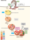
Cues and facilitators of Ebola virus entry and uncoating. Ebola virus binds to the receptors TIM‐1 and AXL and enters cells through classical apoptotic mimicry 31, 236, 237, 284. Endosome maturation, low pH, cathepsin L and cathepsin B activity prime the virus glycoprotein GP for fusion, an event that is also dependent on cellular factors PIKfyve and HOPS 250. Cathepsin activity is enhanced by low pH of endolysosomes (as shown by H+ with arrows) 285. Once the virus reaches NPC1/TPC2‐positive endosomes TIM‐1 binds NPC1, which directly or indirectly activates fusion and penetration 244, 245, 246. TPC2 activity is regulated by NAADP, which is a highly potent intracellular calcium‐mobilizing agent that stimulates intracellular calcium channels to release Ca2+ from endosomes and lysosomes, influencing the trafficking and maturation of endosomes 248, 286. Receptor cues (green), enzymatic cues (pink), chemical cues (blue), facilitators (brown), viral protein/process (gray) and endosomal markers (light blue) are indicated. Abbreviations: AXL, AXL receptor tyrosine kinase; GP, glycoprotein; HOPS, homotypic fusion and vacuole protein sorting; LAMP1, lysosome associated membrane protein 1; NAADP, nicotinic acid adenine dinucleotide phosphate; NPC1, Niemann‐Pick Disease, Type C1; PIKfyve, FYVE finger‐containing phosphoinositide kinase; PS, phosphatidylserine; TIM‐1, T‐cell immunoglobulin and mucin domain 1; TPC2, two pore Ca2+ channel 2; FACIL, facilitator; VIRAL, viral protein/process; MARK, endosomal marker, RECEP, receptor cue; ENZYM, enzymatic cue; CHEM, chemical cue.
Figure 5
Figure 5
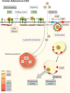
Cues and facilitators of adenovirus entry and uncoating. HAdV‐C2/C5 binds to CAR and integrins on the cell surface, and the actomyosin‐dependent drifting motions of CAR trigger fiber shedding 2, 11, 12, 40. Virus binding to integrins induces signaling and virus uptake into endosomes in a dynamin‐dependent manner 259, 260, Protein VI is dislocated from the inside of the virus and binds to the plasma membrane, forming small pores that allow influx of Ca2+ into the cytosol 272, 287. This danger signal induces rapid lysosomal secretion of ASM to the cell surface. ASM converts SM into CER, which enhances endocytic uptake of the virus. Protein VI is recruited to CER on the internal surface of endosomes, inducing endosomal leakage and rupture, and thereby enables escape of viral particles into the cytosol 260, 272, 288. Low pH is not required for virus penetration, but required to maintain functional secretory lysosomes 147. Receptor cues (green), enzymatic cues (pink), chemical cues (blue), facilitators (brown), viral protein/process (gray) and endosomal markers (light blue) are indicated. Abbreviations: ASM, acid sphingomyelinase; CAR, coxsackie and adenovirus receptor; CER, ceramide; ITGN, integrin; SM, sphingomyelin; FACIL, facilitator; VIRAL, viral protein/process; RECEP, receptor cue; ENZYM, enzymatic cue; CHEM, chemical cue.

Similar articles

Cited by

References

    1. Mercer J, Schelhaas M, Helenius A. Virus entry by endocytosis. Annu Rev Biochem 2010;79:803–833. - PubMed
    1. Greber UF, Willetts M, Webster P, Helenius A. Stepwise dismantling of adenovirus 2 during entry into cells. Cell 1993;75:477–486. - PubMed
    1. Greber UF, Singh I, Helenius A. Mechanisms of virus uncoating. Trends Microbiol 1994;2:52–56. - PubMed
    1. Yamauchi Y, Helenius A. Virus entry at a glance. J Cell Sci 2013;126:1289–1295. - PubMed
    1. Wolfrum N, Greber UF. Adenovirus signalling in entry. Cell Microbiol 2013;15:53–62. - PubMed

Publication types

LinkOut - more resources