. 2016 Feb 9:14:5.
doi: 10.1186/s12964-016-0128-z.

Feedback activation of neurofibromin terminates growth factor-induced Ras activation

Affiliations

Feedback activation of neurofibromin terminates growth factor-induced Ras activation

Anne Hennig et al. Cell Commun Signal. .

Abstract

Background: Growth factors induce a characteristically short-lived Ras activation in cells emerging from quiescence. Extensive work has shown that transient as opposed to sustained Ras activation is critical for the induction of mitogenic programs. Mitogen-induced accumulation of active Ras-GTP results from increased nucleotide exchange driven by the nucleotide exchange factor Sos. In contrast, the mechanism accounting for signal termination and prompt restoration of basal Ras-GTP levels is unclear, but has been inferred to involve feedback inhibition of Sos. Remarkably, how GTP-hydrolase activating proteins (GAPs) participate in controlling the rise and fall of Ras-GTP levels is unknown.

Results: Monitoring nucleotide exchange of Ras in permeabilized cells we find, unexpectedly, that the decline of growth factor-induced Ras-GTP levels proceeds in the presence of unabated high nucleotide exchange, pointing to GAP activation as a major mechanism of signal termination. Experiments with non-hydrolysable GTP analogues and mathematical modeling confirmed and rationalized the presence of high GAP activity as Ras-GTP levels decline in a background of high nucleotide exchange. Using pharmacological and genetic approaches we document a raised activity of the neurofibromatosis type I tumor suppressor Ras-GAP neurofibromin and an involvement of Rsk1 and Rsk2 in the down-regulation of Ras-GTP levels.

Conclusions: Our findings show that, in addition to feedback inhibition of Sos, feedback stimulation of the RasGAP neurofibromin enforces termination of the Ras signal in the context of growth-factor signaling. These findings ascribe a precise role to neurofibromin in growth factor-dependent control of Ras activity and illustrate how, by engaging Ras-GAP activity, mitogen-challenged cells play safe to ensure a timely termination of the Ras signal irrespectively of the reigning rate of nucleotide exchange.


Figures
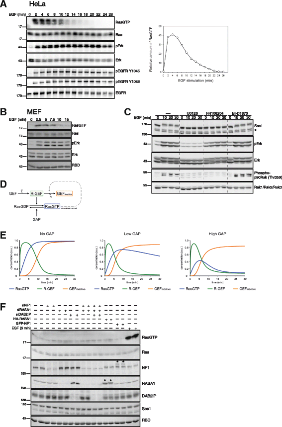
Fig. 1
Fig. 1
EGF induces transient Ras activation and Sos phosphorylation. a Transient Ras activation in HeLa cells. Serum-starved HeLa cells were challenged with 10 ng/ml EGF and Ras activation was determined via Ras-GTP affinity pulldowns. EGFR and Erk phosphorylation were determined using phosphosite-selective antibodies. A quantification of the Ras-GTP kinetics is shown on the right. RBD: Coomassie stain of Ras binding domain used for collecting Ras-GTP. b MEF cells challenged with EGF were processed for Ras and Erk activity assays as in (a). c EGF induces a mobility shift in Sos. HeLa cells were treated with inhibitors for MEK (10 μM U0126), Erk (50 μM FR108204) or Rsk (10 μM BI-D1870) prior to stimulation with EGF. Extracts were processed via western blotting using the indicated antibodies. Asterisk marks an unspecific doublet band. d Minimal Ras model describing Ras deactivation as induced by Ras-GTP-dependent feedback inhibition of Sos. R-GEF: receptor-GEF complex. See experimental section for details. e Simulations of Ras activation/deactivation using the model from (d) in a background of absent, low or high basal GAP activity. f Biochemical analysis of Ras-GTP levels following manipulation of Ras-GAP levels. The indicated Ras-GAP species were knocked down by siRNA (siNF1, siRASA1, siDAB2IP) or transiently overexpressed in HeLa cells (GFP-NF1: GFP-neurofibromin fusion construct; HA-RASA1: HA-tagged RASA1; asterisks mark overexpressed polypeptides). 5 min EGF stimulation is shown as positive control
Fig. 2
Fig. 2
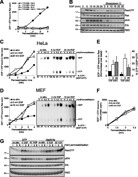
EGF induces transient Ras-GTP accumulation but sustained up-regulation of nucleotide exchange. a Specificity of the Ras nucleotide exchange assay in permeabilized cells. Serum-starved HeLa cells were permeabilized or mock permeabilized by omitting SLO in the presence of [α-32P]GTP. A 100fold molar excess of unlabeled GTP was included where indicated. Cell extracts prepared at the indicated time points were subjected to Ras-IPs or mock IPs lacking the Y13-259 Ras-antibody. Precipitates were washed and associated radioactivity evaluated by cerenkow counting. b Biochemical assay of time-dependent EGF-induced Ras and Erk activation performed in the absence or presence of the permeabilizing agent SLO. SLO was added simultaneously with EGF. c Nucleotide exchange assay in permeabilized HeLa cells before and 5 min or 20 min after EGF administration. Nucleotides associated to Ras-IPs were additionally eluted from Ras and separated via thin layer chromatography (TLC, on the right). %GTP/(GDP + GTP) values were determined by densitometry and plotted under the panel. Of note, initial values start off high and level off only at later time points. This pattern is owed to the different time required for single Ras proteins versus the whole Ras population to achieve steady-state nucleotide turnover. d Same experiment as in C performed in MEF cells. e Quantification of nucleotides bound to Ras-IPs. On the left, the amount of GDP + GTP bound to Ras at the 6 min assay point (as recorded in (c)) was plotted as the fold increase of radioactivity bound to Ras in EGF-stimulated versus unstimulated cells. On the right, the amount of GTP/(GDP + GTP) in the same assay points was plotted as % GTP/(GDP + GTP). Shown are means ± S.E.M. for three independent experiments. f [α-32P]GTP associated to total cellular protein from untreated or EGF-challenged permeabilized cells determined by a filter binding assay. Shown here is the means ± S.E.M. for three independent experiments. g GppNHp but not GTP promotes strong Ras activation in permeabilized cells at late time points of EGF stimulation. HeLa cells were permeabilized for the indicated time frames in the presence of GTP or GppNHp before (no stim.), 5 min or 20 min after EGF stimulation. Reactions were stopped by cell lysis and cell extracts were subjected to biochemical analysis of Ras and Erk activation
Fig. 3
Fig. 3
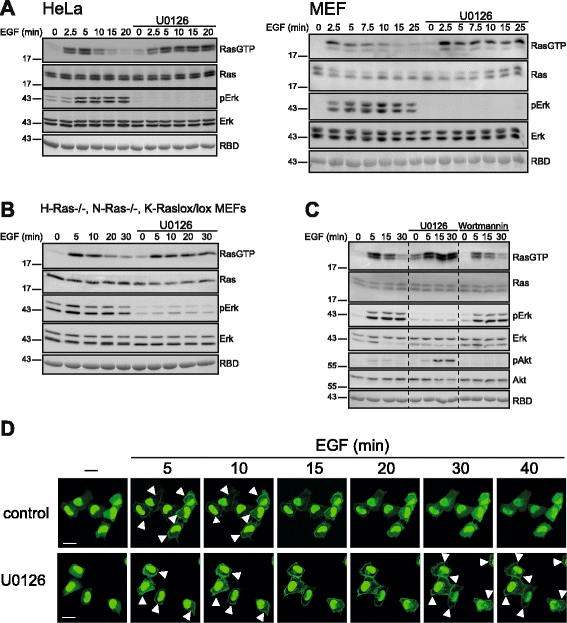
Inhibition of the MEK/Erk/Rsk pathway prolongs Ras activation. a Resting HeLa or MEF cells were left untreated or treated with the MEK inhibitor U0126 (10 μM), followed by EGF stimulation and analysis of Ras and Erk activity. b Same experiment as in A performed in H-Ras-/-, N-Ras-/-, K-Raslox/lox MEFs expressing only K-Ras. c HeLa cells pretreated with the MEK inhibitor U0126 or the PI3K inhibitor Wortmannin (30 min,100 nM) were challenged with EGF and subjected to a biochemical Ras activation assay. d HeLa cells expressing the trivalent affinity probe for Ras-GTP E3-R3(A/D) (see experimental section and Ref.[51]) were treated with U0126 or left untreated prior to stimulation with 10 ng/ml EGF. The time-dependent re-distribution of E3-R3(A/D) was imaged alive by confocal laser scanning microscopy. Probe relocation to the plasma membrane (marked by arrowheads) illustrates Ras activation. Over 30 cells monitored in 3-5 individual experiments responded with the same redistribution kinetics
Fig. 4
Fig. 4
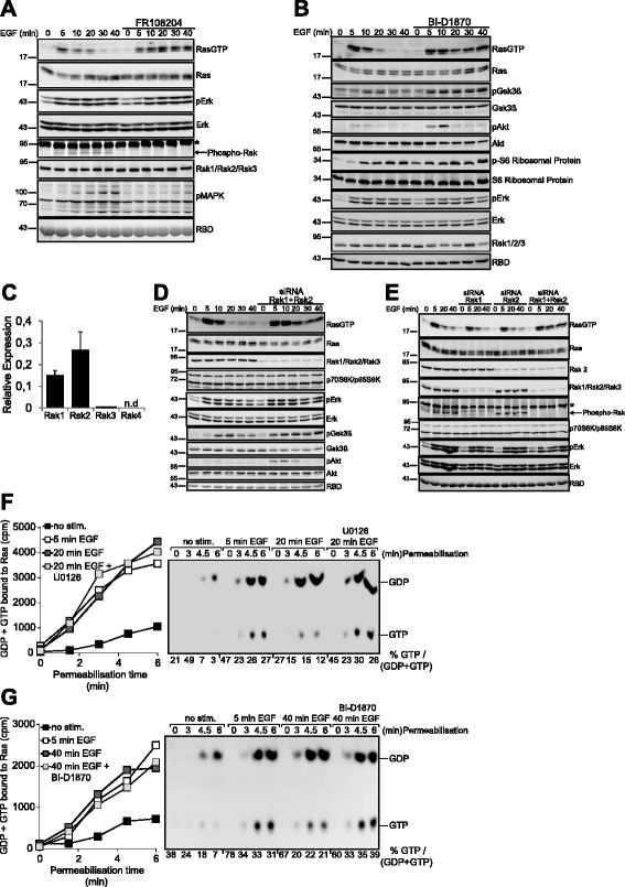
Rsk1 and Rsk2 mediate the feedback deactivation of Ras. a HeLa cells treated or not with the Erk inhibitor FR108204 were challenged with EGF for the indicated periods of time and subjected to a Ras-GTP pulldown assay. The phosphorylation/activation state of the indicated proteins was determined using phophosite-specific antibodies. Phospho-MAPK Substrates (PXS*P or S*PXR/K): Ab recognizing the phosphorylated Erk consensus motif. Asterisk denotes an unspecific band. b Same experiment in cells pre-treated with the pan-Rsk inhibitor BI-D1870. The activation status of Erk was monitored using phospho-site specific antibodies against Erk. Acute inhibition of Rsk with BI-D1870 did not affect Rsk protein stability as illustrated by the immunodetection of total Rsk1/Rsk2/Rsk3. c Real-time PCR analysis of Rsk isoform expression in HeLa cells. d Rsk1 and Rsk2 were simultaneously silenced via siRNA in HeLa cells followed by stimulation with EGF and biochemical analysis of Ras activation. e Biochemical determination of Ras-GTP levels and Erk activity in HeLa cells previously subjected to single or combined siRNA-mediated knockdown of Rsk1 and Rsk2. Immunodetection of p70S6K/p85S6K was performed as a control of specificity of the siRNA-mediated knockdown of Rsk1/2. Asterisk denotes an unspecific band. f Feedback deactivation of Ras is mediated via GAP up-regulation. HeLa cells were pretreated with U0126 where indicated, challenged with 10 ng/ml EGF and subjected to analysis of Ras nucleotide exchange. g Same experiment as in (a) performed in cells treated with the pan-Rsk inhibitor BI-D1870
Fig. 5
Fig. 5
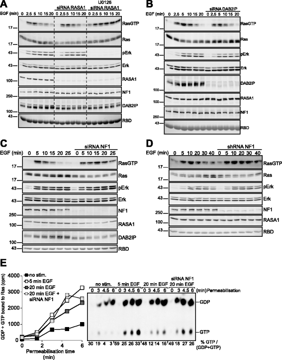
Feedback-mediated stimulation of neurofibromin mediates Ras deactivation. a EGF-induced Ras activation in HeLa cells subjected to previous siRNA-mediated silencing of RASA1. siRNA-transfected cells were additionally treated with the MEK inhibitor U0126 in order to ascertain that siRNA transfections did not distort the feedback mechanism of Ras deactivation. b Same experiment as in (a) in DAB2IP-silenced HeLa cells. c Same experiment as in (b) performed in neurofibromin-silenced HeLa cells. d Time course of EGF-driven Ras activation in HEK293T cells and a derivative line with stable shRNA-mediated knockdown of neurofibromin. e HeLa cells subjected to siRNA mediated silencing of neurofibromin were challenged with EGF. Cells were permeabilized prior to or 5 and 20 min after EGF stimulation and processed for the analysis of nucleotide exchange on Ras
Fig. 6
Fig. 6
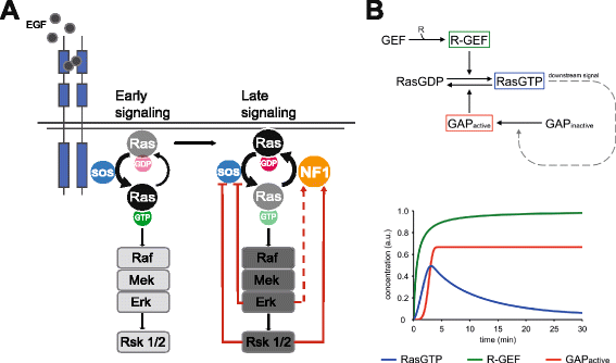
Model of Ras deactivation mediated by the feedback-dependent activation of neurofibromin. a Schematic cartoon of the mechanism of Ras activation/deactivation. The scheme depicts the previously reported Erk and/or Rsk-dependent feedback inhibition of GEF (Sos) activation and the feedback stimulation of neurofibromin reported herein. The dotted line linking Erk to neurofbromin symbolizes the presumptive Rsk-independent feedback loop emanating from Erk. See text for details. b Minimal mathematical model describing Ras activation/deactivation mediated by a positive feedback stimulation of Ras-GAP. R-GEF: receptor-GEF complex. See experimental section for details

Similar articles

Cited by

References

    1. Marshall CJ. Specificity of receptor tyrosine kinase signaling: transient versus sustained extracellular signal-regulated kinase activation. Cell. 1995;80:179–85. doi: 10.1016/0092-8674(95)90401-8. - DOI - PubMed
    1. Crespo P, Leon J. Ras proteins in the control of the cell cycle and cell differentiation. Cell Mol Life Sci. 2000;57:1613–36. doi: 10.1007/PL00000645. - DOI - PMC - PubMed
    1. Muroya K, Hattori S, Nakamura S. Nerve growth factor induces rapid accumulation of the GTP-bound form of p21ras in rat pheochromocytoma PC12 cells. Oncogene. 1992;7:277–81. - PubMed
    1. Heasley LE, Johnson GL. The beta-PDGF receptor induces neuronal differentiation of PC12 cells. Mol Biol Cell. 1992;3:545–53. doi: 10.1091/mbc.3.5.545. - DOI - PMC - PubMed
    1. Traverse S, Gomez N, Paterson H, Marshall C, Cohen P. Sustained activation of the mitogen-activated protein (MAP) kinase cascade may be required for differentiation of PC12 cells. Comparison of the effects of nerve growth factor and epidermal growth factor. Biochem J. 1992;288(Pt 2):351–5. doi: 10.1042/bj2880351. - DOI - PMC - PubMed

Publication types