Arginine ADP-ribosyltransferase 1 promotes angiogenesis in colorectal cancer via the PI3K/Akt pathway
- PMID: 26847718
- PMCID: PMC4771103
- DOI: 10.3892/ijmm.2016.2473
Arginine ADP-ribosyltransferase 1 promotes angiogenesis in colorectal cancer via the PI3K/Akt pathway
Abstract
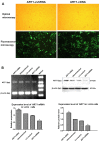
Arginine adenosine diphosphate (ADP)-ribosyl-transferase 1 (ART1) is known to play an important role in many physiological and pathological processes. Previous studies have demonstrated that ART1 promotes proliferation, invasion and metastasis in colon carcinoma. However, it was unclear whether ART1 is involved in angiogenesis in cases of colorectal cancer (CRC). In the present study, lentiviral vector‑mediated ART1‑cDNA or ART1-shRNA were transfected into LoVo cells, and the LoVo cells transfected with ART1-cDNA or ART1-shRNA were co-cultured with human umbilical vein endothelial cells (HUVECs) to determine the influence of ART1 on HUVECs. The proliferation, migration and angiogenesis of HUVECs were monitored using a cell counting kit-8 assay, a Transwell migration assay and immunohistochemical analysis in intrasplenic allograft tumors, respectively. Hypoxia‑inducible factor 1-α (HIF-1α), total (t-)Akt, phosphorylated (p-)Akt, vascular endothelial growth factor (VEGF) and basic fibroblast growth factor (bFGF) expression levels were detected via western blot analysis. Our results revealed that HUVECs which were co-cultured with ART1-cDNA LoVo cells showed higher proliferation, migration and angiogenic abilities, but a reduction was noted in those cultured with ART1-shRNA LoVo cells; p-Akt, HIF-1α, VEGF and bFGF expression was increased in HUVECs cultured with ART1‑cDNA-transfected LoVo cells, but reduced in ART1-shRNA-transfected LoVo cells. In a mouse xenograft model, we noted that the tumor microvessel density (MVD) was significantly increased in intrasplenic transplanted ART1‑cDNA CT26 tumors but decreased in intrasplenic transplanted ART1‑shRNA tumors. These data suggest that ART1 promoted the expression of HIF-1α via the Akt pathway in tumor cells. It also upregulated VEGF and bFGF and enhanced angiogenesis in HUVECs. Thus, we suggest that ART1 plays an important role in the invasion of CRC cells and the metastasis of CRC.
Figures


Similar articles
-
Arginine ADP-ribosyltransferase 1 Regulates Glycolysis in Colorectal Cancer via the PI3K/AKT/HIF1α Pathway.Curr Med Sci. 2022 Aug;42(4):733-741. doi: 10.1007/s11596-022-2606-4. Epub 2022 Jul 7. Curr Med Sci. 2022. PMID: 35798928
-
Effects of β-caryophyllene on arginine ADP-ribosyltransferase 1-mediated regulation of glycolysis in colorectal cancer under high-glucose conditions.Int J Oncol. 2018 Oct;53(4):1613-1624. doi: 10.3892/ijo.2018.4506. Epub 2018 Jul 27. Int J Oncol. 2018. PMID: 30066849
-
Tubb3 regulation by the Erk and Akt signaling pathways: a mechanism involved in the effect of arginine ADP-ribosyltransferase 1 (Art1) on apoptosis of colon carcinoma CT26 cells.Tumour Biol. 2016 Feb;37(2):2353-63. doi: 10.1007/s13277-015-4058-y. Epub 2015 Sep 15. Tumour Biol. 2016. PMID: 26373733
-
Regulation of the RhoA/ROCK/AKT/β-catenin pathway by arginine-specific ADP-ribosytransferases 1 promotes migration and epithelial-mesenchymal transition in colon carcinoma.Int J Oncol. 2016 Aug;49(2):646-56. doi: 10.3892/ijo.2016.3539. Epub 2016 May 27. Int J Oncol. 2016. PMID: 27277835
-
Pull the plug: Anti-angiogenesis potential of natural products in gastrointestinal cancer therapy.Phytother Res. 2022 Sep;36(9):3371-3393. doi: 10.1002/ptr.7492. Epub 2022 Jul 24. Phytother Res. 2022. PMID: 35871532 Review.
Cited by
-
ART1 knockdown decreases the IL-6-induced proliferation of colorectal cancer cells.BMC Cancer. 2024 Mar 19;24(1):354. doi: 10.1186/s12885-024-12120-0. BMC Cancer. 2024. PMID: 38504172 Free PMC article.
-
Uncovering the Invisible: Mono-ADP-ribosylation Moved into the Spotlight.Cells. 2021 Mar 19;10(3):680. doi: 10.3390/cells10030680. Cells. 2021. PMID: 33808662 Free PMC article. Review.
-
Overexpression of the Kininogen-1 inhibits proliferation and induces apoptosis of glioma cells.J Exp Clin Cancer Res. 2018 Aug 2;37(1):180. doi: 10.1186/s13046-018-0833-0. J Exp Clin Cancer Res. 2018. PMID: 30068373 Free PMC article.
-
Expression of the mono-ADP-ribosyltransferase ART1 by tumor cells mediates immune resistance in non-small cell lung cancer.Sci Transl Med. 2022 Mar 16;14(636):eabe8195. doi: 10.1126/scitranslmed.abe8195. Epub 2022 Mar 16. Sci Transl Med. 2022. PMID: 35294260 Free PMC article.
-
Identification of key pathways and genes influencing prognosis in bladder urothelial carcinoma.Onco Targets Ther. 2017 Mar 20;10:1673-1686. doi: 10.2147/OTT.S131386. eCollection 2017. Onco Targets Ther. 2017. PMID: 28356754 Free PMC article.
References
Publication types
MeSH terms
Substances
LinkOut - more resources
Full Text Sources
Other Literature Sources
Medical
Molecular Biology Databases

