Review
. 2016 Jan 27;80(1):161-86.
doi: 10.1128/MMBR.00056-15. Print 2016 Mar.

Biological Nanomotors with a Revolution, Linear, or Rotation Motion Mechanism

Affiliations
Review

Biological Nanomotors with a Revolution, Linear, or Rotation Motion Mechanism

Peixuan Guo et al. Microbiol Mol Biol Rev. .

Abstract

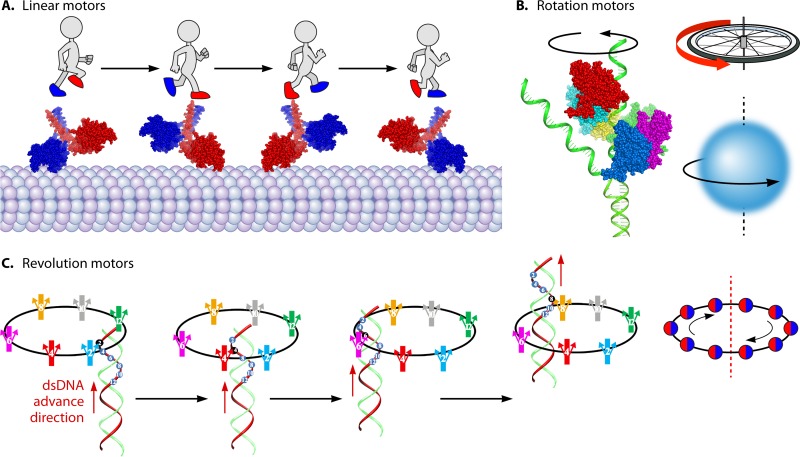
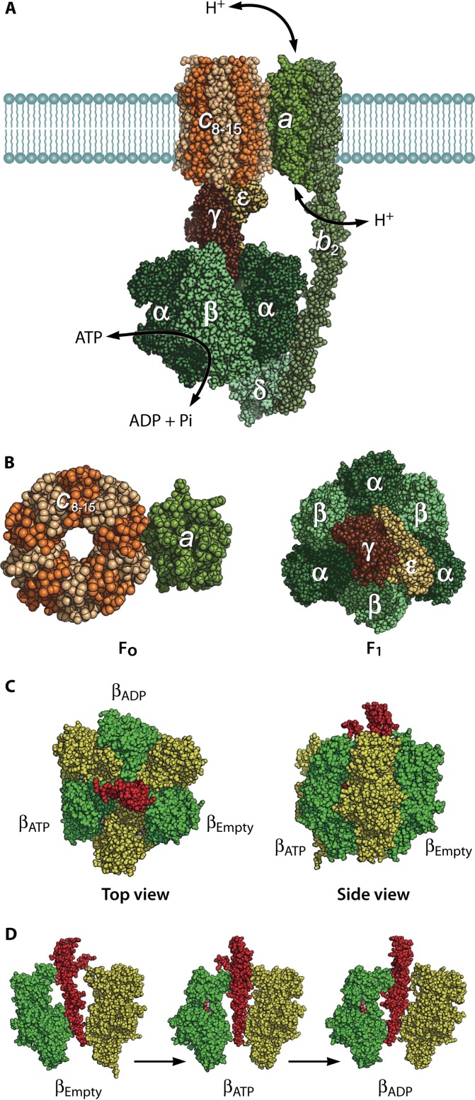
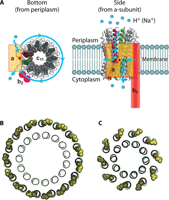
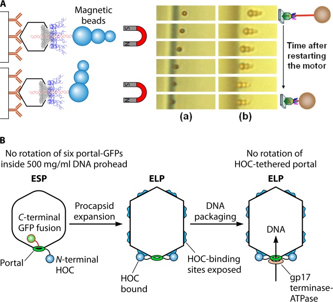
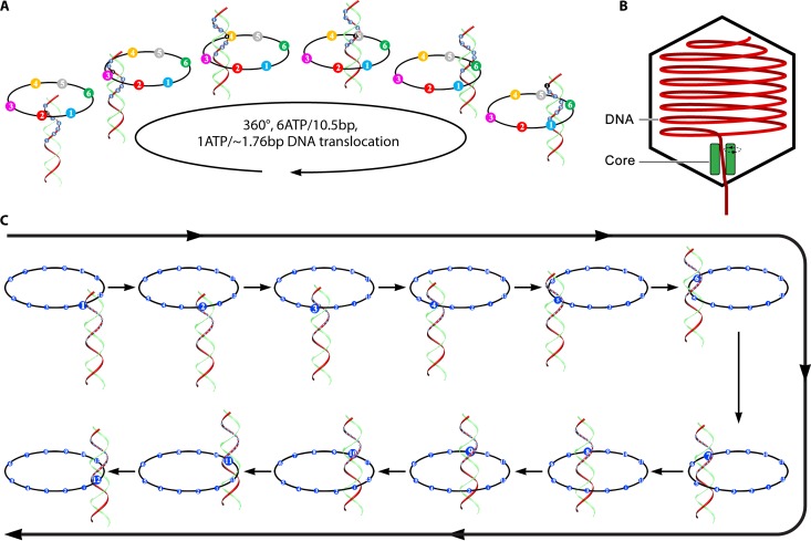
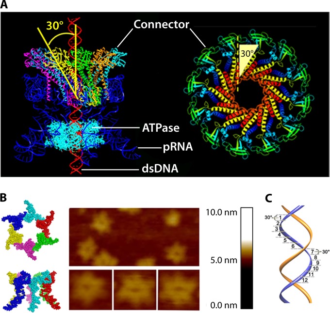
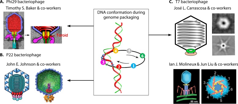
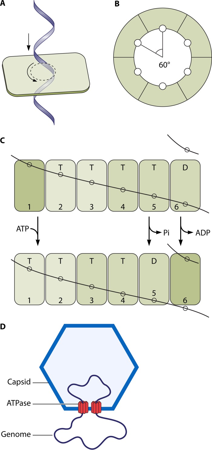
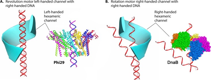
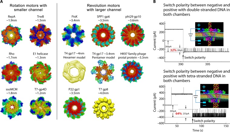
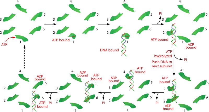
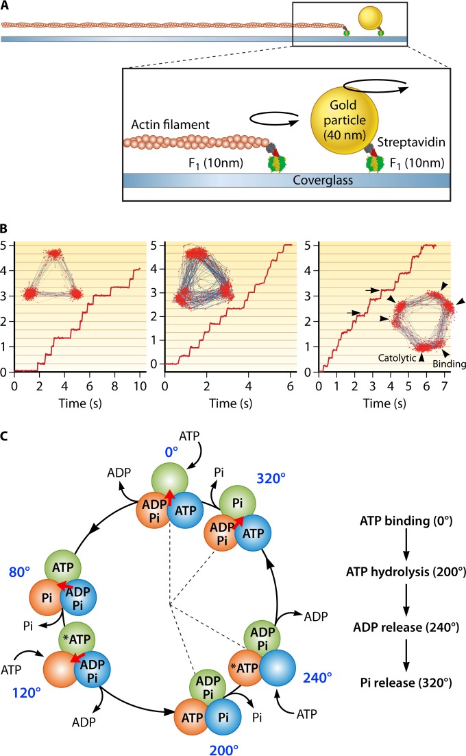
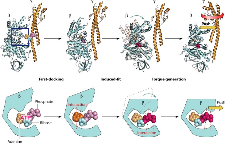
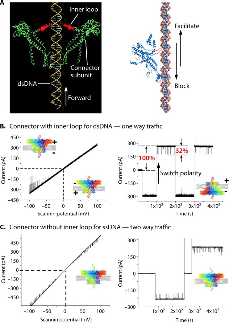
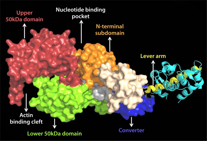
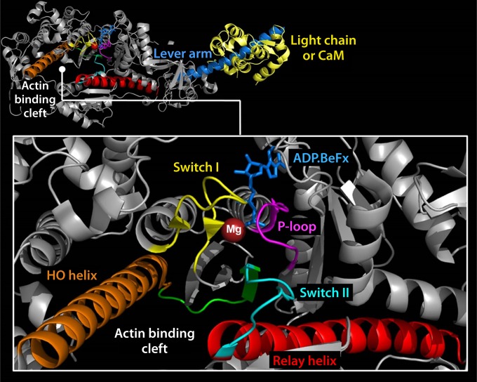
The ubiquitous biological nanomotors were classified into two categories in the past: linear and rotation motors. In 2013, a third type of biomotor, revolution without rotation (http://rnanano.osu.edu/movie.html), was discovered and found to be widespread among bacteria, eukaryotic viruses, and double-stranded DNA (dsDNA) bacteriophages. This review focuses on recent findings about various aspects of motors, including chirality, stoichiometry, channel size, entropy, conformational change, and energy usage rate, in a variety of well-studied motors, including FoF1 ATPase, helicases, viral dsDNA-packaging motors, bacterial chromosome translocases, myosin, kinesin, and dynein. In particular, dsDNA translocases are used to illustrate how these features relate to the motion mechanism and how nature elegantly evolved a revolution mechanism to avoid coiling and tangling during lengthy dsDNA genome transportation in cell division. Motor chirality and channel size are two factors that distinguish rotation motors from revolution motors. Rotation motors use right-handed channels to drive the right-handed dsDNA, similar to the way a nut drives the bolt with threads in same orientation; revolution motors use left-handed motor channels to revolve the right-handed dsDNA. Rotation motors use small channels (<2 nm in diameter) for the close contact of the channel wall with single-stranded DNA (ssDNA) or the 2-nm dsDNA bolt; revolution motors use larger channels (>3 nm) with room for the bolt to revolve. Binding and hydrolysis of ATP are linked to different conformational entropy changes in the motor that lead to altered affinity for the substrate and allow work to be done, for example, helicase unwinding of DNA or translocase directional movement of DNA.


Figures

FIG 1
FIG 1
Illustration of different categories of motors. (A) Linear motors are like people walking (PDB code 3KIN). (B) Rotation motors are like a wheel and like Earth rotating on its own axis. (Adapted from reference .) (C) Revolution motors resemble Earth revolving around the sun without self-rotation. (Adapted from reference with permission from Elsevier.)
FIG 2
FIG 2
Structures of FoF1 ATP synthase and the α3β3γ subcomplex of F1. (A) Reconstituted structure of FoF1 ATP synthase from crystal structures of isolated subunit or subcomplexes: a3b3γε subcomplex (PDB code 3OAA), δ (PDB code 1ABV), b dimer (PDB codes 1B9U, 2KHK, and 1L2P), c ring (PDB code 3UD0), and putative structure of the a subunit (PDB code 1C17). Green parts represent the stator complex, including the peripheral stalk (δ-b2 subcomplex) that holds the a3b3 stator ring of F1 and ab2 stator of Fo. Brown parts represent the rotor complex (γε–c-ring subcomplex). (B) Fo and F1 (Fig. 1A), both viewed from the top. (C) Original crystal structure of F1 from bovine mitochondria (PDB code 1BMF). Sphere representations of the α, β, and γ subunits are shown in yellow, green, and red, respectively. Each β subunit carries either AMP-PNP, ADP, or neither and is designated βATP, βADP, or βEmpty, respectively. (D) Conformational states of 3 β subunits viewed from the side. α-β pairs are shown in green and yellow with the central γ subunit in red. α and β subunits are composed of the N-terminal domain, nucleotide binding domain, and C-terminal domain (from bottom to top). βEmpty has an open conformation in which the α-helical C-terminal domain rotates upward, opening the cleft of the nucleotide binding pocket. Both βATP and βADP have a closed conformation entrapping the nucleotide within the closed pocket. All α subunits represent the open conformation.
FIG 3
FIG 3
Structure of Fo. (A) Crystal structure of the c11 ring of Na+-transporting Fo from Ilyobacter tartaricus (PDB code 1YCE). The blue spheres in the middle of the c11 ring represent bound Na+ ions. The stator ab2 complex is shown in the schematic drawing. The a subunit has 2 hemichannels, each open to the periplasmic space or the cytoplasmic space. A proton transferring between the a and c subunits accompanies the rotation of the c ring. Two c-subunit monomers at the interface of the a subunit are shown in red and green, respectively. (B) “Ion-locked” conformation of cGlu62 (yellow sphere representation) in the crystal structure of the H+-transporting c15 ring from Spirulina platensis (PDB code 2WIE). (C) “Ion-unlocked” conformation of cGlu59 (yellow sphere representation) in the crystal structure of the H+-transporting c10 ring from yeast mitochondria (PDB code 3U2F).
FIG 4
FIG 4
Experiment with the phi29 and T4 motors revealing that neither connector nor dsDNA rotation is required for active DNA packaging. (A) Direct observation of DNA packaging horizontally using a dsDNA with its end linked to a cluster of magnetic beads for stretching the DNA. Panels a and b, real-time sequential images of DNA-magnetic bead complexes. (Adapted from reference and reprinted from reference with permission from AIP Publishing LLC.) (B) Experiment revealing that the T4 motor connector does not rotate during packaging. The packaging activity is not inhibited with the N terminus of the motor connector protein fused and tethered to its protease immune binding site on the capsid. GFP, green fluorescent protein; ESP, empty small particle; ELP, empty large particle; HOC, highly antigenic outer capsid protein. (Adapted from reference and adapted from reference with permission from John Wiley and Sons.)
FIG 5
FIG 5
Schematic showing the sequential revolution motion in translocating dsDNA. (A) Revolution of dsDNA inside the ATPase hexameric ring. (Adapted from reference .) (B) Diagram of cryo-EM results showing offset of dsDNA in the channel of the bacteriophage T7 DNA-packaging motor. The dsDNA did not appear in the center of the channel; instead, the dsDNA tilted toward the wall of the motor channel. (Adapted from reference with permission of the publisher.) (C) Revolution of dsDNA along the 12 subunits of the connector channel. (Adapted from reference .)
FIG 6
FIG 6
Depiction of the structure and function of the phi29 DNA-packaging motor. (A) Side view of phi29 dsDNA-packaging motor (left) and top view of phi29 connector (right). (B) Hexameric pRNA generated from crystal structures of its 3WJ core and AFM images of loop-extended hexameric pRNA. (C) DNA revolving inside the connector channel by contact with each connector subunit in a 30° transition step for each contact. (Adapted from references and .)
FIG 7
FIG 7
Examples of spooling of DNA within capsids of phages to support the revolution mechanism. (A) Bacteriophage phi29. (Adapted from reference with permission from Elsevier.) (B) Bacteriophage P22. (Adapted from reference with permission from AAAS.) (C) Bacteriophage T7. (Adapted from reference and adapted from reference with permission from the American Society for Biochemistry and Molecular Biology.) The toroid formed at the phi29 portal position might be an accumulation of the images of the revolution motion during packaging, as shown in the image in the center. (Adapted from reference with permission from Elsevier.)
FIG 8
FIG 8
Illustration showing how FtsK may undergo the revolution mechanism. (A) One strand of dsDNA contacts with the inner-channel wall of the hexameric ATPase. The continuous contact between DNA and ATPase does not require any rotation of the ATPase or DNA. (B) Each DNA contact is expected to be separated by 60° along the inner surface of the ATPase hexameric channel. (C) Sequential action of dsDNA translocation. DNA is shown as a line. T, ATP-bound; D, ADP-bound. (Adapted from reference with permission of John Wiley and Sons [copyright 2010 WILEY-VCH Verlag GmbH & Co. KGaA, Weinheim].) (D) Speculation on the segregation and translocation of the mimivirus genome into the capsid via a revolution mechanism similar to that for FtsK. (Adapted from reference with permission from Elsevier.)
FIG 9
FIG 9
The FtsK motor protein. (A) E. coli FtsK protein domain structure. The N-terminal domain is in red, with each transmembrane domain represented by a black box. The numbers represent the FtsK amino acid numbers. The C terminus is subdivided into α, β, and γ domains. (B) Two views of the hexameric FtsK motor protein structure. On the left is a side on view emphasizing the α and β domains. Bound nucleotide (ADP) is shown in black in a space-filling model. On the right is a view down the symmetry axis, viewed from the β-domain side. (C) Structure of three γ domains bound to a KOPS DNA, seen along the DNA axis (left) and from the side (right).
FIG 10
FIG 10
Model of the FtsK motor loaded at a KOPS site. The N termini of the γ domains are located on one side of the complex, where they would connect to the motor domains of FtsK. This leads to loading of the motor to one side of the KOPS site so that the motor is pointing in a defined direction. This gives the motor its subsequent directional translocation (the arrow denotes the direction the motor would move along the DNA).
FIG 11
FIG 11
Simplified schematic representation of the ATPase cycle of myosin, showing the proposed mechanism of how the motor is primed (recovery stroke) and generates force (power stroke) by coupling movement of the lever to key steps in the ATPase cycle. (Reprinted from reference with permission.)
FIG 12
FIG 12
Single-molecule photobleaching assay confirming the presence of six copies of phi29 motor pRNA. (A) pRNA dimer design constructed with Cy3- and Cy5-pRNA. (B) Comparison of empirical photobleaching steps with theoretical prediction of Cy3-pRNA in procapsids bound with dually labeled dimers. (C) Photobleaching steps for procapsids reconstituted with the dimer. (Adapted from reference with permission of the publisher [copyright 2007 European Molecular Biology Organization].)
FIG 13
FIG 13
Stoichiometric assays showing the formation of the phi29 ATPase hexamer. (A) A native gel reveals six oligomeric states of the ATPase; the hexamer formation increases as the concentration of protein is increased. (B) A slab gel showing the binding of ATPase to dsDNA in a 6:1 ratio, imaged in GFP (upper panel) and Cy3 (lower panel) channels for ATPase and dsDNA, respectively. (C) Quantification by varying the [ATPase]/[DNA] molar ratio. The concentration of bound DNA plateaus at a molar ratio of 6:1. (Adapted from reference .)
FIG 14
FIG 14
Four steps of pauses for each cycle during the packaging of phi29 dsDNA. (A) The presence of four lysine residues of motor channel protein leads to the formation of four positively charged rings within the negatively charged channels of different motors. (Adapted from reference and adapted from reference with permission from Elsevier.) (B) Diagram showing DNA revolution inside the phi29 connector channel with four steps of pauses due to the interaction of four positively charged lysine rings with the negatively charged dsDNA phosphate backbone. DNA revolution across the 12 connector channel subunits is shown. (Adapted from reference with permission from Elsevier.) PDB codes: phi29 gp10, 1H5W; SPP1 gp6, 2JES; P22 gp1, 3LJ5.
FIG 15
FIG 15
Use of channel chirality to distinguish revolution motors from rotation motors. (A) In revolution motors, the right-handed DNA revolves within a left-handed channel. (B) In rotation motors, the right-handed DNA rotates through a right-handed channel via the parallel thread, with DnaB shown as an example (130). (Adapted from reference .) PDB codes: phi29 gp10, 1H5W; DnaB, 4ESV.
FIG 16
FIG 16
Quaternary structures showing the presence of the left-handed 30° tilting of the connector channels of different bacteriophages. External (A) and cross-sectional (B) views of the motor are shown, showing the antiparallel configuration between the left-handed connector subunits and the right-handed dsDNA helices. The 30° tilt of the helix (highlighted) relative to the vertical axis of the channel can be seen in a cross-sectional internal view of the connector channel and the view of its single subunit in panel B. (Adapted from reference and adapted from reference with permission from Elsevier.) PDB codes: phi29 gp10, 1H5W; HK97 family portal protein, 3KDR; SPP1 gp6, 2JES; P22 gp1, 3LJ5. T7 gp8 EM code: EMD-1231.
FIG 17
FIG 17
Comparison of channel sizes of rotation and revolution motors. (A) Channel sizes of different biomotors that utilize the rotation mechanism (left panel) and the revolution mechanism (right panel). During DNA translocation, the rotation motors use smaller channels (<2 nm), while revolution motors use larger channels (>3 nm in diameter). (B) The larger size of revolution motors has also been proved by the single-pore conductance assay with the phi29 connector, showing the one-way traffic property of the channel with double-stranded or quadruple-stranded DNA. (Adapted from reference .) PDB codes: RepA, 1G8Y; TrwB, 1E9R; ssoMCM, 2VL6; Rho, 3ICE.; E1, 2GXA; T7 gp4D, 1E0J; FtsK, 2IUU; phi29 gp10, 1H5W; HK97 family portal protein, 3KDR; SPP1 gp6, 2JES; P22 gp1, 3LJ5. T7 gp8 EM ID: EMD-1231.
FIG 18
FIG 18
Model of the sequential mechanism of the sequence action of the phi29 DNA-packaging motor. Binding of ATP to the conformationally disordered ATPase subunit stimulates an entropic and conformational change of the ATPase, thus fastening the ATPase at a less random configuration. This lower-entropy conformation enables the ATPase subunit to bind dsDNA and prime ATP hydrolysis. ATP hydrolysis triggers the second entropic and conformational change, which renders the ATPase low affinity for dsDNA, thus pushing the DNA to the next subunit that has already bound ATP. These sequential actions promote the movement and revolution of the dsDNA around the hexameric ATPase ring. (Adapted from reference and adapted from reference with permission from Elsevier.)
FIG 19
FIG 19
Single-molecule rotation of F1. (A) Schematic image of the experimental setup. The α3β3 ring is fixed on the glass surface. A probe (fluorescently labeled actin filament or 40-nm colloidal gold) is attached to the γ subunit. (B) Left, rotation of F1 with 3 binding pauses separated 120°, which is caused by slow ATP binding at 200 nM. The inset shows the trajectory of the rotation. Center, rotation of a mutant F1 (βE190D) with 3 catalytic pauses at 2 mM ATP. Each pause is caused by the extremely slow ATP hydrolysis by the mutant. Right, rotation of mutant F1 (βE190D) at 2 μM ATP. Due to slow ATP binding and hydrolysis, 6 pauses are observed. The pauses before the 80° (arrowheads) and 40° (arrows) substeps correspond to binding and catalytic pauses, respectively. (C) Chemomechanical coupling scheme. Each circle indicates the chemical state of the catalytic sites. One catalytic site is highlighted in dark green. The central arrow (red) represents the angular position of the γ subunit. Each catalytic site retains the bound nucleotide as ATP until the γ subunit rotates 200° from the binding angle (0°). After a 200° rotation, the catalytic site executes the hydrolysis of ATP into ADP and Pi, each of which is released at 240° and 320°, respectively.
FIG 20
FIG 20
The open-to-closed transition of the β subunit of MF1. The accompanying swing motion of the C-terminal domain of the β subunit would push the γ subunit to induce the rotation.
FIG 21
FIG 21
Role of the flexible inner channel loop of phage portal proteins in DNA one-way traffic. (A) Flexible loops within the phi29 (left) and SPP1 (right) connector channels function to interact with dsDNA, facilitating DNA to move forward but blocking reversal of DNA during DNA packaging. (B) Demonstration of one-way traffic of dsDNA through wild-type connectors using a ramping potential or a switching polarity (right). (C) ssDNA is translocated via two-way traffic with a loop-deleted connector. (Adapted from reference .)
FIG 22
FIG 22
Effect of DNA chemistry and structure on its packaging of various bacteriophage dsDNA-packaging motors. (A) Design (upper panel) and results (lower panel) demonstrating the blockage of dsDNA packaging by single-stranded gaps. When a single-stranded gap is present, only the left-end fragment of phi29 genomic DNA is packaged. (Adapted from reference with permission from Elsevier.) (B) Chemical modification of the negatively charged phosphate backbone on DNA packaging. Modification on the 3′→5′ strand does not block dsDNA packaging, but alteration on the other direction seriously affects DNA packaging, evidenced by its traversal probability. The insertion of the modified DNA with up to 10 bp can be tolerated, while further increasing the length will result in a 2-fold reduction of the traversal probability. The results support the finding of the revolution mechanism, showing that only one strand of the dsDNA interacts with the motor channel during revolution. (Adapted from reference with permission from Macmillan Publishers Ltd.)
FIG 23
FIG 23
Crystal structure showing the different subdomains of myosin along with the actin, nucleotide binding, and lever arm regions (PDB code 1W7J). (Reprinted from reference with permission.)
FIG 24
FIG 24
Crystal structure of myosin showing the key structural elements involved in the coordination of ATP and the energy transduction mechanism, as discussed in the text (PDB code 1W7J). (Reprinted from reference with permission.)
None
None
None
None
None

Similar articles

Cited by

References

    1. McCluskey R. 2013. The Sixth Scottish University. The Scots colleges abroad: 1575 to 1799. Catholic Histor Rev 99:360–361.
    1. Hendrix RW. 1978. Symmetry mismatch and DNA packaging in large bacteriophages. Proc Natl Acad Sci U S A 75:4779–4783. doi:10.1073/pnas.75.10.4779. - DOI - PMC - PubMed
    1. Baumann RG, Mullaney J, Black LW. 2006. Portal fusion protein constraints on function in DNA packaging of bacteriophage T4. Mol Microbiol 61:16–32. doi:10.1111/j.1365-2958.2006.05203.x. - DOI - PubMed
    1. Hugel T, Michaelis J, Hetherington CL, Jardine PJ, Grimes S, Walter JM, Faik W, Anderson DL, Bustamante C. 2007. Experimental test of connector rotation during DNA packaging into bacteriophage phi29 capsids. PLoS Biol 5:558–567. - PMC - PubMed
    1. Chang C, Zhang H, Shu D, Guo P, Savran C. 2008. Bright-field analysis of phi29 DNA packaging motor using a magnetomechanical system. Appl Phys Lett 93:153902–153903. doi:10.1063/1.3000606. - DOI - PMC - PubMed

Publication types

MeSH terms

LinkOut - more resources