Blocking protein phosphatase 2A signaling prevents endothelial-to-mesenchymal transition and renal fibrosis: a peptide-based drug therapy
- PMID: 26805394
- PMCID: PMC4726189
- DOI: 10.1038/srep19821
Blocking protein phosphatase 2A signaling prevents endothelial-to-mesenchymal transition and renal fibrosis: a peptide-based drug therapy
Abstract
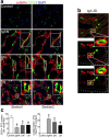
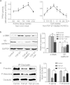
Endothelial-to-mesenchymal transition (EndMT) contributes to the emergence of fibroblasts and plays a significant role in renal interstitial fibrosis. Protein phosphatase 2A (PP2A) is a major serine/threonine protein phosphatase in eukaryotic cells and regulates many signaling pathways. However, the significance of PP2A in EndMT is poorly understood. In present study, the role of PP2A in EndMT was evaluated. We demonstrated that PP2A activated in endothelial cells (EC) during their EndMT phenotype acquisition and in the mouse model of obstructive nephropathy (i.e., UUO). Inhibition of PP2A activity by its specific inhibitor prevented EC undergoing EndMT. Importantly, PP2A activation was dependent on tyrosine nitration at 127 in the catalytic subunit of PP2A (PP2Ac). Our renal-protective strategy was to block tyrosine127 nitration to inhibit PP2A activation by using a mimic peptide derived from PP2Ac conjugating a cell penetrating peptide (CPP: TAT), termed TAT-Y127WT. Pretreatment with TAT-Y127WT was able to prevent TGF-β1-induced EndMT. Administration of the peptide to UUO mice significantly ameliorated renal EndMT level, with preserved density of peritubular capillaries and reduction in extracellular matrix deposition. Taken together, these results suggest that inhibiting PP2Ac nitration using a mimic peptide is a potential preventive strategy for EndMT in renal fibrosis.
Figures


Similar articles
-
Blocking Tyr265 nitration of protein phosphatase 2A attenuates nitrosative stress-induced endothelial dysfunction in renal microvessels.FASEB J. 2019 Mar;33(3):3718-3730. doi: 10.1096/fj.201800885RR. Epub 2018 Dec 6. FASEB J. 2019. PMID: 30521379
-
Blocking protein phosphatase 2A with a peptide protects mice against bleomycin-induced pulmonary fibrosis.Exp Lung Res. 2020 Sep;46(7):234-242. doi: 10.1080/01902148.2020.1774823. Epub 2020 Jun 25. Exp Lung Res. 2020. PMID: 32584210
-
A novel role of kallikrein-related peptidase 8 in the pathogenesis of diabetic cardiac fibrosis.Theranostics. 2021 Feb 20;11(9):4207-4231. doi: 10.7150/thno.48530. eCollection 2021. Theranostics. 2021. PMID: 33754057 Free PMC article.
-
Transcriptional regulation of endothelial-to-mesenchymal transition in cardiac fibrosis: role of myocardin-related transcription factor A and activating transcription factor 3.Can J Physiol Pharmacol. 2017 Oct;95(10):1263-1270. doi: 10.1139/cjpp-2016-0634. Epub 2017 Jul 7. Can J Physiol Pharmacol. 2017. PMID: 28686848 Review.
-
MicroRNAs in kidney fibrosis and diabetic nephropathy: roles on EMT and EndMT.Biomed Res Int. 2013;2013:125469. doi: 10.1155/2013/125469. Epub 2013 Sep 8. Biomed Res Int. 2013. PMID: 24089659 Free PMC article. Review.
Cited by
-
Endothelial to Mesenchymal Transition: Role in Physiology and in the Pathogenesis of Human Diseases.Physiol Rev. 2019 Apr 1;99(2):1281-1324. doi: 10.1152/physrev.00021.2018. Physiol Rev. 2019. PMID: 30864875 Free PMC article. Review.
-
Epoxyeicosatrienoic acid activation moderates endothelial mesenchymal transition to reduce obstructive nephropathy.Kidney Res Clin Pract. 2017 Dec;36(4):299-301. doi: 10.23876/j.krcp.2017.36.4.299. Epub 2017 Dec 31. Kidney Res Clin Pract. 2017. PMID: 29285421 Free PMC article. No abstract available.
-
Puerarin Protects against Cardiac Fibrosis Associated with the Inhibition of TGF-β1/Smad2-Mediated Endothelial-to-Mesenchymal Transition.PPAR Res. 2017;2017:2647129. doi: 10.1155/2017/2647129. Epub 2017 May 30. PPAR Res. 2017. PMID: 28638404 Free PMC article.
-
The therapeutic potential of targeting the endothelial-to-mesenchymal transition.Angiogenesis. 2019 Feb;22(1):3-13. doi: 10.1007/s10456-018-9639-0. Epub 2018 Aug 3. Angiogenesis. 2019. PMID: 30076548 Free PMC article. Review.
-
Redox inhibition of protein phosphatase PP2A: Potential implications in oncogenesis and its progression.Redox Biol. 2019 Oct;27:101105. doi: 10.1016/j.redox.2019.101105. Epub 2019 Jan 14. Redox Biol. 2019. PMID: 30686777 Free PMC article. Review.
References
-
- Zeisberg M. & Neilson E. G. Mechanisms of tubulointerstitial fibrosis. J Am Soc Nephrol 21, 1819–1834 (2010). - PubMed
-
- Ohashi R. Peritubular Capillary regression during the progression of experimental obstructive nephropathy. J Am Soc Nephrol 13, 1795–1805 (2002). - PubMed
-
- Zeisberg E. M., Potenta S., Xie L., Zeisberg M. & Kalluri R. Discovery of Endothelial to Mesenchymal Transition as a Source for Carcinoma-Associated Fibroblasts. Cancer Res 67, 10123–10128 (2007). - PubMed
Publication types
MeSH terms
Substances
LinkOut - more resources
Full Text Sources
Other Literature Sources

