

Broadly Neutralizing Antibodies against HIV-1 As a Novel Aspect of the Immune Response
- PMID: 26798488
- PMCID: PMC4717246
Broadly Neutralizing Antibodies against HIV-1 As a Novel Aspect of the Immune Response
Abstract
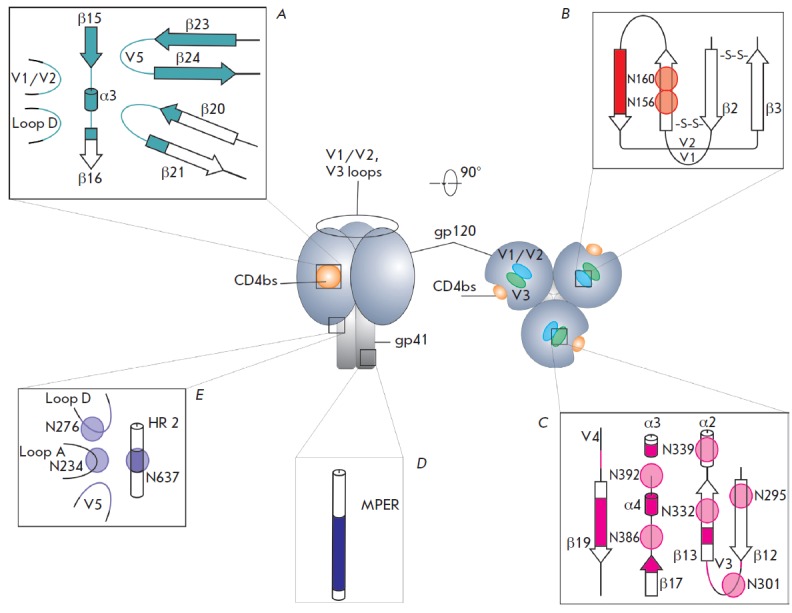
The human immunodeficiency virus-1 (HIV-1) has the ability to evade the adaptive immune response due to high mutation rates. Soon after the discovery of HIV-1, it was originally proposed that neutralizing of antibodies to the virus occurs rarely or cannot be elicited at all. In the 1990s, there appeared reports that sera of select HIV-1-infected individuals contained antibodies capable of neutralizing different virus subtypes. Such antibodies were named broadly neutralizing antibodies (bNAbs). Since 2009, the development of new cell technologies has intensified research efforts directed at identifying new bNAbs with a neutralization potency of over 90% of primary HIV-1 isolates. These antibodies have unique characteristics which include high levels of somatic mutations and unusually long variable loops that penetrate through the glycan shield of HIV-1 Env to contact the protein surface. In this review, we will attempt to summarize the latest data on bNAbs against HIV-1 in terms of their interactions with the sites of vulnerability on HIV-1 glycoproteins.
Keywords: Broadly neutralizing antibodies; HIV-1; bNAbs; gp120; gp41.
Figures


Similar articles
-
Hyperglycosylated stable core immunogens designed to present the CD4 binding site are preferentially recognized by broadly neutralizing antibodies.J Virol. 2014 Dec;88(24):14002-16. doi: 10.1128/JVI.02614-14. Epub 2014 Sep 24. J Virol. 2014. PMID: 25253346 Free PMC article.
-
A Rare Mutation in an Infant-Derived HIV-1 Envelope Glycoprotein Alters Interprotomer Stability and Susceptibility to Broadly Neutralizing Antibodies Targeting the Trimer Apex.J Virol. 2020 Sep 15;94(19):e00814-20. doi: 10.1128/JVI.00814-20. Print 2020 Sep 15. J Virol. 2020. PMID: 32669335 Free PMC article.
-
Development of Broadly Neutralizing Antibodies and Their Mapping by Monomeric gp120 in Human Immunodeficiency Virus Type 1-Infected Humans and Simian-Human Immunodeficiency Virus SHIVSF162P3N-Infected Macaques.J Virol. 2016 Mar 28;90(8):4017-4031. doi: 10.1128/JVI.02898-15. Print 2016 Apr. J Virol. 2016. PMID: 26842476 Free PMC article.
-
Strategies for induction of HIV-1 envelope-reactive broadly neutralizing antibodies.J Int AIDS Soc. 2021 Nov;24 Suppl 7(Suppl 7):e25831. doi: 10.1002/jia2.25831. J Int AIDS Soc. 2021. PMID: 34806332 Free PMC article. Review.
-
Development of broadly neutralizing antibodies in HIV-1 infected elite neutralizers.Retrovirology. 2018 Sep 5;15(1):61. doi: 10.1186/s12977-018-0443-0. Retrovirology. 2018. PMID: 30185183 Free PMC article. Review.
Cited by
-
Signal peptide exchange alters HIV-1 envelope antigenicity and immunogenicity.Front Immunol. 2024 Sep 24;15:1476924. doi: 10.3389/fimmu.2024.1476924. eCollection 2024. Front Immunol. 2024. PMID: 39380992 Free PMC article.
-
Glycosylation of the core of the HIV-1 envelope subunit protein gp120 is not required for native trimer formation or viral infectivity.J Biol Chem. 2017 Jun 16;292(24):10197-10219. doi: 10.1074/jbc.M117.788919. Epub 2017 Apr 26. J Biol Chem. 2017. PMID: 28446609 Free PMC article.
-
Evolution of Neutralization Response in HIV-1 Subtype C-Infected Individuals Exhibiting Broad Cross-Clade Neutralization of HIV-1 Strains.Front Immunol. 2018 Mar 27;9:618. doi: 10.3389/fimmu.2018.00618. eCollection 2018. Front Immunol. 2018. PMID: 29662494 Free PMC article.
-
Mapping of Neutralizing Antibody Epitopes on the Envelope of Viruses Obtained from Plasma Samples Exhibiting Broad Cross-Clade Neutralization Potential Against HIV-1.AIDS Res Hum Retroviruses. 2019 Feb;35(2):169-184. doi: 10.1089/AID.2018.0224. Epub 2018 Dec 20. AIDS Res Hum Retroviruses. 2019. PMID: 30328700 Free PMC article.
-
Employing Broadly Neutralizing Antibodies as a Human Immunodeficiency Virus Prophylactic & Therapeutic Application.Front Immunol. 2021 Jul 20;12:697683. doi: 10.3389/fimmu.2021.697683. eCollection 2021. Front Immunol. 2021. PMID: 34354709 Free PMC article.
References
-
- Deyev S.M., Lebedenko E.N., Petrovskaya L.E.E., Dolgikh D.A., Gabibov A.G., Kirpichnikov M. P., Russian Chemical Reviews. 2015;84(1):1–26.
LinkOut - more resources
Full Text Sources
Other Literature Sources
Research Materials