. 2016 Jun;159(6):585-97.
doi: 10.1093/jb/mvw002. Epub 2016 Jan 19.

Protein purification and cloning of diacylglycerol lipase from rat brain

Affiliations

Protein purification and cloning of diacylglycerol lipase from rat brain

Chizu Aso et al. J Biochem. 2016 Jun.

Abstract

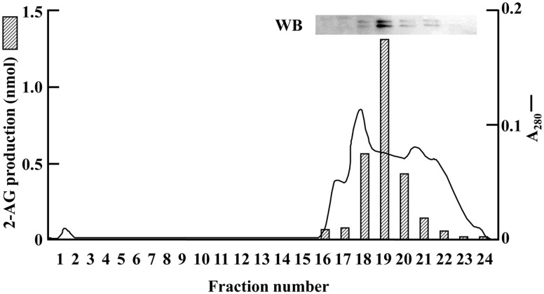
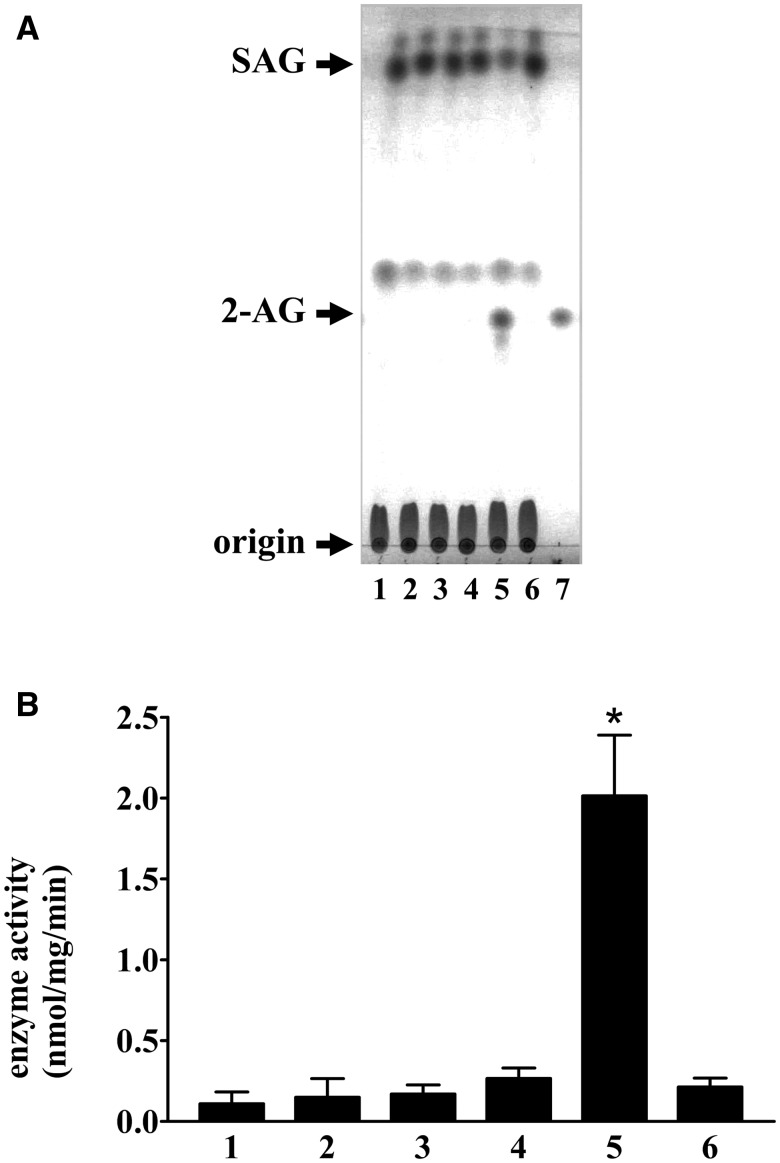
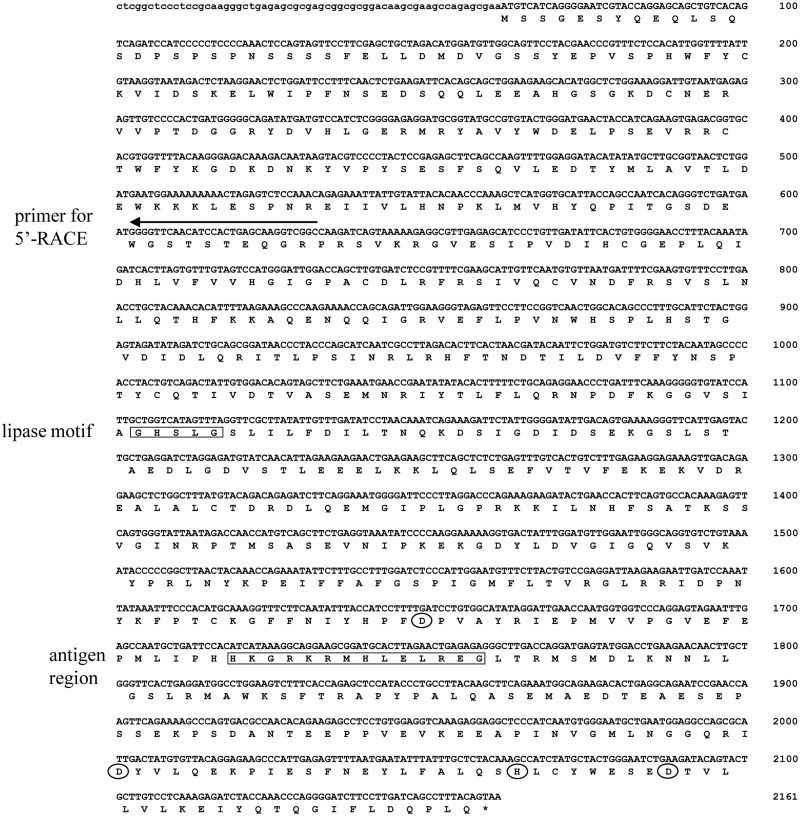
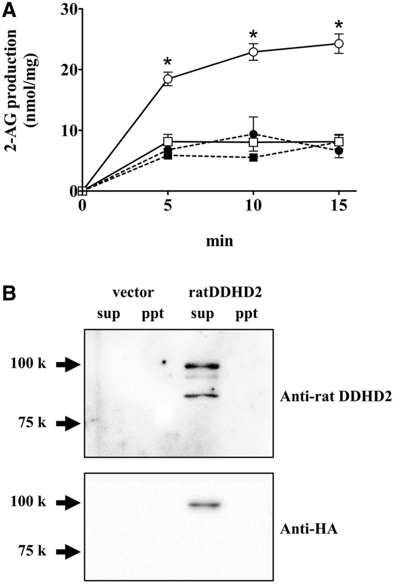
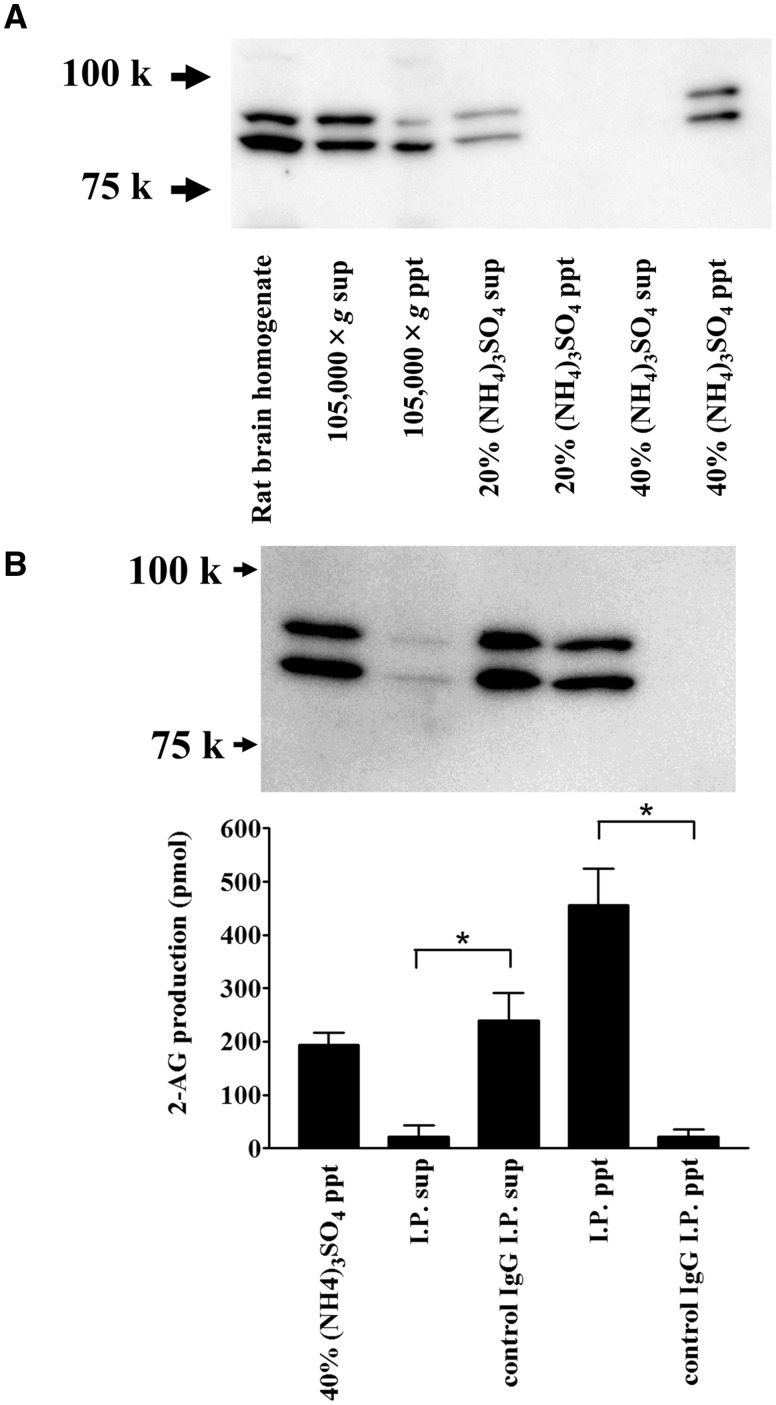
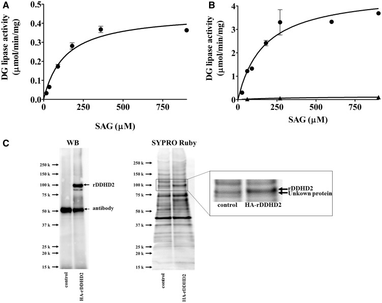
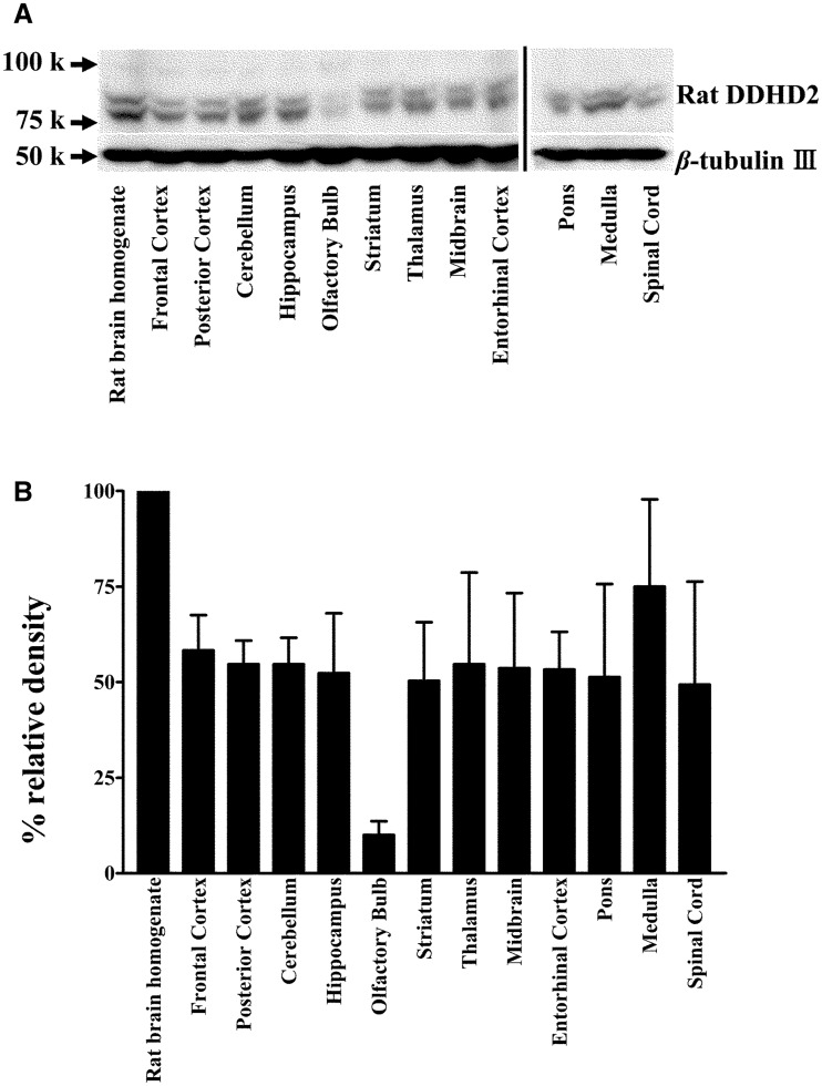
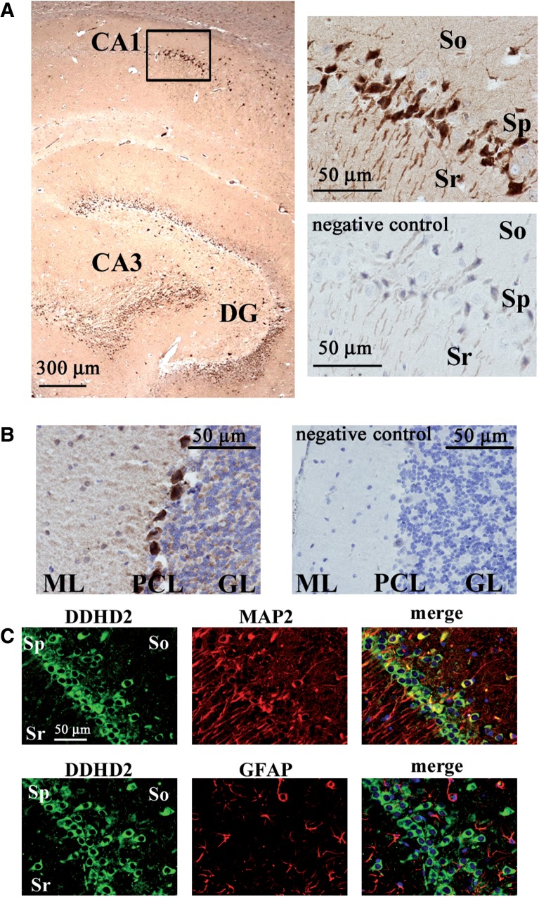
Diacylglycerol (DG) lipase, which hydrolyses 1-stearoyl-2-arachidonyl-sn-glycerol to produce an endocannabinoid, 2-arachidonoylglycerol, was purified from the soluble fraction of rat brain lysates. DG lipase was purified about 1,200-fold by a sequential column chromatographic procedure. Among proteins identified by mass spectrometry analysis in the partially purified DG lipase sample, only DDHD domain containing two (DDHD2), which was formerly regarded as a phospholipase A1, exhibited significant DG lipase activity. Rat DDHD2 expressed in Chinese hamster ovary cells showed similar enzymatic properties to partially purified DG lipase from rat brain. The source of DG lipase activity in rat brain was immunoprecipitated using anti-DDHD2 antibody. Thus, we concluded that the DG lipase activity in the soluble fraction of rat brain is derived from DDHD2. DDHD2 is distributed widely in the rat brain. Immunohistochemical analysis revealed that DDHD2 is expressed in hippocampal neurons, but not in glia.

Keywords: 2-arachidonoylglycerol; diacylglycerol; endocannabinoid; lipase; phospholipase.


Figures

Fig. 1
Fig. 1
Anion exchange column chromatography for the purification of DG lipase from rat brain. A typical elution profile of POROS HQ column chromatography in the purification step of rat brain DG lipase is shown. Western blot of fractions 16–23 using anti-rat DDHD2 antibody is also shown. Absorbance at 280 nm is shown as a solid line. The enzyme activity (hatched bars) of each fraction was determined as described in Materials and Methods. Data are representative of three independent experiments.
Fig. 2
Fig. 2
Screening of DG lipase activity. (A) DG lipase activity in the soluble fraction of CHO cells expressing the candidate proteins (100 µg) was measured using 40 µM cold SAG as a substrate. The assay mixture was incubated at 37°C for 30 min. Lipids were separated using TLC and visualized using iodine vapour. Lane 1: acyl-peptide hydrolase, lane 2: anti-depressant-related protein ARG123, lane 3: dihydropyrimidinase-like 2, lane 4: TUC-4b, lane 5: human DDHD2, lane 6: vector control, lane 7: 5 nmol 2-AG. (B) The amount of 2-AG shown in panel (A) was quantified using ImageJ software. Data are representative of three independent experiments. Data are the means of triplicate measurements. Error bars represent standard errors of the mean. *P < 0.05 for human DDHD2 versus vector control.
Fig. 3
Fig. 3
Nucleic acid sequence of rat DDHD2 cDNA. The arrow indicates the nucleotides for 5′-RACE. The region of the antigen peptide used for the generation of the anti-DDHD2 antibody is enclosed by a square. The lipase motif is also enclosed by a square. The four amino acids contained in the DDHD domain are surrounded by circles.
Fig. 4
Fig. 4
DG lipase activity of rat DDHD2 expressed in CHO cells. (A) The DG lipase activity was measured using 9 µM [14C]SAG as a substrate. The protein samples used for the enzyme assay were the soluble (open circles) and the membrane (closed circles) fractions of CHO cells expressing HA-rDDHD2, and were the soluble (open squares) and the membrane (closed squares) fraction of vector-transfected CHO cells. (B) The soluble fraction (25 µg) and membrane fraction (25 µg) from the CHO cells were used for western blotting. An anti-rat DDHD2 antibody (upper panel) and an anti-HA antibody (lower panel) were used. Data are the means of triplicate measurements. Error bars represent standard error of the mean. Data are representative of three independent experiments. *P < 0.01 for the soluble fraction of CHO cells expressing HA-rDDHD2 versus the soluble fraction of vector-transfected CHO cells.
Fig. 5
Fig. 5
Ammonium sulphate precipitation and immunoprecipitation of rat brain DDHD2. (A) Ammonium sulphate fractionation of rat brain DDHD2. The soluble fraction of rat brain homogenates (105,000 × g supernatant) was further fractionated by ammonium sulphate precipitation. The same proportion of each sample was loaded onto an SDS-PAGE and analysed by western blotting using an anti-rat DDHD2 antibody. (B) Immunoprecipitation of DDHD2 in rat brain. Forty percent ammonium sulphate precipitate was used for the experiment. Ten micrograms of the anti-DDHD2 or control IgG was used. Aliquots of the samples were loaded onto an SDS-PAGE gel and used for the DG lipase assay. DG lipase activity in the immunoprecipitated beads or in the supernatant was measured. [14C]SAG was used as a substrate. Data are the means of triplicate measurements. Error bars represent standard error of the mean. Data are representative of three independent experiments. *P < 0.01 for the anti-DDHD2 IgG samples versus control IgG samples.
Fig. 6
Fig. 6
Substrate concentration-dependence. (A) Various concentrations of [14C]SAG were incubated with the partially purified rat brain DG lipase from the CHT 5-I column (15 µg per assay). Except for the substrate concentrations, the assay conditions were the same as those for the standard assay. (B) Various concentrations of [14C]SAG were incubated with HA-rDDHD2 (closed circles) prepared using anti-rat DDHD2 antibody. Control (closed triangles) means the DG lipase activity using the same aliquot of protein sample obtained from vector transfected cells. (C) Control sample and HA-rDDHD2 sample were analysed by western blotting using anti-rat DDHD2 antibody and SYPRO Ruby staining following SDS-PAGE. The rectangular area in SYPRO Ruby staining is magnified in the right panel. Data are the means of triplicate measurements. Error bars represent standard errors of the mean. Data are representative of three independent experiments.
Fig. 7
Fig. 7
Effects of inhibitors on the partially purified rat brain DG lipase and HA-rDDHD2 expressed in CHO cells. DG lipase activity was measured in the presence of various concentrations of RHC80267 (closed circles for rat brain DG lipase, open circles for CHO-rDDHD2) and THL (closed squares for rat brain DG lipase, open squares for CHO-rDDHD2). The lipase inhibitors were added to the mixture before preincubation. Data are the means of triplicate measurements. Error bars represent standard error of the mean. Data are representative of three independent experiments.
Fig. 8
Fig. 8
pH dependency of the partially purified DG lipase. DG lipase activity in the active fractions (20 µg per assay) from the CHT 5-I column was measured at various pH levels. Buffers used for the experiments were MOPS-NaOH (closed triangles), Tris-HCl (open circles) and glycine-NaOH (closed circles). Except for the buffer used, assay conditions were the same as those for the standard assay. Data are the means of triplicate measurements. Error bars represent standard errors of the mean. Data are representative of three independent experiments.
Fig. 9
Fig. 9
Distribution of DDHD2 in rat brain. (A) Fifty micrograms of protein of homogenates from normal rat brain region tissues was loaded onto each lane. The expression of DDHD2 was analysed by western blotting using anti-rat DDHD2 antibody. The same membrane was re-probed with an anti-β-tubulin III antibody. (B) Histograms showing relative DDHD2 ratios against β-tubulin. The DDHD2 expression was quantified using ImageJ software. Data are the means of triplicate measurements. Error bars represent standard errors of the mean. Data are representative of three independent experiments.
Fig. 10
Fig. 10
Immunostaining of DDHD2 in rat brain. (A) The distribution of DDHD2 is shown using a sagittal section of the hippocampus in a rat brain. DDHD2 was stained by the anti-DDHD2 antibody with DAB (brown). Nuclei were stained by with hematoxylin (blue). The rectangular area is magnified in the upper right panel. The lower right panel is from a negative control experiment using the excessive peptides used for the immunization (16 µg/ml) to absorb primary antibody. (B) The distribution of DDHD2 is shown using a sagittal section of the cerebellum in a rat brain. The right panel is from a negative control experiment. ML, molecular layer; PCL, Purkinje cell layer; GL, granule cell layer. (C) The distribution of DDHD2 is shown using a coronal section of a rat brain. Double staining of DDHD2 and MAP2 (upper panels) or DDHD2 and GFAP (lower panels) at the CA1 region in the hippocampus shows that DDHD2 is localized in the cell body of hippocampal neurons. Sr, stratum radiatum; So, stratum oriens; Sp, stratum pyramidale. Data are representative of three independent experiments.

Similar articles

Cited by

References

    1. Basavarajappa B.S. (2007) Critical enzymes involved in endocannabinoid metabolism. Protein Pept. Lett. 14, 237–246 - PMC - PubMed
    1. Wang J., Ueda N. (2008) Role of the endocannabinoid system in metabolic control. Curr. Opin. Nephrol. Hypertens. 17, 1–10 - PubMed
    1. Sugiura T., Kishimoto S., Oka S., Gokoh M. (2006) Biochemistry, pharmacology and physiology of 2-arachidonoylglycerol, an endogenous cannabinoid receptor ligand. Prog. Lipid Res. 45, 405–446 - PubMed
    1. Matsuda L.A., Bonner T.I., Lolait S.J. (1993) Localization of cannabinoid receptor mRNA in rat brain. J. Comp. Neurol. 327, 535–550 - PubMed
    1. Tsou K., Brown S., Sanudo-Pena M.C., Mackie K., Walker J.M. (1998) Immunohistochemical distribution of cannabinoid CB1 receptors in the rat central nervous system. Neuroscience 83, 393–411 - PubMed

MeSH terms