Protein unfolding as a switch from self-recognition to high-affinity client binding
- PMID: 26787517
- PMCID: PMC4735815
- DOI: 10.1038/ncomms10357
Protein unfolding as a switch from self-recognition to high-affinity client binding
Abstract
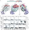
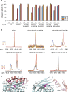
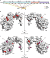
Stress-specific activation of the chaperone Hsp33 requires the unfolding of a central linker region. This activation mechanism suggests an intriguing functional relationship between the chaperone's own partial unfolding and its ability to bind other partially folded client proteins. However, identifying where Hsp33 binds its clients has remained a major gap in our understanding of Hsp33's working mechanism. By using site-specific Fluorine-19 nuclear magnetic resonance experiments guided by in vivo crosslinking studies, we now reveal that the partial unfolding of Hsp33's linker region facilitates client binding to an amphipathic docking surface on Hsp33. Furthermore, our results provide experimental evidence for the direct involvement of conditionally disordered regions in unfolded protein binding. The observed structural similarities between Hsp33's own metastable linker region and client proteins present a possible model for how Hsp33 uses protein unfolding as a switch from self-recognition to high-affinity client binding.
Figures


Similar articles
-
Order out of disorder: working cycle of an intrinsically unfolded chaperone.Cell. 2012 Mar 2;148(5):947-57. doi: 10.1016/j.cell.2012.01.045. Cell. 2012. PMID: 22385960 Free PMC article.
-
A Role of Metastable Regions and Their Connectivity in the Inactivation of a Redox-Regulated Chaperone and Its Inter-Chaperone Crosstalk.Antioxid Redox Signal. 2017 Nov 20;27(15):1252-1267. doi: 10.1089/ars.2016.6900. Epub 2017 Apr 10. Antioxid Redox Signal. 2017. PMID: 28394178
-
Activation of the redox-regulated molecular chaperone Hsp33--a two-step mechanism.Structure. 2001 May 9;9(5):377-87. doi: 10.1016/s0969-2126(01)00599-8. Structure. 2001. PMID: 11377198
-
Redox-regulated molecular chaperones.Cell Mol Life Sci. 2002 Oct;59(10):1624-31. doi: 10.1007/pl00012489. Cell Mol Life Sci. 2002. PMID: 12475172 Free PMC article. Review.
-
Novel insights into the mechanism of chaperone-assisted protein disaggregation.Biol Chem. 2005 Aug;386(8):739-44. doi: 10.1515/BC.2005.086. Biol Chem. 2005. PMID: 16201868 Review.
Cited by
-
The Cys Sense: Thiol Redox Switches Mediate Life Cycles of Cellular Proteins.Biomolecules. 2021 Mar 22;11(3):469. doi: 10.3390/biom11030469. Biomolecules. 2021. PMID: 33809923 Free PMC article. Review.
-
Local unfolding of the HSP27 monomer regulates chaperone activity.Nat Commun. 2019 Mar 6;10(1):1068. doi: 10.1038/s41467-019-08557-8. Nat Commun. 2019. PMID: 30842409 Free PMC article.
-
Mechanical properties of BiP protein determined by nano-rheology.Protein Sci. 2018 Aug;27(8):1418-1426. doi: 10.1002/pro.3432. Protein Sci. 2018. PMID: 29696702 Free PMC article.
-
Stress-Activated Chaperones: A First Line of Defense.Trends Biochem Sci. 2017 Nov;42(11):899-913. doi: 10.1016/j.tibs.2017.08.006. Epub 2017 Sep 8. Trends Biochem Sci. 2017. PMID: 28893460 Free PMC article. Review.
-
Maintaining a Healthy Proteome during Oxidative Stress.Mol Cell. 2018 Jan 18;69(2):203-213. doi: 10.1016/j.molcel.2017.12.021. Mol Cell. 2018. PMID: 29351842 Free PMC article. Review.
References
-
- Hsu W. L. et al. Intrinsic protein disorder and protein-protein interactions. Pac. Symp. Biocomput. 116–127 (2012) . - PubMed
-
- Dunker A. K., Cortese M. S., Romero P., Iakoucheva L. M. & Uversky V. N. Flexible nets. The roles of intrinsic disorder in protein interaction networks. FEBS J. 272, 5129–5148 (2005) . - PubMed
Publication types
MeSH terms
Substances
Grants and funding
LinkOut - more resources
Full Text Sources
Other Literature Sources
Molecular Biology Databases
Miscellaneous

