. 2016 Jan 19;44(1):73-87.
doi: 10.1016/j.immuni.2015.11.011. Epub 2016 Jan 5.

The Neutrophil Btk Signalosome Regulates Integrin Activation during Sterile Inflammation

Affiliations

The Neutrophil Btk Signalosome Regulates Integrin Activation during Sterile Inflammation

Stephanie Volmering et al. Immunity. .

Abstract

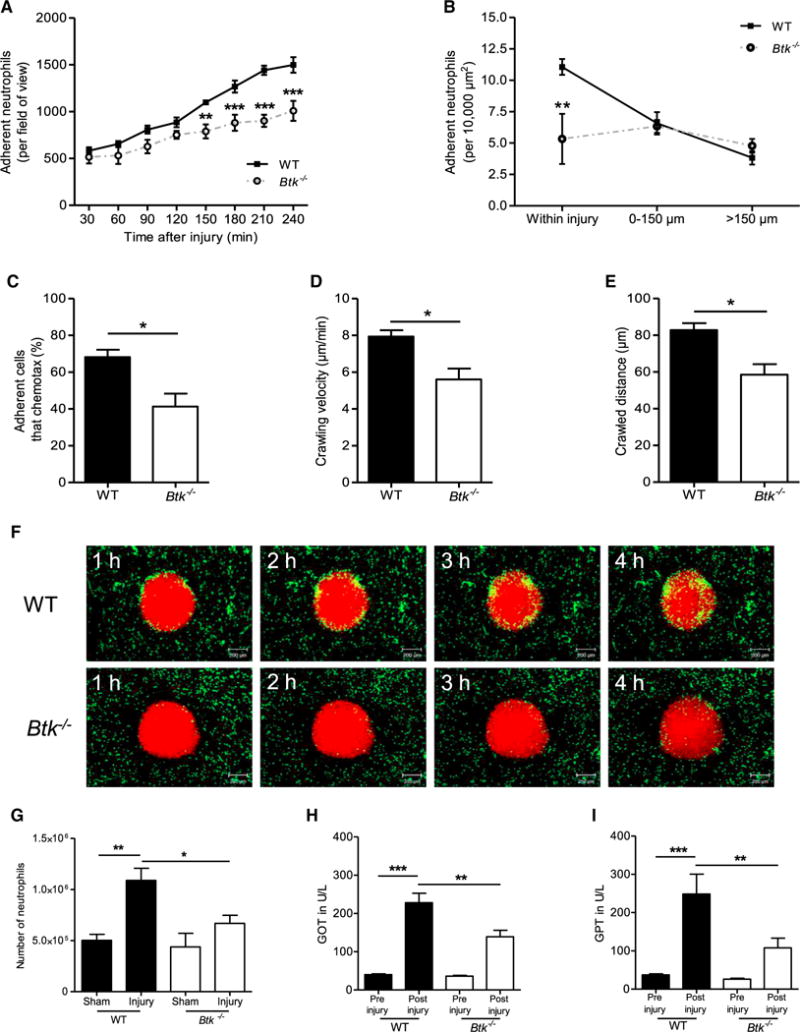
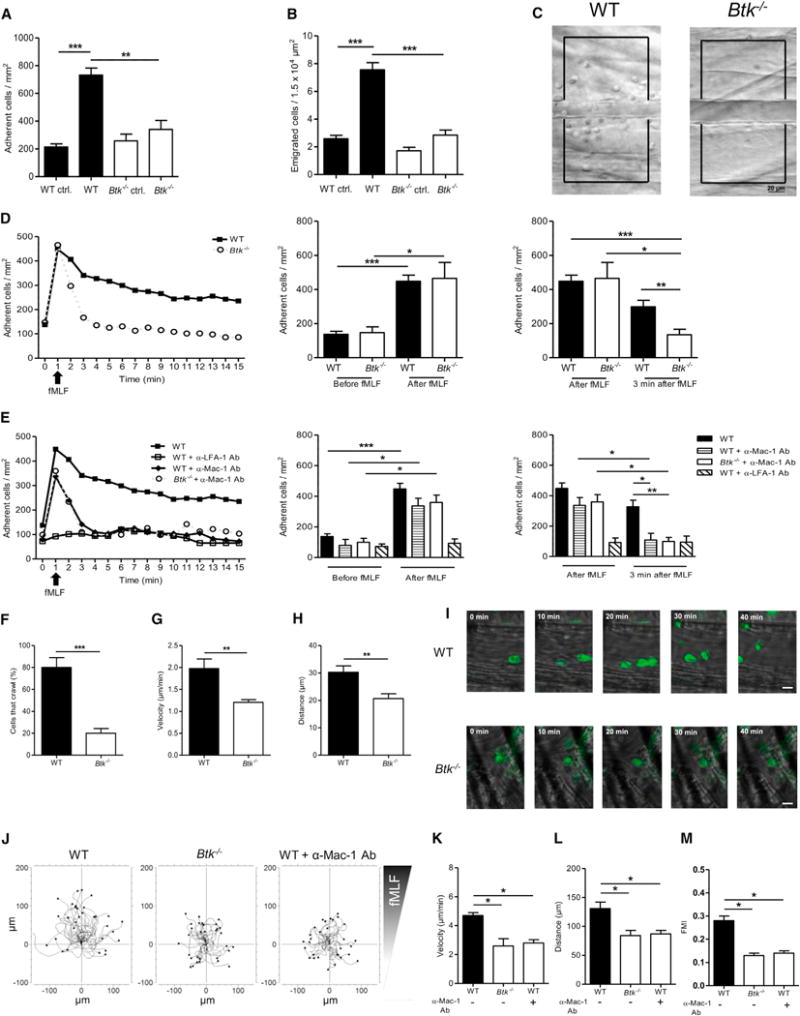
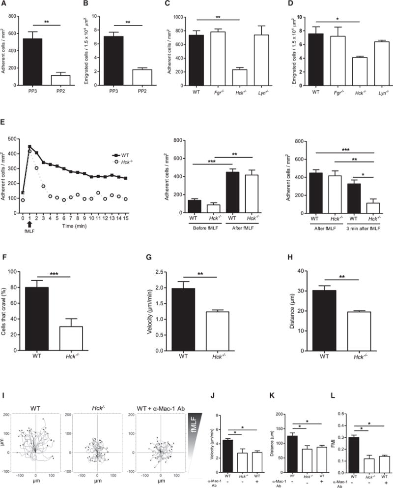
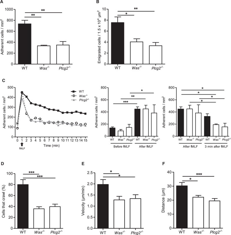
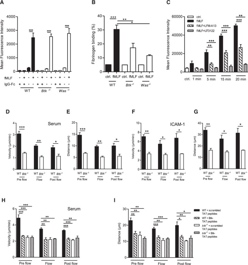
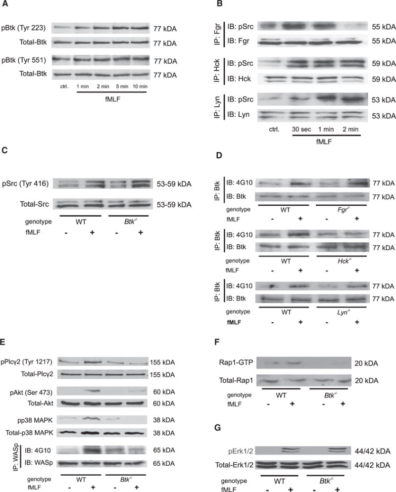
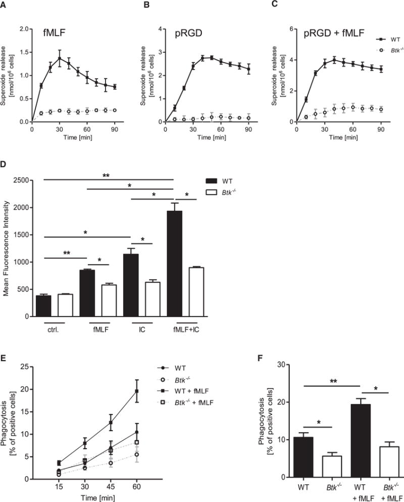
Neutrophils are recruited from the blood to sites of sterile inflammation, where they are involved in wound healing but can also cause tissue damage. During sterile inflammation, necrotic cells release pro-inflammatory molecules including formylated peptides. However, the signaling pathway triggered by formylated peptides to integrin activation and leukocyte recruitment is unknown. By using spinning-disk confocal intravital microscopy, we examined the molecular mechanisms of leukocyte recruitment to sites of focal hepatic necrosis in vivo. We demonstrated that the Bruton's tyrosine kinase (Btk) was required for multiple Mac-1 activation events involved in neutrophil recruitment and functions during sterile inflammation triggered by fMLF. The Src family kinase Hck, Wiskott-Aldrich-syndrome protein, and phospholipase Cγ2 were also involved in this pathway required for fMLF-triggered Mac-1 activation and neutrophil recruitment. Thus, we have identified a neutrophil Btk signalosome that is involved in a signaling pathway triggered by formylated peptides leading to the selective activation of Mac-1 and neutrophil recruitment during sterile inflammation.

Keywords: Btk; Src family kinases; WASp; fMLF; integrin; phospholipase C; signaling.


Figures

Figure 1
Figure 1. Btk Is Required for Neutrophil Recruitment during Sterile Inflammation Induced by Focal Hepatic Necrosis
(A) Number of adherent neutrophils per field of view in WT and Btk−/− mice in response to focal hepatic necrosis for all indicated time points (30–240 min). (B) Number of adherent neutrophils per 10,000 μm2 within indicated regions around foci of necrosis 4 hr after sterile injury in WT and Btk−/− mice. (C–E) Percentage of neutrophils that chemotax toward the injury site (C), neutrophil crawling velocity (D), and crawled distance 2.5 hr after injury (E). (F) Representative time-lapse images from SD-IVM demonstrating the recruitment of neutrophils (green) to the injury area (red, propidium iodide) in WT and Btk−/− mice. Scale bars represent 200 μm. (G) Number of Gr-1-labeled neutrophils per liver derived from WT or Btk−/− mice normalized to the weight of the liver 4 hr after focal hepatic necrosis or sham surgery as determined by flow cytometry. (H and I) Levels of GOT (H) and GPT (I) in U/l in serum before and 4 hr after liver injury in WT and Btk−/− mice. Depicted are mean + SEM; n ≥ 3 individual mice/group; *p < 0.05; **p < 0.01; ***p < 0.001. See also Figures S1 and S7 and Movies S1 and S2.
Figure 2
Figure 2. Btk Is Required for fMLF-Mediated Neutrophil Extravasation in the Murine Cremaster Muscle and Chemotaxis In Vitro
(A–H) Intravital microscopy of postcapillary venules in the murine cremaster was performed in WT and Btk−/− mice. (A and B) Number of adherent cells per mm2 (A) and number of transmigrated cells per 1.5 × 104 μm2 (B) 4 hr after intrascrotal fMLF or vehicle injection. (C) Representative images of inflamed WT (left) and Btk−/− (right) cremasteric venules visualized by reflected light oblique transillumination microscopy. Scale bar represents 20 μm. (D and E) GPCR-induced arrest of neutrophils in postcapillary venules of WT (filled square) and Btk−/− (open circle) mice after fMLF injection (i.v.) (D) or after injection of blocking antibodies against LFA-1 (open square) or Mac-1 (filled diamond) prior to fMLF injection (E). Respective statistics are presented in related bar graphs. (F–H) Intravascular crawling of Gr-1-labeled neutrophils during superfusion with fMLF. Percentage of adherent cells that crawled (F), crawling velocity (G), and crawled distance (H). (I) Representative images of extravasated WT and Btk−/−. Scale bars represent 10 μm. (J) Representative trajectory plots (30 cells from 3 independent experiments) of chemotaxing WT and Btk−/− neutrophils toward indicated fMLF gradient in vitro. (K–M) Velocity (K), migration distance (L), and forward migration index (FMI) (M) of WT and Btk−/− neutrophils. Depicted are mean + SEM; n ≥ 3 individual mice/group; *p < 0.05; **p < 0.01; ***p < 0.001 for all panels except (C), (I), and (J). See also Figures S2, S3, and S7 and Movies S3 and S4.
Figure 3
Figure 3. Hck, but Not Other Src Family Kinases, Is Required for fMLF-Mediated Neutrophil Recruitment
(A–H) Intravital microscopy of murine cremaster muscle venules after treatment with fMLF for 4 hr in WT mice after i.v. injection of the SFK inhibitor (PP2) or the inactive control (PP3) (A, B) and in WT, Fgr−/−, Hck−/−, and Lyn−/− (C, D) mice. (A–D) Number of adherent cells per mm2 (A, C) and number of transmigrated cells per 1.5 × 104 μm2 (B, D) 4 hr after intrascrotal injection of fMLF. (E) GPCR-induced arrest of neutrophils in postcapillary venules of WT (filled square) and Hck−/− (open circle) mice after fMLF injection (i.v.). Respective statistics are presented in related bar graphs. (F–H) Intravascular crawling of Gr-1-labeled neutrophils in WT and Hck−/− mice during superfusion with fMLF. Percentage of adherent cells that crawled (F), crawling velocity (G), and crawled distance (H). (I) Representative trajectory plots (30 cells from 3 independent experiments) of chemotaxing WT and Hck−/− neutrophils toward indicated fMLF gradient in vitro. (J–L) Velocity (J), migration distance (K), and forward migration index (FMI) (L) of WT and Hck−/− neutrophils. Depicted are mean + SEM; n ≥ 3 individual mice/group; *p < 0.05; **p < 0.01; ***p < 0.001 for all panels except (I). See also Figures S3 and S7 and Movies S3 and S4.
Figure 4
Figure 4. WASp and PLCγ2 Are Required for fMLF-Mediated Neutrophil Recruitment
Intravital microscopy of postcapillary venules in the murine cremaster was performed in WT, Was−/−, and Plcg2−/− mice. (A and B) Number of adherent cells per mm2 (A) and number of transmigrated cells per 1.5 × 104 μm2 (B) 4 hr after intrascrotal fMLF injection. (C) GPCR-induced arrest of neutrophils in postcapillary venules of WT (filled square), Was−/− (open circle), and Plcg2−/− (filled triangle) after fMLF injection (i.v.). Respective statistics are presented in related bar graphs. (D–F) Intravascular crawling of Gr-1-labeled neutrophils in WT, Was−/−, and Plcg2−/− mice during superfusion with fMLF. Percentage of adherent cells that crawled (D), crawling velocity (E), and crawled distance (F). Depicted are mean + SEM; n ≥ 3 individual mice/group; *p < 0.05; **p < 0.01; ***p < 0.001. See also Figure S7.
Figure 5
Figure 5. Crosstalk between Btk and WASp Is Required for fMLF-Mediated Mac-1, but Not LFA-1, Activation
(A) LFA-1 binding of ICAM-1 by unstimulated and fMLF-stimulated WT, Btk−/−, and Was−/− neutrophils and respective IgG-Fc control. (B) Mac-1-dependent fibrinogen binding in unstimulated and fMLF-stimulated WT, Btk−/−, and Was−/− neutrophils. (C) Binding of Mac-1 on isolated human neutrophils by an activation-dependent reporter antibody for Mac-1 (CBRM1/5) with or without fMLF stimulation for indicated time points and with or without preincubation with the Btk inhibitor LFM-A13 and the phospholipases C inhibitor U73122. (D–I) Parallel plate flow chamber crawling assay of WT and Btk−/− neutrophils stimulated with fMLF. Crawling velocities (D, F, H) and accumulated crawled distance (E, G, I) before (pre flow), during (flow), and after (post flow) applying flow (2 dyn/cm2, 5 min) of untreated WT and Btk−/− neutrophils on murine blood serum (D, E) or ICAM-1 (F, G) and scrambled or Btk-TAT peptide-pretreated cells on murine blood serum (H, I). Depicted are mean + SEM of n ≥ 3 independent performed experiments; *p < 0.05; **p < 0.01; ***p < 0.001. See also Figures S4 and S7.
Figure 6
Figure 6. Btk Regulates fMLF-Triggered Intracellular Signaling
(A) Lysates of WT neutrophils were immunoblotted with a p-Btk (Tyr223 or Tyr551) antibody or total-Btk antibody (n = 4). (B) Lysates of WT neutrophils were immunoprecipitated (IP) with an antibody against Fgr, Hck, or Lyn followed by immunoblotting with a p-Src (Tyr416) antibody or total Fgr, Hck, or Lyn antibodies. (C) Lysates of WT and Btk−/− neutrophils were immunoblotted with a p-Src (Tyr416) antibody or total-Src antibody. (D) Lysates of WT, Fgr−/−, Hck−/−, and Lyn−/− neutrophils were immunoprecipitated (IP) with an antibody against Btk followed by immunoblotting with a general phosphotyrosine (4G10) antibody or total Btk antibody. (E) Lysates of WT and Btk−/− neutrophils were immunoblotted or immunoprecipitated, demonstrating the phosphorylation of PLCγ2, Akt, p38 MAPK, or WASp. Total lysates were immunoblotted with antibodies to p-PLCγ2 (Tyr1217), total-PLCγ2, p-Akt (Ser473), total-Akt, p-p38 MAPK, or total-p38 MAPK. Lysates were immunoprecipitated with a general phosphotyrosine (4G10) antibody or total-WASp antibody. (F) GTP-bound Rap1 (Rap1-GTP) was precipitated from whole cell lysates using immobilized GST effector fusion protein. Lysates were immunoblotted with an antibody against Rap1. (G) Lysates of WT and Btk−/− neutrophils were immunoblotted with a p-p44 and 42 MAPK (Erk1 and 2) antibody or total-p44 and 42 MAPK (Erk1 and 2) antibody. Depicted are representative immunoblots of n ≥ 3 independent performed experiments. See also Figures S5–S7.
Figure 7
Figure 7. Btk Is Involved in Integrin-Mediated Outside-In Signaling and FcRγ-Mediated Functions
(A–C) Superoxide release of WT and Btk−/− neutrophils stimulated with 3 μM fMLF (A), plated on a polyvalent integrin ligand-coated surface (pRGD) without stimulus (B) or with 3 μM fMLF (C). Control values were subtracted from stimulated values. (D) Quantitative binding of fibrinogen-coated fluorescent beads to unstimulated WT and Btk−/− neutrophils, stimulated with 1 μM fMLF, 10 μg/ml IgG immune complexes, or a combination of both as determined by flow cytometry. (E) Phagocytosis of IgG-coated fluorescent beads by WT and Btk−/− neutrophils incubated at 37°C with or without 1 μM fMLF for indicated time points. (F) Respective statistics of phagocytosis after 1 hr. Depicted are mean + SEM of n ≥ 3 independent performed experiments; *p < 0.05; **p < 0.01; ***p < 0.001. See also Figure S7.

Similar articles

Cited by

References

    1. Abram CL, Lowell CA. Convergence of immunoreceptor and integrin signaling. Immunol Rev. 2007;218:29–44. - PubMed
    1. Afar DE, Park H, Howell BW, Rawlings DJ, Cooper J, Witte ON. Regulation of Btk by Src family tyrosine kinases. Mol Cell Biol. 1996;16:3465–3471. - PMC - PubMed
    1. Block H, Zarbock A. The role of the tec kinase Bruton’s tyrosine kinase (Btk) in leukocyte recruitment. Int Rev Immunol. 2012;31:104–118. - PubMed
    1. Cheng G, Ye ZS, Baltimore D. Binding of Bruton’s tyrosine kinase to Fyn, Lyn, or Hck through a Src homology 3 domain-mediated interaction. Proc Natl Acad Sci USA. 1994;91:8152–8155. - PMC - PubMed
    1. Dorward DA, Lucas CD, Chapman GB, Haslett C, Dhaliwal K, Rossi AG. The role of formylated peptides and formyl peptide receptor 1 in governing neutrophil function during acute inflammation. Am J Pathol. 2015;185:1172–1184. - PMC - PubMed

Publication types

Substances